Weekend Course
The Heart in the Right Place
Joint Annual Meeting ISMRM-ESMRMB & ISMRT 31st Annual Meeting • 07-12 May 2022 • London, UK

| Off the Wall | |||
| 13:00 | 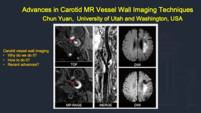 |
Advances in Carotid MR Vessel Wall Imaging Techniques
Chun Yuan
Carotid vessel wall imaging complements angiographic techniques, which are based on measurements of luminal stenosis, has the ability to identify atherosclerotic plaque features associated with increased risk for ischemic events. These features include luminal morphology, fibrous cap rupture, and ulcerations, intraplaque hemorrhage, lipid rich necrotic core, neovasculature and inflammation, and overall plaque burden. There are many imaging techniques developed for vessel wall imaging and quantitative analysis in recent years.This talk will summarize the current expert consensus on carotid vessel wall imaging and introduce recent advances in MRI based techniques.
|
|
| 13:25 | Clinical Applications of 3D MR Vessel Wall Imaging
Huilin Zhao
3D MR vessel wall imaging (3D MR VWI) can characterise vessel wall pathology affecting extra- and intracranial arteries and helps in differentiating vasculopathies. 3D MR VWI has emerged as an invaluable technique for understanding and evaluating cerebrovascular diseases. In this talk, technical considerations of 3D MR VWI are discussed and current applications of 3D MR VWI in cerebrovascular atherosclerosis, aneurysm and dissection are reviewed.
|
||
| It Runs in the Blood | |||
| 13:50 | Exercise Cardiac MR Techniques
Oliver Wieben
Exercise cardiovascular magnetic resonance (CMR) is an emerging technique with great clinical potential. Advances in device design and availability and image acquisition and reconstruction and MR methodology continue to improve the image quality and robustness and enable novel research approaches such as realtime MRI or complex quantitative hemodynamic analysis with 4D Flow MRI under exercise conditions.
|
||
| 14:15 | Clinical Applications of Exercise Cardiac MR Video Unavailable |
||
| 14:40 | Break & Meet the Teachers |
||
| Eat Your Heart Out | |||
| 15:05 | Cardiac MR Spectroscopy: Techniques & Applications
Adrianus Bakermans
Metabolic derangements have been identified in various cardiovascular pathologies, and many metabolic disorders may lead to a cardiac phenotype. Magnetic resonance spectroscopy (MRS) has the unique capability to provide quantitative data on in vivo tissue content of relevant metabolites, and can therefore be of added value to cardiac MRI protocols. Human anatomy and physiology pose substantial challenges to adequately localized MRS of the in vivo heart. This educational will discuss localization strategies for MRS of the in vivo heart, consider aspects for a meaningful quantitative interpretation of acquired spectra, and highlight various applications of proton, phosphorus-31 and carbon-13 MRS.
|
||
| 15:30 | Hyperpolarized Cardiac MR: Opportunities & Challenges
Jessica Bastiaansen
Hyperpolarization methods have enabled metabolic imaging of the heart with unprecedented signal gains. Hyperpolarized metabolic imaging following dissolution DNP made its way to the clinic within 10 years of its discovery. Despite its fast progress, the field relies on the proximity of dedicated hardware and fast imaging approaches to get around the rapid T1 induced signal loss. In this educational talk we will introduce the topic of hyperpolarization and its application to cardiac metabolic imaging. We will compare its strengths and weaknesses with other imaging modalities, and discuss the current challenges and remaining opportunities of this promising field.
|
||
| Young at Heart | |||
| 15:55 | 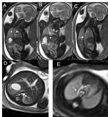 |
Fetal Cardiac MR
Su-Zhen Dong, Ming Zhu
The objective of this presentation is to provide an overview of fetal cardiac MR imaging methods and their applications in congenital heart disease.
|
|
| 16:20 |  |
Motion Management in Pediatric Cardiac MR
Eric Schrauben
Pediatric cardiac patients have high heart rates and fast and often irregular breathing. Aperiodic and sporadic bulk motion is also an issue, especially for very young patients. Mitigation, compensation, or correction of motion is therefore paramount. This educational talk will describe patient preparation, hardware, acquisition, and reconstruction strategies for motion. Simple routine approaches will be discussed, such as: sedation versus general anesthesia and breath-holding versus averaging. More advanced and emerging approaches, which include real-time imaging, resolving cardiorespiratory motion, and the application of machine learning, will also be addressed. Examples from literature and ISMRM will be highlighted to exemplify these approaches.
|
|
 Back to Meeting Home
 Back to the Program-at-a-Glance
The International Society for Magnetic Resonance in Medicine is accredited by the Accreditation Council for Continuing Medical Education to provide continuing medical education for physicians.
 Back to Meeting Home
 Back to the Program-at-a-Glance
 View the Presentation