Weekend Course
MRI Contrast to Measure Vascular Function
Joint Annual Meeting ISMRM-ESMRMB & ISMRT 31st Annual Meeting • 07-12 May 2022 • London, UK

Weekend Course
MRI Contrast to Measure Vascular Function
| Vascular Function: Back to Basics | |||
| 13:00 | MR Angiography: Basics
TBD
|
||
| Vascular Function: New Horizons in Clinical | |||
| 13:25 | MR Angiography: Translational Potential to Combine High-Resolution MRA with single-vessel BOLD, CBV and CBF-velocity fMRI
Xin Yu, David Hike
There are two ongoing challenges when specifying vascular function underlying vascular cognitive impairment in patients. One issue is the size of vessels that altered anatomy and function can be detected in the brain. The other issue is the location of the affected vessels to be detected, e.g. gray matter vs. white matter, or cortical vs. subcortical cerebral vasculature. MR Angiography(MRA) provides the key anatomical vasculature measurement when studying cerebrovascular diseases or impaired cerebral blood flow related to vascular dementia. Here, we will discuss the linkage of high-resolution MRA with single-vessel fMRI to characterize vessel-specific oxygenation(BOLD), cerebral volume, and flow changes.
|
||
| Vascular Function: Back to Basics | |||
| 13:50 |  |
Dynamic Contrast: Basics
Ho-Ling Anthony Liu
Dynamic susceptibility contrast (DSC) and dynamic contrast enhanced (DCE) MRI are two widely used methods in the clinic to evaluate perfusion and microvasculature of the tissue. They acquire dynamic T2*- and T1-weighted images, respectively, before and after a bolus injection of the Gd-based contrast agent (GBCA). The measured signal time curves can be converted to concentration time curves which are then used to quantify physiological parameters, such as blood flow, blood volume, transit times, vessel permeability, and volume fraction of the extravascular extracellular space. This lecture will cover basic principles, image acquisition, and quantitative analysis of the two MRI methods.
|
|
| Vascular Function: New Horizons in Clinical | |||
| 14:15 | Dynamic Contrast: State of Clinical Applications
TBD
|
||
| 14:40 | Break & Meet the Teachers |
||
| Vascular Function: Back to Basics | |||
| 15:05 | ASL: Basics
Hanzhang Lu
Arterial spin labeling (ASL) MRI is a non-invasive technique to measure cerebral blood flow (CBF) and other hemodynamic parameters. In 2015, an ASL white paper was published on the clinical implementation of ASL, which largely harmonized the acquisition and processing of ASL MRI in basic science and clinical application. More recently, there are several ongoing efforts that aim to provide further recommendations on the use of ASL MRI. There is also an international initiative to standardize and enable open-source processing of ASL MRI data. This presentation will provide a summary of these latest progress.
|
||
| Vascular Function: New Horizons in Clinical | |||
| 15:30 | ASL/VASO: State of Clinical Applications
Lirong Yan
Vascular imaging has been widely used in clinical routine practice. Arterial spin labeling (ASL) and vascular space occupancy (VASO) are two non-invasive vascular MRI techniques that enable measuring cerebral blood flow (CBF) and cerebral blood volume (CBV), respectively, without the use of exogenous tracers. Given the complete non-invasive profile, ASL and VASO have been demonstrated as useful vascular imaging tools in some selected clinical applications, such as stroke, vascular malformation, tumor, and neurodegenerative diseases, etc. This talk will review the current state of clinical applications using ASL and VASO.
|
||
| 15:55 | 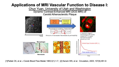 |
Applications of MRI Vascular Function to Disease I
Chun Yuan
MRI contrast agent application has played a key role in vascular disease diagnosis from its early adoption in dynamic MR angiography to recent applications in a wide range of vascular disease: such as atherosclerosis, aneurysm, and vasculitis. Imaging techniques including analysis tools have also advanced to improve data acquisition, quantitation, and analysis.This talk will introduce several contrast agent applications in vascular disease and the technical advancement.
|
|
| 16:20 | Applications of MRI Vascular Function to Disease II Video Unavailable |
||
 Back to Meeting Home
 Back to the Program-at-a-Glance
The International Society for Magnetic Resonance in Medicine is accredited by the Accreditation Council for Continuing Medical Education to provide continuing medical education for physicians.
 Back to Meeting Home
 Back to the Program-at-a-Glance
 View the Presentation