Weekend Course
Cardiovascular Anatomy & Function with CMR
Joint Annual Meeting ISMRM-ESMRMB & ISMRT 31st Annual Meeting • 07-12 May 2022 • London, UK

Weekend Course
Cardiovascular Anatomy & Function with CMR
| On Tubes & Flow | |||
| 12:30 | MR Angiography: The Ins & Outs
Giles Roditi
|
||
| 12:55 | 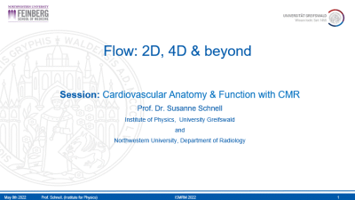 |
Flow: 2D, 4D & Beyond
Susanne Schnell
Phase-contrast MRI uses bipolar gradients to encode tissue or fluid motion into the MRI signal phase. If measured in 2D with 1-directional encoding this is termed 2D Phase-Contrast MRI. Extended to a time-resolved heart-or pulse rate triggered 3D sequence with 3-directional velocity encoding, this is termed 4D flow MRI. Error sources will be described throughout the course as well as data analysis and quantification approaches. Further extensions such as dual- and multi-venc, and 5D flow MRI will be described. Finally applications in the thorax, head, and abdomen will be introduced.
|
|
| A Map of Your Heart | |||
| 13:20 | T1 & ECV Mapping
Vanessa Ferreira
|
||
| 13:45 |  |
T2, T2* & T1ρ Mapping
Graham Wright
MRI measures of signal decay without refocusing, with intermittent refocusing and with continuous refocusing reflected by time constants T2*, T2, and T1ρ can yield important clinical information about cardiac tissue damage and response to injury. Reduced T2* reflects localized susceptibility effects as seen in hemorrhage and iron overload. T2 can depict changes in blood oxygenation reflecting ischemia and changes in water mobility reflecting inflammation. T1ρ can highlight chemical and spin exchange effects, increasing contrast between healthy and infarcted myocardium. Recently, higher dimensional and multi-parameter imaging methods have been developed for improved differentiation of myocardial pathophysiologies.
|
|
| 14:10 | Break & Meet the Teachers |
||
| Drawing Fewer Circles | |||
| 14:35 |  |
Deep Learning for CMR Reconstruction and Super-Resolution
Guang Yang
Cardiovascular Magnetic Resonance (CMR) is a safe technique, which can provide non-invasive gold-standard assessment of cardiac structure and function in patients with cardiovascular disease. While standard CMR imaging is relatively robust, some CMR techniques are inherently less reliable and image quality can be reduced. Besides, for high-resolution or 3D imaging, the acquisition duration is long and image quality may be further compromised. Recently, deep learning based methods have gained performance dividends in medical image analysis. In this talk, I will introduce the basic ideas of deep learning and its development and applications in CMR reconstruction and super-resolution towards future perspectives.
|
|
| 15:00 | Radiomics for CMR Image Analysis
Bettina Baeßler
|
||
| Harder, Better, Faster, Stronger | |||
| 15:25 | Deep Learning for CMR Image Analysis
Avan Suinesiaputra
Deep learning has become an ubiquitous tool for image analysis, including CMR. The method is capable to learn specific human tasks from a large amount of data, provided sufficient computational power. Image segmentation and recognition are the two most used applications, but there are more creative solutions. Deep learning can learn to reconstruct MRI, leading to a faster MR acquisition. It can generate realistic contrast-enhanced MRI without using the actual contrast agent. In this course, we are going to learn how deep learning can be applied to solve CMR image analysis to derive cardiac function and anatomy of the heart.
|
||
| 15:50 | Translating AI to the Clinic
Steffen Petersen
Research and development into artificial intelligence (AI) algorithms for the classification, analysis and prediction of disease using medical data carries with it the expectation of transformative benefits to healthcare. The increasing activity in this field is, in part, driven by the improved accessibility of machine learning frameworks and skilled practitioners but translation to clinical use is restrained by data access, regulatory, ethical and privacy concerns. This talk highlights the key milestones in this path from concept to AI prototype to clinical decision-making tool.
|
||
 Back to Meeting Home
 Back to the Program-at-a-Glance
The International Society for Magnetic Resonance in Medicine is accredited by the Accreditation Council for Continuing Medical Education to provide continuing medical education for physicians.
 Back to Meeting Home
 Back to the Program-at-a-Glance
 View the Presentation