Weekend Course
Facilitating Technique Translation
Joint Annual Meeting ISMRM-ESMRMB & ISMRT 31st Annual Meeting • 07-12 May 2022 • London, UK

Weekend Course
Facilitating Technique Translation
| Widely Used Sequence Examples | |||
| 12:30 | Everyday Sequences: How & Why They Are in the Protocol
C. C. Tchoyoson Lim
Clinical MRI protocols comprise individual MR pulse sequences put together to help radiologists answer a clinical question. Which sequences to select necessitates trade-offs between highest resolution spatial or function biomarkers and safety, reliability, speed. Standard Brain MRI protocols include T1, T2, flow, diffusion sequences: there is a high bar for replacing or adding to current protocols. The need for reproducibility & QIBA best practice to enhance the value of MRI will be discussed.
|
||
| 12:50 | 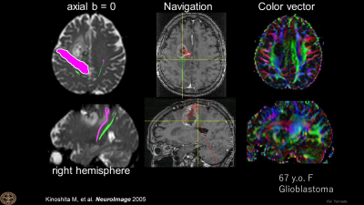 |
DWI & DTI: Where, Why & How It Is & Is Not Used
Kei Yamada
Diffusion-weighted imaging (DWI)and diffusion-tensor imaging (DTI) has become one of the essential research/clinical tools in analyzing the brain in both normal and pathological states. In this presentation, I will first cover the brief history of DWI, and then explain how this tool has become an essential part of our daily practice.
|
|
| Quantitative Imaging Examples | |||
| 13:10 | DENSE: A Technique in Translation
Xiaoying Cai
Myocardial strain imaging is an important approach for quantifying cardiac function. Cine DENSE is a dedicated MR technique for strain imaging. By encoding tissue displacement into the phase of the imaging signal, cine DENSE allow rapid and comprehensive quantification of myocardial mechanics. This talk gives an overview of about cine DENSE imaging from data acquisition to reproducibility and its translation.
|
||
| 13:30 | 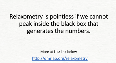 |
Relaxometry: Where, Why & How It Is & Is Not Used
Nikola Stikov
The promise of relaxometry is making MR measurements that reproduce across sites. The qMRI community has not delivered on this promise, and clinicians are losing patience. The only way forward is through standardization, data sharing, and opening up the black boxes that generate the numbers.
More at: http://qmrlab.org/relaxometry |
|
| 13:50 | Break & Meet the Teachers |
||
| Rapid Imaging & Reconstruction Examples | |||
| 14:10 | Parallel Imaging: How & Why It Moved So Quickly to Product
Rita Nunes
The aim of this talk is to introduce the basic concepts of Parallel Imaging and to discuss what made it such a successful technique. Parallel imaging took advantage of the availability of multichannel coil arrays to accelerate exams. It has had a major impact in MRI, quickly becoming available on standard scanners and being used in every day routine clinical practice.
|
||
| 14:30 |  |
Spirals: History, Opportunities, & What It Will Take to Get It into Routine Clinical MRI
James Pipe
Spiral MRI was invented by Ann and Cho in 1986, and was greatly enabled by the Stanford MRSL in the early 1990's. This method continues to evolve, and beyond just being a "fast" MRI method, it has many unique favorable properties, including low gradient moments, very low minimum TE, incoherent underdamping artifacts, and the capacity for high SNR efficiency. Despite all of this, Spiral MRI is not widely used clinically. This talk will outline some of the remaining obstacles to clinical adoption, with high confidence that they can be overcome.
|
|
| 14:50 | 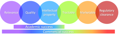 |
CS & AI: Will They Tread the Same Path to Translation?
R. Marc Lebel
This course covers aspects related to translation and commercialization of new MR techniques. We will identify and explain factors paving the perilous road to commercial success with a specific focus on AI applications. The aim is to provide insight into the common reasons why novel applications falter and to provide design principles that improve chances of successful translation from bench to bedside.
|
|
| Addressing the Challenges | |||
| 15:10 | 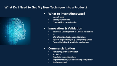 |
What Do I Need to Get My New Technique into a Product?
Maggie Fung
Just invented the next breakthrough in MRI technology? That’s only part of the equation. This presentation will cover what it takes to develop and productize new MRI technology, starting from identifying the unmet needs, innovation, down-selection and clinical validation, to commercialization and regulatory aspects. We will walk through some successful examples and summarize the key ingredients to a successful product.
|
|
| 15:30 | From Product to Routine Clinical Practice
Christopher Filippi
This talk discusses implementation science in MRI, which promotes the uptake of research and other evidence-based imaging studies into routine, clinical practice. Barriers to adaptation of emerging imaging technology include variability across scanners and sites such as magnetic field strengths, protocols, MR vendor and model types. Linearity, bias, and precision of measurements are needed for consistent, patient-specific management according to Radiology Society of North America’s Quantitative Imaging Biomarker Alliance (QIBA). (1-3) Quantitative imaging markers are needed for precision health, but determination of “ground truth” and lack of a clear road map limit impact currently.
|
||
| 15:50 | Live demos: Systems and Components | ||
 Back to Meeting Home
 Back to the Program-at-a-Glance
The International Society for Magnetic Resonance in Medicine is accredited by the Accreditation Council for Continuing Medical Education to provide continuing medical education for physicians.
 Back to Meeting Home
 Back to the Program-at-a-Glance
 View the Presentation