Combined Educational & Scientific Session
Non-Ischemic Cardiomyopathies: From Microstructure & Tissue Characterization to Function & Outcomes
ISMRM & ISMRT Annual Meeting & Exhibition • 03-08 June 2023 • Toronto, ON, Canada

Combined Educational & Scientific Session
Non-Ischemic Cardiomyopathies: From Microstructure & Tissue Characterization to Function & Outcomes
| 16:00 | Non-Ischemic Cardiomyopathy: Predicting Prognosis with CMR Jeremy Collins | |
| 16:20 | Magnetic Resonance-Based Characterization of Myocardial Architecture Stefan Zimmerman | |
| 16:40 | 1427.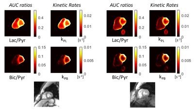 |
Regional quantification of cardiac metabolism with hyperpolarized [1-13C]-pyruvate MRI
Peder Eric Zufall Larson1, Shuyu Tang2, Xiaoxi Liu1, Avantika Sinha1, Nicholas Dwork3, Sanjay Sivalokanathan4, Jing Liu1, Robert Bok1, Karen G Ordovas5, James Slater1, Jeremy W Gordon1, and M. Roselle Abraham1
1Radiology and Biomedical Imaging, University of California - San Francisco, San Francisco, CA, United States, 2HeartVista, Palo Alto, CA, United States, 3University of Colorado Anschutz, Aurora, CO, United States, 4University of Pennsylvania, Philadelphia, PA, United States, 5University of Washington, Seattle, WA, United States Keywords: Myocardium, Metabolism Hyperpolarized (HP) 13C-pyruvate MRI is a promising new tool for non-invasive quantification of myocardial glycolytic and Krebs cycle metabolism. In this study we evaluated whole-heart imaging and metabolism quantification methods in 7 healthy volunteers under a fasted and fed state. We observed that the 13C-pyruvate-to-bicarbonate conversion rate, kPB, a measure of PDH flux, had the highest, statistically significant correlation with blood glucose levels, with smaller changes in the 13C-lactate/pyruvate ratio and 13C-pyruvate-to-lactate conversion rate, kPL. 13C-pyruvate and 13C-lactate were detected simultaneously in the RV blood pool, immediately after intravenous injection, reflecting LDH activity in blood. |
16:48 |
1428.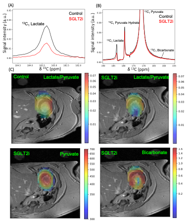 |
Diabetic Treatment and Oral Ketone Supplement effect on Cardiac Function and Metabolism in Heart Failure Model by Cardiac and hyperpolarized MRSI
David O. Guarin Bedoya1,2, Salva Yurista1,3,4, Jonah P Weigan Witthier1, Shi Chen1,3,4, Robert Eder1,3,4, William Jiang1,3,4, Feiyang Liu 1,3,4, Atsushi M. Takahashi5, Christopher Nguyen1,3,4, and Yi-Fen Yen1 1Athinoula A. Martinos Center for Biomedical Imaging,Radiology Department, Massachusetts General Hospital, Charlestown, MA, United States, 2Polarize ApS, Frederiksberg, Denmark, 3Corrigan Minehan Heart Center, Division of Cardiology, Massachusetts. General Hospital, Harvard Medical School, Boston, MA, United States, 4Cardiovascular Innovation Research Center, Heart, Vascular, and Thoracic Institute, Cleveland Clinic, Cleveland, OH, United States, 5Department of Brain and Cognitive Sciences, Massachusetts Institute of Technology, Cambridge, MA, United States Keywords: Cardiomyopathy, Hyperpolarized MR (Non-Gas), Heart Failure, Contrast Agent, Contrast Mechanisms, Metabolism Using hyperpolarized [1-13C]pyruvate MR spectroscopy imaging and cine MRI, we show that targeting cardiometabolic dysregulation with metabolic treatment, such as ketone ester supplementation and/or sodium–glucose cotransporter 2 inhibitor, was effective in improving cardiac function and ameliorating cardiac remodeling in a preclinical model of HFpEF. These results provide a rationale for the assessment of metabolic interventions for patients with HFpEF. |
| 16:56 | 1429.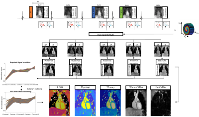 |
3D whole-heart joint T1/T1ρ/T2 mapping and water-fat imaging for contrast-agent free myocardial tissue characterization at 1.5T
Michael G Crabb1, Karl P Kunze1,2, Camila Munoz1, Donovan Tripp1, Anastasia Fotaki1, Carlos Velasco1, Radhouene Neji1,2, Claudia Prieto1,3, and Rene M Botnar1,3,4
1School of Biomedical Engineering and Imaging Sciences, King's College London, London, United Kingdom, 2MR Research Collaborations, Siemens Healthcare Limited, Camberley, United Kingdom, 3Institute for Biological and Medical Engineering, Pontificia Universidad Catolica de Chile, Santiago, Chile, 4School of Engineering, Pontificia Universidad Catolica de Chile, Santiago, Chile Keywords: Myocardium, Tissue Characterization Native T1 and T1ρ mapping has shown promising results for the detection of focal and diffuse myocardial fibrosis without the need of contrast agents, whereas T2 mapping enables characterisation of inflammation and edema. However, conventional myocardial maps are acquired in sequential 2D breath-hold scans with limited heart coverage. Here, we propose a novel free-breathing, 3D joint T1/T1ρ/T2 mapping sequence with Dixon encoding to provide whole-heart T1, T1ρ and T2 maps and co-registered water/fat volumes with isotropic spatial resolution for comprehensive contrast-agent free myocardial tissue characterization. Preliminary results demonstrate good agreement with reference values in phantoms and promising results in-vivo. |
| 17:04 | 1430.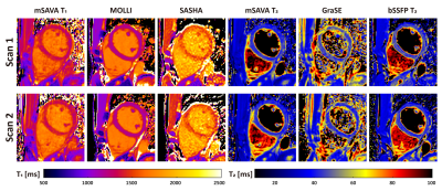 |
Accuracy, Precision, and Reproducibility of 3D Whole Heart Simultaneous T1 and T2 Mapping Based on Multi-parametric SAVA: A Comparison Study
Dongyue Si1, Rui Guo2, Daniel A. Herzka3, and Haiyan Ding1
1Center for Biomedical Imaging Research, Department of Biomedical Engineering, Tsinghua University, Beijing, China, 2School of Medical Technology, Beijing Institute of Technology, Beijing, China, 3Department of Radiology, Case Western Reserve University, Cleveland, OH, United States Keywords: Myocardium, Tissue Characterization MR parametric mapping including T1 and T2 enabled quantitative evaluation of changes of myocardium. We previously proposed a time-efficient technique for 3D free-breathing simultaneous T1 and T2 mapping based on multi-parametric SAturation recovery and Variable flip Angle (mSAVA). This study evaluated the accuracy, precision, and reproducibility of mSAVA in comparison with conventional 2D sequences. mSAVA achieved good accuracy, between that of MOLLI and SASHA, and better precision and reproducibility than SASHA for T1 measurements. T2 measured by mSAVA had better precision and reproducibility than both GraSE and bSSFP T2. mSAVA offers a promising option for myocardial tissue characterization. |
| 17:12 | 1431.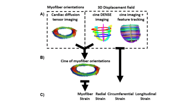 |
Myofiber strain estimation using cDTI, DENSE, and feature tracking.
Kevin Moulin1,2, Luigi E. Perotti3, Magalie Viallon1,2, and Pierre Croisille1,2
1University of Lyon, UJM-Saint-Etienne, INSA, CNRS UMR 5520, INSERM U1206, CREATIS, F-42023, Saint-Etienne, France, 2Department of Radiology, University Hospital Saint-Etienne, Saint-Etienne, France, 3Department of Mechanical and Aerospace Engineering, University of Central Florida, Orlando, FL, United States Keywords: Myocardium, Cardiovascular, Cardiac Function, cDTI, DENSE, Feature Tracking, cardiac microstructure Myofiber strain (MS) is a promising biomarker of cardiac function, but it requires the combination of cDTI and of a 3D cardiac displacement field. Displacement fields can be measured using DENSE imaging, but with low spatial resolution and limited spatial coverage. Feature tracking (FT) allows the estimation of the displacement directly from cine imaging. In this study, myofiber strains estimated using DENSE and FT were compared on thirty healthy volunteers. The magnitude of myofiber strain calculated with DENSE was higher than with FT (MSDENSE=-0.15[-0.16;-0.14] vs MSFT=-0.11[-0.14;-0.06], p<0.001) but no correlation was found between MSDENSE and MSFT (r=0.14 p=0.47). |
| 17:20 |
1432.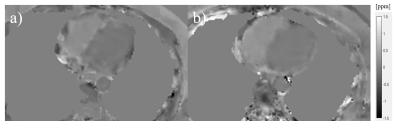 |
Accelerated 3D Stack-of-Spiral Cardiac Quantitative Susceptibility Mapping: Noninvasive Measurement of Heart Oxygenation in a Breath-Hold
Jiahao Li1,2, Pablo Villar-Calle3, Hannah Agoglia3, Nicole Liberman3, Thanh D. Nguyen2, Yi Wang1,2, Jiwon Kim3, Jonathan W. Weinsaft3, and Pascal Spincemaille2
1Biomedical Engineering, Cornell University, Ithaca, NY, United States, 2Radiology, Weill Cornell Medicine, New York, NY, United States, 3Medicine, Weill Cornell Medicine, New York, NY, United States Keywords: Heart, Oxygenation A breath-holding non-cardiac gated 3D stack-of-spiral data acquisition scheme was developed to continuously sample the data for cardiac quantitative susceptibility mapping. Compared to the previously proposed navigator-based prospective Cartesian acquisition, the accelerated spiral sequence can be done within 20 seconds breath-holds, leading to over a 20-fold reduction in scan time. The spiral QSM as well as the navigator QSM were performed on cohorts of healthy volunteers and COVID-19 survivors, showing well aligned quantification results on the differential blood oxygenation between the right and left heart. |
| 17:28 | 1433.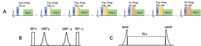 |
Free-breathing simultaneous myocardial T2 and T1ρ mapping for non-contrast assessment of uremic cardiomyopathy
Qinfang Miao1,2, Zhenfeng Lv1,2, Sha Hua3, Zhongqi Zhang4, Jian Xu4, Peng Hu1,2, and Haikun Qi1,2
1School of Biomedical Engineering, ShanghaiTech University, Shanghai, China, 2Shanghai Clinical Research and Trial Center, Shanghai, China, 3Department of Cardiovascular Medicine, Ruijin Hospital Lu Wan Branch, Shanghai Jiao Tong University School of Medicine, Shanghai, China, 4UIH America, Inc., Houston, TX, United States Keywords: Myocardium, Cardiomyopathy Uremic cardiomyopathy is the adverse cardiac remodeling that commonly occurs in patients with chronic kidney disease. Previous studies have indicated increased intramyocardial fluid and myocardial fibrosis in uremic patients, which makes native myocardial T2 and T1ρ mapping ideal imaging biomarkers to characterize these changes. Therefore, we propose a free-breathing simultaneous T2 and T1ρ mapping technique to provide co-registered T2 and T1ρ maps. The proposed technique was firstly evaluated in phantoms and ten healthy subjects, which achieved similar performance to the conventional separate T2 and T1ρ mapping methods. The preliminary validation in four hemodialysis patients showed promising results. |
| 17:36 | 1434.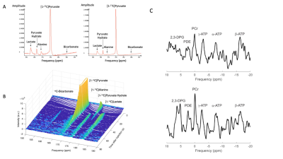 |
A Multi-Nuclear MRI/MRS Study Exploring the Impact of a Novel Cardiac Mitotrope in the Treatment of Diabetic Cardiomyopathy
Damian Tyler1, Moritz Hundertmark1, Adrienne Siu1, Violet Matthews2, Andrew Lewis1, James Grist1,2, Jai Patel3, Paul Chamberlin3, Rizwan Sarwar1, Arash Yavari1,3, Hakim-Moulay Dehbi4, Michael Frenneaux5, Ladislav Valkovič1,6, Jack Miller1,7, Stefan Neubauer1, and Oliver Rider1
1University of Oxford, Oxford, United Kingdom, 2Oxford University Hospitals NHS Foundations Trust, Oxford, United Kingdom, 3Imbria Pharmaceuticals, Boston, MA, United States, 4University College London, London, United Kingdom, 5Hamad Medical Corporation, Doha, Qatar, 6Slovak Academy of Sciences, Bratislava, Slovakia, 7Aarhus University, Aarhus, Denmark Keywords: Heart, Diabetes, Multi-nuclear Spectroscopy Patients with Type 2 Diabetes (T2D) are at a significantly increased risk of heart failure (HF) with the development of HF driven by energetic, metabolic, structural, and functional cardiac changes. In this study we have used a multi-nuclear MRI/MRS approach to demonstrate that the novel metabolic modulator, ninerafaxstat, can significantly improve myocardial energetics, cardiac steatosis and diastolic function in patients with T2D and obesity. In addition, using cardiac hyperpolarized [1-13C]pyruvate MRS, we have identified a potential signal for increased pyruvate dehydrogenase flux upon treatment with ninerafaxstat. |
| 17:44 | 1435.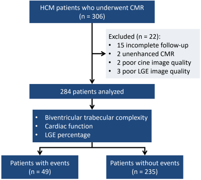 |
Fractal Analysis in Cardiovascular MR: Prognostic Value of Biventricular Trabecular Complexity in Hypertrophic Cardiomyopathy
Wen-yi Jiang1, Weibo Chen2, Yan Zhou1, Lei Zhao3, and Lian-Ming Wu1
1Renji Hospital affiliated to Shanghai Jiao Tong University, Shanghai, China, 2Philips Healthcare, Shanghai, China, 3Beijing Anzhen Hospital affiliated to Capital Medical University, Beijing, China Keywords: Cardiomyopathy, Heart Trabecular complexity of left and right ventricles can be quantified as fractal dimension (FD), a number between 1 to 2, by fractal analysis on short axis cine images of cardiovascular magnetic resonance. We found that biventricular FDs provide significant prognostic value for sudden cardiac death and composited adverse events including rehospitalization due to heart failure in patients with hypertrophic cardiomyopathy (HCM). LV maximal apical FD and RV global FD are independent prognostic factors in HCM. In addition, they provide incremental prognostic value to the conventional predictors including European Society of Cardiology predictors and late gadolinium enhancement percentage. |
| 17:52 | 1436.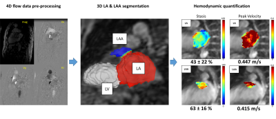 |
Post-ablation Changes in Left Atrial Hemodynamics and Volume in Patients with Atrial Fibrillation
David Dushfunian1, Anthony Maroun1, Justin Baraboo1, Maurice Pradella1,2, Daniel C. Lee1,3, Philip Greenland4, Rod Passman3, Daniel Kim1, and Michael Markl1 1Department of Radiology, Northwestern University Feinberg School of Medicine, Chicago, IL, United States, 2Department of Radiology, University of Basel, Basel, Switzerland, 3Division of Cardiology, Department of Medicine, Northwestern University Feinberg School of Medicine, Chicago, IL, United States, 4Department of Preventive Medicine, Northwestern University Feinberg School of Medicine, Chicago, IL, United States Keywords: Flow, Velocity & Flow Atrial fibrillation (AF) leads to detrimental changes in the left atrial (LA) and left atrial appendage’s (LAA) hemodynamics and volumes. When indicated, AF ablation is used to reduce AF burden. We recruited 17 post-ablation patients, 10 of whom had no documented AF recurrences. 4D flow MRI was acquired, immediately before and 6-12months after ablation, and quantified for LA and LAA stasis, volume, and peak velocities. Our findings support the notion that restoration of sinus rhythm corrects the deleterious changes of AF. |
 Back to Meeting Home
 Back to the Program-at-a-Glance
The International Society for Magnetic Resonance in Medicine is accredited by the Accreditation Council for Continuing Medical Education to provide continuing medical education for physicians.
 Back to Meeting Home
 Back to the Program-at-a-Glance
 View Presentation Video