Combined Educational & Scientific Session
New Currents: New Contrast Mechanisms to Image Neuronal Activity
ISMRM & ISMRT Annual Meeting & Exhibition • 03-08 June 2023 • Toronto, ON, Canada

Combined Educational & Scientific Session
New Currents: New Contrast Mechanisms to Image Neuronal Activity
| 13:30 | Invited Talk #1 Fernando Boada | |
| 13:50 | In vivo direct imaging of neuronal activity at high temporal and spatial resolution
Jang-Yeon Park
Keywords: Contrast mechanisms: fMRI There has been a longstanding demand for noninvasive neuroimaging with high spatiotemporal resolution. Recently, an approach has been proposed that enables Direct Imaging of Neuronal Activity (DIANA) with milliseconds temporal resolution, demonstrated by in vivo mouse brain imaging at 9.4T. DIANA showed high correlations with neuronal spikes, capturing neuronal-activity propagation along the thalamocortical pathway. The DIANA contrast mechanism may be attributed to changes in membrane potential-associated T2 relaxation time. Finally, DIANA may require different considerations in data acquisition and analysis than BOLD-fMRI, as DIANA is directly related to neuronal activity including spontaneous ongoing activity as well as responses to stimuli. |
|
| 14:10 | 0914.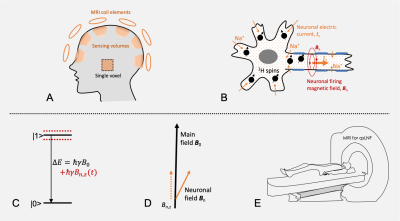 |
Quantum-Sensing MRI for Non-Invasive Detection of Neuronal Firing in Human Brain: Initial Demonstration via Finger-Tapping Task
Yongxian Qian1, Xingye Chen1, Ying-Chia Lin1, Simon Henin2,3, Nahbila-Malikha Kumbella1, Liz Aguilera1, Zena Rockowitz3, Ashley Clayton3, James Babb1, Yulin Ge1, Arjun Masurkar2,3, Anli Liu2,3, Yvonne W. Lui1,4, and Fernando E. Boada1,5
1Radiology, NYU Grossman School of Medicine, New York, NY, United States, 2Neurology, NYU Grossman School of Medicine, New York, NY, United States, 3Neurology, NYU Langone Health, New York, NY, United States, 4Radiology, NYU Langone Health, New York, NY, United States, 5Radiology, Stanford University, Stanford, CA, United States Keywords: Nerves, Neuro, fMRI (task) Neuronal firing is the electrophysiological basis of brain function. BOLD-based functional MRI indirectly detects neuronal activity through an uncertain neuro-hemodynamic mechanism and is also limited to the detection of slow neuronal activity such as postsynaptic potentials due to its relatively low temporal resolution. Recent efforts have improved temporal resolution to milliseconds or even sub-milliseconds, under the assumption that neuronal activity is exactly repeatable in time, though unlikely to be practical. Here we demonstrate the potential of quantum-sensing MRI to directly detect neuronal firing using a finger-tapping task. |
| 14:18 | 0915.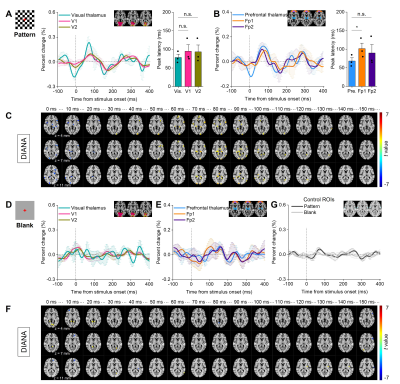 |
Observing visually evoked responses in human thalamus and cortex at millisecond temporal resolution: Preliminary study at 3 T
Phan Tan Toi1,2, Sehong Oh3, and Jang-Yeon Park1,2
1Department of Biomedical Engineering, Sungkyunkwan University, Suwon, Korea, Republic of, 2Department of Intelligent Precision Healthcare Convergence, Sungkyunkwan University, Suwon, Korea, Republic of, 3Division of Biomedical Engineering, Hankuk University of Foreign Studies, Yongin, Korea, Republic of Keywords: fMRI, Brain There has been interested in observing neural activity dynamics in humans at high temporospatial resolution using noninvasive neuroimaging. Here, we report preliminary results of direct imaging of neuronal activity (DIANA) in humans at 3T using checkerboard pattern stimulation. The results show sequential temporospatial dynamics of activities in visual and prefrontal areas evoked by checkerboard pattern stimulation with a signal change of 0.1~0.3%. In contrast, DIANA signals were distinct from control experiments compared with checkerboard pattern stimulation ones. Our preliminary observations suggest that DIANA is feasible for human fMRI studies. |
| 14:26 | 0916.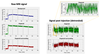 |
Investigating the Direct Imaging of Neuronal Activity (DIANA) contrast mechanism using human brain organoids
Sean Morrison1, Ewald Webber2, Carl Dixon3, Donald Maillet3, Ernst Wolvetang1, and Martijn A. Cloos3
1Australian Institute for Bioengineering and Nanotechnology, University of Queensland, St Lucia, Australia, 2School of Information Technology and Electrical Engineering, University of Queensland, St Lucia, Australia, 3Centre for Advanced Imaging, University of Queensland, St Lucia, Australia Keywords: Contrast Mechanisms, fMRI, DIANA Here we describe our initial results investigating DIANA’s ability to capture neuronal activation in humans brain organoids. Following drug delivery a 3min long window with 0.5% increased signal was observed. Although tantalising, further experiments are needed to confirm these results. |
| 14:34 | 0917.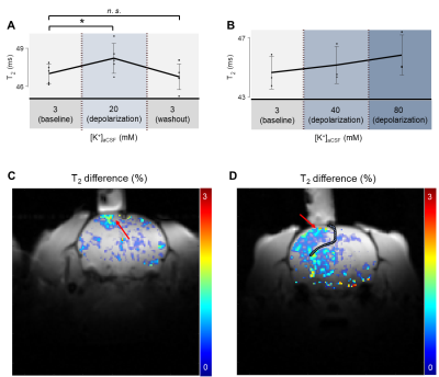 |
In vivo Detection of Changes in Membrane Potential in Rat Cortex by T2 mapping
Kyeongseon Min1, Sungkwon Chung2, Seung‐Kyun Lee3, Jongho Lee1, Tan Toi Phan4,5, Daehong Kim6, and Jang-Yeon Park4,5 1Department of Electrical and Computer Engineering, Seoul National University, Seoul, Korea, Republic of, 2Department of Physiology, Sungkyunkwan University School of Medicine, Suwon, Korea, Republic of, 3GE Global Research, Niskayuna, NY, United States, 4Department of Biomedical Engineering, Sungkyunkwan University, Suwon, Korea, Republic of, 5Department of Intelligent Precision Healthcare Convergence, Sungkyunkwan University, Suwon, Korea, Republic of, 6National Cancer Center, Goyang, Korea, Republic of Keywords: fMRI, Contrast Mechanisms Membrane potential is the crucial element of neuronal activation. We investigated the possibility of using MRI to detect the depolarization of membrane potential in rat cortex. To directly access brain cells, a craniotomy was performed to make a burr hole on the skull. T2 change in the exposed cortex was measured while depolarizing it by directly perfusing artificial cerebrospinal fluid of high potassium concentration. Our findings showed that T2 value of depolarized cortex increased by +2.7% at [K+] = 80 mM. This observation demonstrates that changes in membrane potential are detectable with MRI by T2 contrast in vivo. |
| 14:42 | 0918.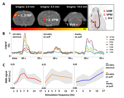 |
Correlation of zero echo time MB-SWIFT fMRI with neuronal activity during sensory stimulation in anesthetized and awake rats
Juha Valjakka1, Jaakko Paasonen1, Raimo A. Salo1, Ekaterina Paasonen1, Irina Gureviciene1, Mikko Kettunen1, Djaudat Idiyatullin2, Shalom Michaeli2, Silvia Mangia2, Heikki Tanila1, and Olli Gröhn1
1A. I. Virtanen Institute for Molecular Sciences, University of Eastern Finland, Kuopio, Finland, 2Center for Magnetic Resonance Research, University of Minnesota, Minneapolis, MN, United States Keywords: fMRI, Brain, Zero echo time Zero echo time (ZTE) fMRI with MB-SWIFT poses an artefact free alternative to traditional EPI-based BOLD fMRI approaches. However, the exact origin of the fMRI signal captured with ZTE is unknown. In this work, we investigated the correlation between electrical brain activity and the ZTE fMRI signal during sensory stimulation in anesthetized and awake rats. It is shown that a linear model explains the relationship between the fMRI signal and electrical activity in anesthetized but not in awake rats. We conclude that MB-SWIFT fMRI provides a good proxy for neuronal activity under conventional experimental conditions. |
| 14:50 | 0919.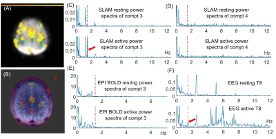 |
Direct detection of neural activity in humans by ultrafast high-sensitivity SLAM MRI at 3T
Yi Zhang1,2, Peiying Liu2, Wenqi Wang1, Deng Mao3, and Paul Bottomley2 1Key Laboratory for Biomedical Engineering of Ministry of Education, Department of Biomedical Engineering, College of Biomedical Engineering & Instrument Science at Zhejiang University; and the Department of Radiology, Children’s Hospital, Zhejiang University, Hangzhou, China, 2Division of MR Research, Department of Radiology, Johns Hopkins University, Baltimore, MD, United States, 3Philips Healthcare, Cleveland, OH, United States Keywords: fMRI, fMRI The direct MRI detection of neural activity in humans is a long-sought goal that has been attempted by many. We posit that a key reason for prior failures is inadequate sensitivity. Spectroscopy with linear algebraic modeling (SLAM) is a compartmental imaging method that provides enormous gains in speed and signal-to-noise ratio (SNR). Here we use it to directly obtain MRI signals from arbitrarily-shaped brain regions with arguably the highest possible SNR efficiency. We successfully detect neural responses elicited by rhythmic finger-tapping at the tapping frequency in humans at 3T, as confirmed by simultaneously-acquired EEG data. |
| 14:58 | 0920.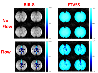 |
Blood volume based functional MRI using velocity selective pulses and SERIOS readout
Luis Hernandez-Garcia1, David Frey1, Jon-Fredrik Nielsen1, and Douglas Clare Noll1 1Radiology, University of Michigan, Ann Arbor, MI, United States Keywords: New Signal Preparation Schemes, Brain, fmri We investigate a new fast readout sequence to collect blood volume image time series in using two different velocity selective pulses. We compare two classes of pulses that target the blood or the stationary tissue. The two approaches are demonstrated and compared in a simple viso-motor blood volume based functional MRI experiment. |
| 15:06 | 0921.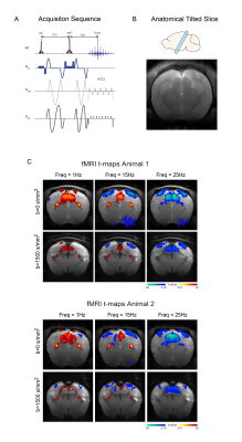 |
Fast Diffusion fMRI (dfMRI) along the visual pathway closely tracks electrophysiological signals in the negative BOLD regime
Rita Gil1, Ana Mafalda Valente1, and Noam Shemesh1 1Champalimaud Research, Champalimaud Foundation, Lisbon, Portugal Keywords: fMRI, Multimodal, Preclinical The underlying sources of negative BOLD responses (NBRs) are still debated. We have recently shown that Positive BOLD response (PBR) to NBR transitions can be induced in the visual pathway by modulating the visual frequency of stimulation, reflecting neural activation/suppression, respectively. Here, we investigate how diffusion functional MRI (dfMRI) signals, which suffer less from vessel contamination, correspond to these activation and suppression regimes. Our results show that dfMRI signals are sharper and more sensitive to suppression induced at high visual stimulation frequencies. Furthermore, striking electrophysiology characteristics such as onsets and offset peaks are more prominent in the dfMRI signals. |
| 15:14 | 0922.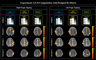 |
Investigation of electrical conductivity changes during functional activity of the brain via phase-based MR-EPT: Preliminary observation
Kyu-Jin Jung1, Chuanjiang Cui1, Jae-Hun Lee1, Jun-Hyeong Kim1, Kyoung-Jin Park1,2, SooHyoung Lee1, SunYoung Jung3, and Dong-Hyun Kim1
1Department of Electrical and Electronic Engineering, Yonsei University, Seoul, Korea, Republic of, 2Department of Radiology, Asan Medical Center, University of Ulsan College of Medicine, Seoul, Korea, Republic of, 3Department of Biomedical Engineering, Yonsei University, Wonju, Korea, Republic of Keywords: Electromagnetic Tissue Properties, Electromagnetic Tissue Properties BOLD-fMRI is measured from time-series of magnitude information, whereas the phase contrast information, which is related to the electrical properties, is excluded from process. In this study, we focus on the phase information and attempt to expose the relationship between BOLD signal and conductivity activation. In addition, we investigate the potential of functional EPT relative to standard BOLD fMRI. |
| 15:22 | 0923.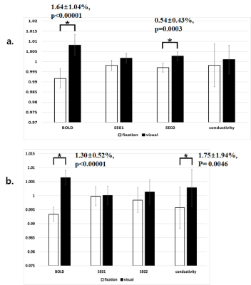 |
High-frequency Conductivity Signal Changes Measured with Functional MREPT during Visual Stimulation
Geon-Ho Jahng1, Jeonin Jeong2, Mun Bae Lee3, Jiyoon Lee2, and Oh In Kwon3
1Radiology, Kyung Hee University Hospital at Gangdong, Seoul, Korea, Republic of, 2Biomedical Engineering, Kyung Hee University, Yongin-si, Korea, Republic of, 3Mathematics, Konkuk University, Seoul, Korea, Republic of Keywords: Electromagnetic Tissue Properties, fMRI To investigate the neuronal response of conductivity during visual stimulation and compare that with BOLD, 30 young healthy volunteers were recruited from the local community. We performed two independent experiments of functional magnetic resonance electrical properties tomography (MREPT) MRI. We found that the conductivity value was increased during visual stimulation to some brain areas, indicating that functional MREPT MRI can be used to measure neuronal activity; therefore, conductivity-based fMRI signals may be helpful for measuring neuronal activation during stimulation. |
 Back to Meeting Home
 Back to the Program-at-a-Glance
The International Society for Magnetic Resonance in Medicine is accredited by the Accreditation Council for Continuing Medical Education to provide continuing medical education for physicians.
 Back to Meeting Home
 Back to the Program-at-a-Glance
 View Presentation Video