Combined Educational & Scientific Session
Musculoskeletal Combined Session
ISMRM & ISMRT Annual Meeting & Exhibition • 03-08 June 2023 • Toronto, ON, Canada

Combined Educational & Scientific Session
Musculoskeletal Combined Session
| 08:15 | Spectroscopic Imaging of Muscle Disease Hermien Kan | |
| 08:35 | Non-Spectroscopic Quantitative Muscle Imaging
Dimitrios Karampinos
Keywords: Musculoskeletal: Muscular The present educational lecture will provide an overview of the technical aspects of the most important quantitative skeletal muscle imaging techniques and will discuss recent applications and current trends. Special focus will be given to techniques to measure the proton density fat fraction and relaxation parameters of skeletal muscle and to the application of the techniques in the context of neuromuscular diseases and orthopedic imaging. |
|
| 08:55 | 0734.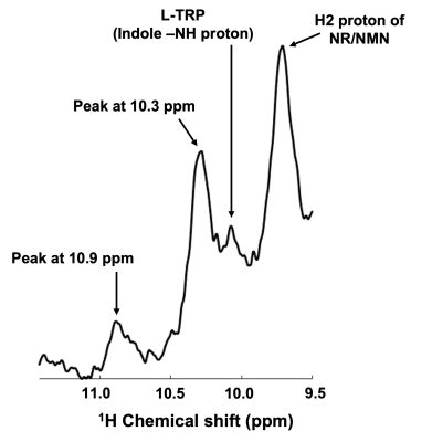 |
Detection of new resonances in the down-field 1H MRS of human calf muscle in vivo at 7T
Ravi Prakash Reddy Nanga1, Mark A. Elliott1, Anshuman Swain2, Neil E. Wilson1, Sophia Swago2, Narayan Datt Soni1, Walter Witschey1, and Ravinder Reddy3 1CAMIPM, Department of Radiology, Perelman School of Medicine at The University of Pennsylvania, Philadelphia, PA, United States, 2Bioengineering, University of Pennsylvania, Philadelphia, PA, United States, 3Radiology, Perelman School of Medicine at The University of Pennsylvania, Philadelphia, PA, United States Keywords: MSK, Spectroscopy, Down-field 1H MRS We have detected new resonances that were not reported previously with chemical shifts of ~10.1, 10.3, and 10.9 ppm occurring in the down-field 1H MRS (DFMRS) from the human calf muscle in vivo. Based on the phantom and literature data, we speculate that the contribution to the 10.1 ppm peak might be from L-Tryptophan whereas the peaks at 10.3 and 10.9 ppm are yet to be assigned. This is for the first time that these peaks are observed and reported from the down-field 1H MRS of the human calf muscle. |
| 09:03 | 0735. |
MR elastography-based slip interface imaging (SII) to assess the mobility of the myofascial interface in extremities: A feasibility study
Ziying Yin1, Yi Sui1, Keni Zheng1, Xiang Shan1, Philips Rossman1, Armando Manduca2, Brent A. Bauer3, and Richard L. Ehman1
1Radiology, Mayo Clinic, Rochester, MN, United States, 2Physiology and Biomedical Engineering, Mayo Clinic, Rochester, MN, United States, 3General Internal Medicine, Mayo Clinic, Rochester, MN, United States Keywords: Muscle, Elastography, slip interface imaging, myofascial interface, myofascial pain syndrome Myofascial pain syndrome (MPS) is a common chronic pain disorder that can cause disability. Efforts to understand the MPS pathology have focused on myofascial connective tissue and the function of fascial plane mobility. Slip interface imaging (SII) offers a unique opportunity to assess fascial plane mobility noninvasively. Here, we investigated the feasibility of SII to visualize the mobility of the intermuscular myofascial interface in the upper leg and the functional intramuscular interface in the forearm flexor muscles in healthy volunteers. This creates a foundation for using MRE/SII to distinguish between a healthy and a dysfunctional fascial plane in MPS patients. |
| 09:11 | 0736.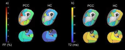 |
Quantitative muscle MRI depicts microstructural abnormalities but no signs of inflammation or dystrophy in Post COVID-19 condition
Lara Schlaffke1, Johannes Forsting1, Marlena Rohm1,2, Peter Schwenkreis1, Martin Tegenthoff1, Christine Meyer-Frießem3, and Elena Enax-Krumova1
1Neurology, University Clinic Bergmannsheil Bochum gGmbH, Bochum, Germany, 2Heimer Institute for Muscle Research, University Clinic Bergmannsheil Bochum gGmbH, Bochum, Germany, 3Anaesthesiology, Intensive Care and Pain Management, University Clinic Bergmannsheil Bochum gGmbH, Bochum, Germany Keywords: Muscle, COVID-19 Patients with post COVID-19 condition (PCC) often suffer from musculoskeletal pain with unknown pathophysiology. qMRI of the lower limbs was used to unravel the underlying mechanisms. 20 PCC were compared to 20 age and gender matched controls with regard to muscle fatfraction (revealed by Dixon imaging) water T2 time (using T2-mapping) and structural alterations (using DTI). Quantitative MRI did not depict any signs of ongoing inflammation or dystrophic process of the skeletal muscles in PCC patients. However, differences observed in muscle DTI depicts microstructural abnormalities, which may reflect potentially reversible fiber hypotrophy due to deconditioning. |
| 09:19 | 0737.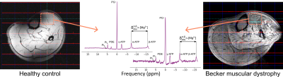 |
Skeletal muscle Mg2+, membrane permeability, and pH are altered in Becker muscular dystrophy: A 31P-MRS and DT-MRI study
Donnie Cameron1, Melissa T. Hooijmans2, Erik H. Niks3, and Hermien E. Kan1
1C.J. Gorter MRI Center, Department of Radiology, Leiden University Medical Center, Leiden, Netherlands, 2Department of Radiology and Nuclear Medicine, Amsterdam UMC, Location AMC, Amsterdam, Netherlands, 3Department of Neurology, Leiden University Medical Center, Leiden, Netherlands Keywords: Muscle, Permeability, Neuromuscular diseases Becker muscular dystrophy (BMD) is an X-linked disorder characterised by variable, progressive muscle damage and loss of function. MR-derived fat fraction is used as a disease-progression marker, but early markers are lacking. Here we compared membrane permeability measures between BMD patients and controls to test whether these are altered in BMD. Phosphorus-(31P)-MRS showed reduced ionised magnesium [Mg2+], and increased phosphodiester/adenosine-triphosphate ratios and pH in the tibialis anterior muscle in patients, while diffusion-tensor-(DT)-MRI-derived permeability showed no inter-group differences. Future studies should determine the predictive value of 31P-MRS-measured [Mg2+], pH, and membrane breakdown for disease progression, to establish their potential as biomarkers. |
| 09:27 | 0738.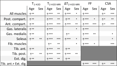 |
Multi-parametric ageing study on 51 subjects in the lower leg by 1H water T1 MR fingerprinting, multi-compartment water T2, fat fraction and 31P MRS
Alfredo Liubomir Lopez Kolkovsky1, Béatrice Matot1, Harmen Reyngoudt1, Benjamin Marty1, Ericky Caldas de Almeida Araujo1, and Yves Fromes1
1NMR Laboratory , Neuromuscular Investigation Center, Institute of Myology, Paris Cedex 13, France Keywords: Muscle, Aging, Multi-contrast, Relaxometry, MR Fingerprinting Aging is a multi-factorial process and studies in the lower leg are scarce. We performed a multi-contrast protocol in 51 volunteers from 20 to 81 y.o. We found an age-related increase of muscle water T1(water-fat separation,MR fingerprinting), fat fraction, water T2 and T2 heterogeneity in the anterior and posterior compartments. Phosphodiesters and mitochondrial stress biomarkers also increased with age. Through bi-compartment water T2 CPMG measures, age-related increases of the long water T2 relative fraction were observed relative to the short water T2 (T2-H2O-CPMG-short) fraction, but T2-H2O-CPMG-short values were unaltered with age, suggesting inflammation with preservation of the intracellular water compartment. |
| 09:35 | 0739.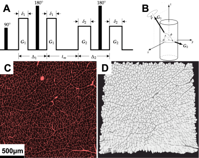 |
Double Pulsed Field Gradient Diffusion MRI of skeletal muscle is sensitive to muscle microstructure
David B Berry1, Vitaly Galinsky1, Elizabeth Hutchinson2, Samuel R Ward1, and Lawrence R Frank1 1UC San Diego, La Jolla, CA, United States, 2University of Arizona, Tuscon, AZ, United States Keywords: Muscle, Diffusion/other diffusion imaging techniques Single pulsed field gradient (sPFG) diffusion MRI (dMRI) is a commonly researched tool to monitor changes in muscle microstructure associated with injury. Double pulsed field gradient (dPFG) dMRI has been used to characterize microstructure of neural tissues, but is not commonly applied to skeletal muscle. This study demonstrates both in silico and in vivo applications of dPFG dMRI to assess changes in muscle microstructure that are related to muscle function (e.g. fiber diameter). Additionally, a novel method for analyzing dPFG data called Diffusion Tensor Subspace Imaging (DiTSI) demonstrates high sensitivity to quantifiable biomarkers of muscle microstructure. |
| 09:43 | 0740.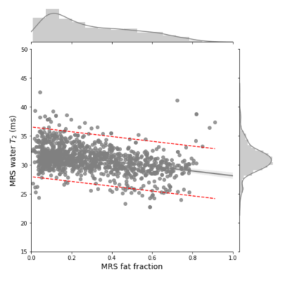 |
Fat fraction dependent decrease of muscle 1H2O T2 in Duchenne muscular dystrophy is steeper in patients with faster disease progression
Eric Baetscher1, William Triplett2, Glenn Walter2, Sean Forbes2, Rebecca Willcocks2, Robert Mueller2, Alison Barnard2, Krista Vandenborne2, and William Rooney3
1Biomedical Engineering, Oregon Health & Science University, Portland, OR, United States, 2University of Florida, Gainesville, FL, United States, 3Oregon Health & Science University, Portland, OR, United States Keywords: Muscle, Relaxometry Fatty tissue replacement of muscle is characteristic of disease progression in Duchenne muscular dystrophy (DMD), and correlates with a decrease in muscle 1H2O T2. We report longitudinal analysis of MRS data from 86 subjects with DMD. We detect a significant effect of μ (age at 50% fat-fraction) on the slope of T2 by FF. The effect of μ on slope is a consequence of lower T2 at high FF in early progressing subjects, not an effect of T2 differences at low FF. The slope of muscle water T2 decline with FF may contain additional information related to pathology in DMD. |
| 09:51 | 0741.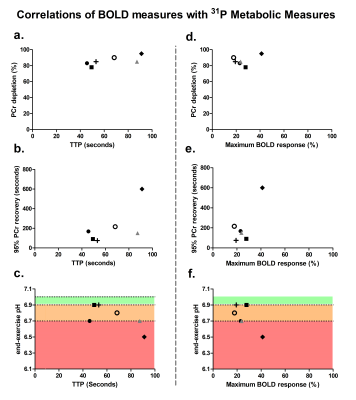 |
Interleaved BOLD and 31P-MRS examination of working human upper arm muscles
Melissa T. Hooijmans1, Jeroen A.L. Jeneson1,2, Sandra van der Berg1, Gustav J. Strijkers3, and Aart J. Nederveen1 1Radiology and Nuclear Medicine, University of Amsterdam, Amsterdam Movement Sciences, Amsterdam UMC, Amsterdam, Netherlands, 2Center for Child Development, Exercise and Physical Literacy, Wilhelmina Children’s Hospital/Division of Child Health, University Medical Center Utrecht, Utrecht, Netherlands, 3Biomedical Engineering and Physics, University of Amsterdam, Amsterdam Movement Sciences, Amsterdam UMC, Amsterdam, Netherlands Keywords: Muscle, Metabolism, capillary function Phosphorous (31P) MRS is frequently used to study alterations in muscle mitochondrial function but works under the assumption that exercise triggers sufficient changes in perfusion to allow for delivery oxygen and energy substrates to the tissue. Here, we used interleaved scanning of 31PMRS and BOLD imaging to obtain an in-debt evaluation of muscle oxidative capacity in the upper arm muscles during arm-cycling exercise. We demonstrated that similar levels of PCr depletion and PCr recovery times resulted in variable Time-To-Peaks (TTPs) and max %BOLD response while a tendency was observed between a lower end-exercise pH and longer TTP. |
| 09:59 | 0742.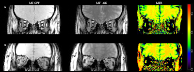 |
Magnetization transfer ratio for characterizing extra-ocular muscles changes in myasthenia gravis: a pilot feasibility study
Qin Zhou1, Pei Chen1, Xiaoxiao Zhao1, Jing Zhao1, Mengzhu Wang2, and Zhiyun Yang1
1The first affiliated hospital of sun yat-sen university, Guangzhou, China, 2MR Scientific Marketing, Siemens Healthineers Ltd, Guangzhou, China Keywords: Muscle, CEST & MT, Myasthenia Gravis,MTR This study looked into the feasibility of using MTI to assess the EOM morphological and pathological changes in MG. The MTR can effectively quantify fibrosis and atrophy of EOM in MG patients, which is significantly related to the poor response to medication and the long duration of the disease, implying that it can be used as a non-invasive auxiliary diagnostic tool for MG patients' prognosis evaluation. |
| 10:07 | 0743.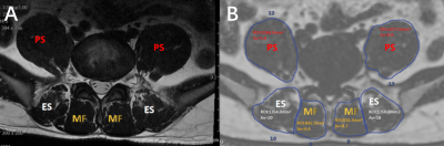 |
Quantitative assessment of the lumbar paraspinal muscle in patients with unilateral lumbar disc herniation by IDEAL-IQ MR sequence
Junrong Chen1, Jiujun Lan1, Ze Li2, Min Fu2, Xiaochun Yuan2, and Huilou Liang3
1Sichuan Province Orthopedic Hospital, Chengdu, China, 2Chengdu Sport University, Chengdu, China, 3GE Healthcare China, Beijing, China Keywords: Muscle, Fat, Quantitative imaging, Lumbar disc herniation The development, progression, treatment and rehabilitation of lumbar disc herniation (LDH) are strongly associated with morphology and structural characteristics of lumbar paraspinal muscle. The IDEAL-IQ MR sequence allows the direct quantification of lumbar paraspinal muscle properties including fat fraction (FF) and cross-sectional area (CSA). The side-to-side differences between quantitative properties of bilateral paraspinal muscles in patients with unilateral LDH remain unclear. Therefore, this study aimed to investigate the possible unilateral changes of the paraspinal muscles in patients with unilateral LDH. Our results found no difference in FF and CSA values of paraspinal muscles between the affected and unaffected sides. |
 Back to Meeting Home
 Back to the Program-at-a-Glance
The International Society for Magnetic Resonance in Medicine is accredited by the Accreditation Council for Continuing Medical Education to provide continuing medical education for physicians.
 Back to Meeting Home
 Back to the Program-at-a-Glance
 View Presentation Video