Combined Educational & Scientific Session
Laminar fMRI
ISMRM & ISMRT Annual Meeting & Exhibition • 03-08 June 2023 • Toronto, ON, Canada

Combined Educational & Scientific Session
Laminar fMRI
| 15:45 |  |
Data Acquisition & Analysis Basics for Laminar fMRI
Renzo Huber
Keywords: Contrast mechanisms: fMRI, Neuro: Brain function, Image acquisition: Sequences Layer-fMRI is a technique that uses functional magnetic resonance imaging (fMRI) to measure activity in specific layers of the cortex. This is done by acquiring high-resolution data and dividing cortical voxels into thin groups of different cortical depth. Layer-fMRI can be used to study the directional information flow between brain areas and see how different parts of the cortex work together to perform different tasks. However, layer-fMRI is still a relatively new method and there are some technical challenges for data acquisition and analysis. In this presentation, I will give an overview of common layer-fMRI acquisition and analysis approaches. |
| 16:05 | Physiological Basis of the Laminar fMRI Signal
Seong-Gi Kim
Keywords: Contrast mechanisms: fMRI, Neuro: Brain function, Neuro: Brain connectivity Laminar fMRI has been increasingly used for determining feedforward and feedback inputs to the cortex. To properly design and interpret laminar-resolution fMRI experiments, it is critical to examine biophysical and physiological sources of hemodynamic and fMRI signals. In this education talk, we will discuss 1) laminar fMRI contrasts such as GE-BOLD, SE-BOLD, CBV and CBF, 2) up-to-dated depth-dependent neurophysiological findings, and 3) relationships between laminar-specific electrophysiology and hemodynamic responses. |
|
| 16:25 | 0614. |
Simultaneous BOLD fMRI and two-photon microscopic imaging on mice brain at 16.4T
Guangle Zhang1, Wei Zhu1, Zongyue Cheng2, Chenmao Wang2, Kamil Ugurbil1, Xiao-Hong Zhu1, Meng Cui2, and Wei Chen1
1Center for Magnetic Resonance Research, Department of Radiology, University of Minnesota, MN, USA, Minneapolis, MN, United States, 2School of Electrical and Computer Engineering, Department of Biological Sciences, Purdue University, West Lafayette, USA, West Lafayette, IN, United States Keywords: Multimodal, fMRI (resting state), two-photon microscopy (TPM); simultaneous fMRI and TPM While BOLD-based fMRI can non-invasively map whole brain activation and connectivity in humans and animals, the BOLD signal provides only an indirect measure of neural activity, and its cellular and neurophysiological origins remain not fully understood. We have developed a multi-scale neuroimaging modality allowing simultaneous fMRI and two-photon microscopic imaging (TPMI) on mice brains at UHF (16.4T). With the virus injection of GCaMP6s into the mouse brain, for the first time, we have successfully obtained functional MR images of the mouse brain and the neuronal calcium signals at layer II-III of the right S1 cortex simultaneously at resting state. |
| 16:33 | 0615.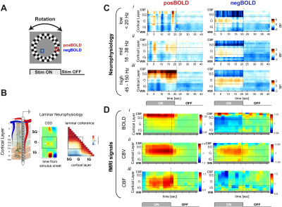 |
Layer dependent changes of neural activity underlying laminar fMRI
Daniel Zaldivar1, Yvette Bohraus2, Nikos Logothetis2,3,4, and Jozien Goense5,6,7
1LN, National Institute of Mental Health, Bethesda, MD, United States, 2Physiology of Cognitive Processes, Max Planck Institute for Biological Cybernetics, Tuebingen, Germany, 3University of Manchester, Manchester, United Kingdom, 4International Center for Primate Brain Research, Shanghai, China, 5Department of Psychology, University of Illinois at Urbana-Champaign, Champaign, IL, United States, 6Department of Bioengineering, University of Illinois at Urbana-Champaign, Urbana, IL, United States, 7Beckman Institute for Advanced Science and Technology, Urbana, IL, United States Keywords: Brain Connectivity, fMRI, Laminar fMRI and neurophysiology How accurately fMRI reflects the underlying laminar differences in neural processing? In the current study we investigated the relationship between neural activity and fMRI signals across different cortical layers. We found layer and frequency dependent differences in neural activity during the presentation of visual stimulus that elicits positive and negative BOLD response. |
| 16:41 | 0616.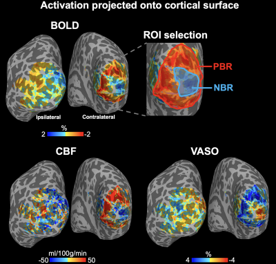 |
Investigating the origin of negative BOLD responses using laminar CBF, CBV, T2 BOLD and CMRO2 fMRI in human visual cortex at 7T
Xingfeng Shao1, Jung Hwan Kim2, David Ress2, Chenyang Zhao1, Qinyang Shou1, Kay Jann1, and Danny JJ Wang1
1Laboratory of FMRI Technology (LOFT), Mark & Mary Stevens Neuroimaging and Informatics Institute, Keck School of Medicine, University of Southern California, Los Angeles, CA, United States, 2Department of Neuroscience, Baylor College of Medicine, Waco, TX, United States Keywords: fMRI (task based), High-Field MRI, multi-contrast, ASL, VASO, T2 BOLD, CMRO2 We proposed a 7T laminar concurrent ASL, VASO and BOLD fMRI sequence to obtain quantitative CBF, CBV, T2-BOLD and CMRO2 measurements with high resolution and specificity to detect layer-dependent vascular and metabolic activities. Ipsilateral visual stimuli (eccentricity of 4°–6°) induced ring-shaped BOLD and CBF signal increase on cortical surface corresponding to the pattern of visual stimulus while decreased BOLD/CBF signals can be seen in adjacent fovea regions. In negative BOLD response (NBR) regions, moderate decrease in T2-BOLD and CBV signals and strong CBF and CMRO2 decreases especially in deep cortical layers were observed, suggesting suppressed neuronal activities in NBR regions. |
16:49 |
0617.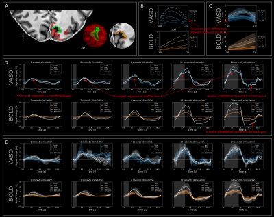 |
Characterisation of laminar spatiotemporal dynamics of CBV and BOLD signals using fast sampling at 7T fMRI in humans
Sebastian Dresbach1,2, Renzo Huber1,3, Omer Faruk Gulban1,4, Alessandra Pizzuti1,4, Robert Trampel2, Dimo Ivanov1, Rainer Goebel1,4, and Nikolaus Weiskopf2,5
1Department of Cognitive Neuroscience, Faculty of Psychology and Neuroscience, Maastricht University, Maastricht, Netherlands, 2Department of Neurophysics, Max-Planck-Institut for Human Cognitive and Brain Science, Leipzig, Germany, 3National institute of Health, Bethesda, DC, United States, 4Brain Innovation B.V., Maastricht, Netherlands, 5Felix Bloch Institute for Solid State Physics, Faculty of Physics and Earth Sciences, Leipzig University, Leipzig, Germany Keywords: fMRI, Neuroscience Characterisation of cortical laminar activity requires detailed knowledge of the spatiotemporal haemodynamic response across vascular compartments. Additionally, laminar models of the BOLD-response are dependent on CBF and CBV responses to neural activity. Therefore, we characterised the depth-dependent CBV- and BOLD-haemodynamic responses across varying stimulus durations with 0.9mm spatial, 0.785s effective temporal resolution. Furthermore, we obtained fine-scale vascular details using ME-GRE data at 0.35mm to investigate signal contributions from different vascular compartments. Our results contribute to the understanding of the laminar VASO response, provide guidance for neuroscientific applications, and support modelling of the laminar haemodynamic responses in humans. |
| 16:57 | 0618.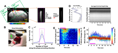 |
Detecting high temporal laminar-specific responses with line-scanning fMRI in awake mice
Sangcheon Choi1, Zeping Xie1,2, Xiaochen Liu1, Bei Zang1, David Hike1, Andy Liu1,3, and Xin Yu1
1Athinoula A. Martinos Center for Biomedical Imaging, Department of Radiology, Harvard Medical School, Massachusetts General Hospital, Charlestown, MA, United States, 2School of Traditional Chinese Medicine, Southern Medical University, Guangzhou, China, 3Department of Neuroscience, Boston University, Boston, MA, United States Keywords: fMRI (task based), fMRI The 2D line-scanning fMRI has been used to map laminar BOLD onset and single-vessel vasodynamic changes. Lately, this method has been applied to map ultra-fast T2*-weighted MRI signal changes, directly coupled to the multi-unit activity in anesthetized mice with 5ms TR. Here, we have adjusted the 1D line-scanning fMRI method to test it for awake mice. The current setup enables the detection of evoked BOLD fMRI signals in the visual cortex following 4s (3 Hz, 20ms) light exposure. It provides the proof-of-concept to further examine the ultra-fast MRI signals directly linked to neuronal activity. |
| 17:05 | 0619.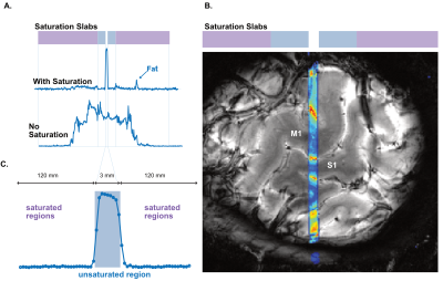 |
Optimized signal saturation for fast functional line-scanning of cortical layers
Nils Nothnagel1, Tyler Morgan2, Alison Symon1, Lars Muckli1, and Jozien Goense3,4,5
1School of Psychology & Neuroscience, University of Glasgow, Glasgow, United Kingdom, 2NIH, Bethesda, MD, United States, 3Beckman Institute for Advanced Science and Technology, Urbana, IL, United States, 4Department of Psychology, University of Illinois at Urbana-Champaign, Champaign, IL, United States, 5Department of Bioengineering, University of Illinois at Urbana-Champaign, Urbana, IL, United States Keywords: fMRI, High-Field MRI, High Resolution fMRI Layer-dependent fMRI is typically performed at sampling rates below 1 Hz and voxel sizes of 0.7-0.9 mm3 containing signal from multiple anatomical layers. Line-scanning can enhance spatial resolution to 0.2-0.4 mm while maintaining sub-second TR. It is therefore an ideal method to study temporal evolution of BOLD responses across cortical layers. However, current saturation schemes for human line-scanning suffer from broad saturation profiles that lead to signal contamination of BOLD responses from adjacent areas. We implemented line-scanning in humans with sharp line-profiles of 3 mm. Using this method, we show that cortical layers of M1 have unique BOLD time courses. |
| 17:13 | 0620.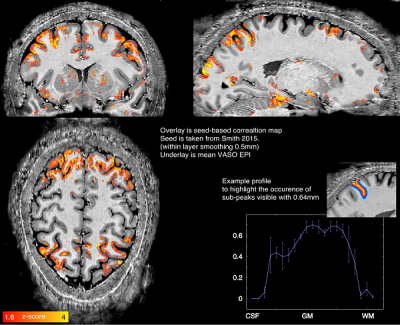 |
Whole brain Layer-fMRI on the NexGen 7T scanner with high performance gradients and 64-channel receiver array.
Alexander JS Beckett1,2, Renzo Huber3, Samantha J Ma4, Suvi Häkkinen1, Shajan Gunamony5,6, and David A Feinberg1,2
1Helen Wills Neuroscience Institute, University of California, Berkeley, CA, United States, 2Advanced MRI Technologies, Sebastopol, CA, United States, 3Faculty of Psychology and Neuroscience, Maastricht University, Maastricht, Netherlands, 4Siemens Medical Solutions USA, Inc., Malvern, PA, United States, 5MR CoilTech Limited, Glasgow, United Kingdom, 6Imaging Centre of Excellence, University of Glasgow, Glasgow, United Kingdom Keywords: fMRI, Data Acquisition Laminar-resolved fMRI has the potential to capture directional information flow within and between cortical areas to inform network neuroscience. However, common layer-fMRI imaging protocols are constrained by:
In this abstract, we use the NexGen 7T scanner to develop a whole-brain functional imaging protocol at 0.6mm resolutions. The aim was to identify and mitigate challenges in protocol optimization of 3D-EPI VASO:
We show whole-brain 0.64mm CBV-based connectivity maps covering the entire neocortex. |
| 17:21 | 0621.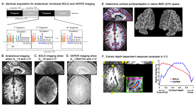 |
Layer-specific functional connectivity with 3D VAPER fMRI
Yuhui Chai1, A. Tyler Morgan2, Hua Xie3, Linqing Li4, Laurentius Huber4, Peter A. Bandettini2,4, and Bradley P. Sutton1
1Beckman Institute for Advanced Science and Technology, University of Illinois at Urbana-Champaign, Urbana, IL, United States, 2Section on Functional Imaging Methods, NIMH, NIH, Bethesda, MD, United States, 3Children's National Hospital, Washington DC, DC, United States, 4Functional MRI Core, NIMH, NIH, Bethesda, MD, United States Keywords: Brain Connectivity, fMRI, fMRI connectivity We introduced a whole-brain 3D VAPER sequence tool and analysis approach for layer-specific resting state fMRI. It allows investigation of the directional connectivity between brain areas and determine whether any given connection is better described as predominantly feedforward vs. feedback driven. To exemplify this, we demonstrated that different directions of the same connection within the default mode network (mPFC <-> PCC/Parietal region) lead to different laminar correlation profile, suggesting different projection types (feedforward/feedback). |
| 17:29 | 0622.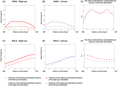 |
Depth Dependent Signal from Human Ocular Dominance Columns with VASO fMRI at 7T
Atena Akbari1,2, Joseph S. Gati1, Peter Zeman1, Brett Liem1,2, and Ravi S. Menon1,2 1Centre for Functional and Metabolic Mapping, Robarts Research Institute, The University of Western Ontario, London, ON, Canada, 2Department of Medical Biophysics, The University of Western Ontario, London, ON, Canada Keywords: fMRI (task based), High-Field MRI, VASO, laminar fMRI In this study, we measured the monocular and binocular responses of ocular dominance columns across cortical depths in human V1 using vascular-space-occupancy (VASO) fMRI at 7T. BOLD and VASO images were acquired from five participants using the DZNE sequence with an isotropic resolution of 0.8 mm. Our results indicate that VASO better differentiates the monocular responses between adjacent columns compared to GRE BOLD. In addition, the binocular modulation of the monocular neurons in V1 could possibly be revealed with VASO contrast. |
| 17:37 | 0623.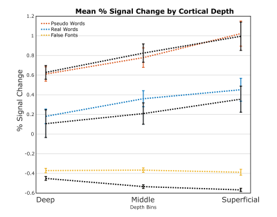 |
Layer-resolved FMRI activation and connectivity of the left inferior frontal cortex during reading
Daniel Sharoh1, Peter Hagoort1,2, and David G Norris1,3,4 1Donders Institute, Radboud University, Nijmegen, Netherlands, 2Max Planck Institute for Psycholinguistics, Nijmegen, Netherlands, 3Erwin L. Hahn Institute for Magnetic Resonance Imaging, University of Duisburg-Essen, Essen, Germany, 4Faculty of Science and Technology, Magnetic Detection and Imaging, University Twente, Enschede, Netherlands Keywords: Brain Connectivity, fMRI (task based), Laminar FMRI We present results which demonstrate simultaneous bottom-up and top-down connectivity from BA44 to regions hierarchically inferior/superior to it in the context of a reading paradigm. BA44 is critical to a number of cognitive functions in humans, notably language. We also demonstrate that the layer dependent signal is sensitive to stimulus length. This effect is contrary to what would be expected in primary sensory cortices, as increased length relates to decreased input. Hence, we also provide insight to how canonical cortico-cortical circuits can lead to different outcomes depending on the nature of the input and the stage of the processing hierarchy. |
 Back to Meeting Home
 Back to the Program-at-a-Glance
The International Society for Magnetic Resonance in Medicine is accredited by the Accreditation Council for Continuing Medical Education to provide continuing medical education for physicians.
 Back to Meeting Home
 Back to the Program-at-a-Glance
 View Presentation Video