Combined Educational & Scientific Session
From Animal to Human: Potential & Challenges of Translating Preclinical Neuroinflammation Studies to the Bedside
ISMRM & ISMRT Annual Meeting & Exhibition • 03-08 June 2023 • Toronto, ON, Canada

Combined Educational & Scientific Session
From Animal to Human: Potential & Challenges of Translating Preclinical Neuroinflammation Studies to the Bedside
| 15:45 | Basics of Animal MRI: What Clinicians Should Know
Noam Shemesh
|
|
| 16:05 | How Does Animal Research Reach Clinical Practice? Diego Martin | |
| 16:25 | 0654.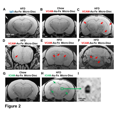 |
High Fat Diet Induced Cerebrovascular Pathology and Immune Cell Recruitment: A MRI Guided Histology Study
Li Liu1, Ryan Hunt1, Hari Rallapalli1, Nikorn Pothayee1, Stephen Dodd1, Nadia Bouraoud1, Dragan Maric1, Gary Zabow2, and Alan P Koretsky1
1National Institute of Neurological Disorders and Stroke, National Institute of Health, Bethesda, MD, United States, 2Magnetic Imaging Group, National Institute of Standards and Technology, Boulder, CO, United States Keywords: Neuroinflammation, Neuroinflammation High fat diet (HFD) causes chronic low-grade inflammation and is associated with an increased risk of cerebrovascular pathology and neurodegenerative disorders. Hypothalamus can sense peripheral signals and has been the major focus of HFD induced neuroinflammation. This work aims to study the activation of BBB endothelial cells, immune cell infiltration, and brain cell inflammation in the whole brain in real time caused by short-term HFD. Molecular MRI, using a new ultrahigh moment microfabricated gold-iron micro-disc, provides non-invasive guidance to the histology study. Our data indicate that HFD causes neuroinflammation in a remarkably short time in many structures of brain. |
| 16:33 | 0655.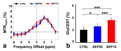 |
Cerebral Mapping of Glutamate-Weighted MR Imaging (GluCEST) in a Rat Model of Lipopolysaccharide-Induced Sepsis
Do-Wan Lee1, Jae-Im Kwon2, Hwon Heo3, Chul‐Woong Woo4, Yeon Ji Chae3, Na Hee Yu2, Seongwon Na5, Yousun Ko1, Nari Kim6, Joongkee Min4, Monica Young Choi4, Kyung Won Kim1, and Dong‐Cheol Woo3,4
1Department of Radiology, Asan Medical Center, University of Ulsan College of Medicine, Seoul, Korea, Republic of, 2QuBEST Bio Co. ltd., Gyeonggi-do, Korea, Republic of, 3Department of Convergence Medicine, Asan Medical Center, University of Ulsan College of Medicine, Seoul, Korea, Republic of, 4Convergence Medicine Research Center, Asan Institute for Life Sciences, Asan Medical Center, Seoul, Korea, Republic of, 5Biomedical Research Center, Asan Institute for Life Sciences, Asan Medical Center, Seoul, Korea, Republic of, 6Department of Medical Science, Asan Medical Institute of Convergence Science and Technology, Asan Medical Center, University of Ulsan College of Medicine, Seoul, Korea, Republic of Keywords: Neuroinflammation, Molecular Imaging Glutamate-weighted chemical exchange saturation transfer (GluCEST) is a useful imaging tool which is used to detect glutamate signal alterations caused by neuroinflammation. The present study quantitatively evaluated glutamate level changes in the hippocampal region of a rat model of sepsis-induced brain injury using GluCEST and proton magnetic resonance spectroscopy (1H-MRS). The GluCEST and 1H-MRS results showed that GluCEST values and glutamate concentrations were significantly higher in the sepsis-induced rats than in the controls. GluCEST imaging could be a helpful technique for defining a biomarker to estimate the glutamate-related metabolism in sepsis-associated diseases. |
| 16:41 | 0656.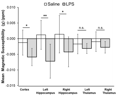 |
Systemic inflammation causes cerebral hypoperfusion, reductions in brain susceptibility and hippocampal hypoxia: a 9.4T MRI animal study
Qandeel Shafqat1, Ying Wu1, Uche J Ohaezukosi1, Ty Makarowski1, Hongfu Sun2, Rehman Tariq1, and Jeff F. Dunn1 1Department of Radiology, University of Calgary, Calgary, AB, Canada, 2School of Information Technology and Electrical Engineering, University of Queensland, Brisbane, Australia Keywords: Multiple Sclerosis, Brain Inflammation is a pathological characteristic of multiple sclerosis (MS). Individuals with MS are reported to experience reductions in cerebral blood flow (CBF) and brain hypoxia (low oxygenation). The cause of these phenomena is not well-known. We studied the possible association between inflammation and hypoxia in an inflammatory mouse model by quantifying CBF (arterial-spin labeling MRI) and magnetic susceptibility (quantitative susceptibility mapping), as well as measuring hippocampal oxygen using oxygen-sensitive probes. We found that inflammation is associated with reductions in CBF, brain susceptibility, and hippocampal oxygenation. This supports the idea that inflammation can induce brain hypoxia and disrupt cerebrovascular autoregulation. |
| 16:49 | 0657.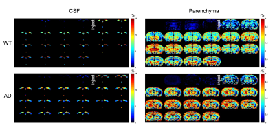 |
DGE MRI detects altered glucose uptake and utilization upon microglia activation in Alzheimer’s disease
Zilin Chen1, Jianpan Huang1, Haoyun Su1,2, Wai Po Chong3, and Kannie W.Y. Chan1,2,4,5,6
1Department of Biomedical Engineering, City University of Hong Kong, Hong Kong, China, 2Hong Kong Centre for Cerebro-Cardiovascular Health Engineering (COCHE), Hong Kong, China, 3School of Chinese Medicine, Hong Kong Baptist University, Hong Kong, China, 4Russell H. Morgan Department of Radiology and Radiological Science, The Johns Hopkins University School of Medicine, Baltimore, MD, United States, 5City University of Hong Kong Shenzhen Research Institute, Shenzhen, China, 6Tung Biomedical Science Centre, City University of Hong Kong, Hong Kong, China Keywords: Alzheimer's Disease, Neuroscience Immune cell activation is a hallmark of Alzheimer’s disease (AD). Microglia activation impairs with glucose metabolism, which may serve as early biomarker for AD. Here, we employed dynamic glucose-enhanced (DGE)-MRI to assess glucose uptake and clearance in AD, and study its relationship with microglia based on our previous study. We observed an increase in microglia in AD than WT, accompanied with relatively high parenchymal glucose uptake and low CSF clearance. We believe further studies to associate the underlying molecular and cellular pathology of AD with DGE-MRI could benefit the identification of AD in early stages. |
| 16:57 | 0658.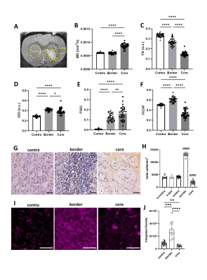 |
Diffusion MRI as a marker of neuroinflammation following intracerebral haemorrhage
Josephine Thomas1,2, Hamied Haroon2,3, Emmanuel Pinteaux1,2, Catherine Lawrence1,2, Stuart M Allan1,2, and Ben R Dickie2,4 1Division of Neuroscience, School of Biological Sciences, Faculty of Biology, Medicine and Health, University of Manchester, Manchester Academic Health Science Centre, Manchester, M13 9PT, Manchester, United Kingdom, 2Geoffrey Jefferson Brain Research Centre, University of Manchester, Manchester Academic Health Science Centre, UK, Manchester, United Kingdom, 3Division of Psychology, Communication & Human Neuroscience, Faculty of Biology, Medicine and Health, The University of Manchester, UK, Manchester, United Kingdom, 4Division of Informatics, Imaging and Data Sciences, Faculty of Biology, Medicine and Health, University of Manchester, UK, Manchester, United Kingdom Keywords: Stroke, Inflammation Diffusion tensor imaging (DTI) and neurite orientation and density imaging (NODDI) is used to observe regional changes in brain microstructure following intracerebral haemorrhage (ICH) in rats. On day 7 following ICH, DTI and NODDI metrics were significantly altered in haemorrhaged sub-regions as well as healthy appearing overlying cortex, compared to contralateral tissue. Histological analysis suggest these changes are driven by altered cell density and populations, with notable regional changes in microglia/macrophages. In summary, DTI and NODDI parameters are altered in ICH and may reflect changes in the immune cell populations in both haemorrhaged and overlying normal-appearing cortex. |
| 17:05 | 0659.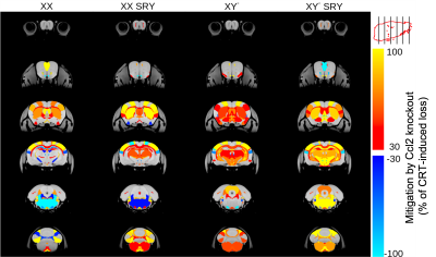 |
Sex chromosomes and sex hormones differentially mitigate radiation-induced neuroanatomic deficits in Ccl2 knockout mice.
Jonas Yeung1,2, Taylor De Young1, Shoshana Spring1, Qi Xiao3, Jason Lerch4, Mark Palmert5, and Brian J. Nieman1,2,6
1Translational Medicine, The Hospital for Sick Children, Toronto, ON, Canada, 2Medical Biophysics, University of Toronto, Toronto, ON, Canada, 3The Hospital for Sick Children, Toronto, ON, Canada, 4Wellcome Centre for Integrative Neuroimaging, University of Oxford, Oxford, United Kingdom, 5Department of Paediatrics, The Hospital for Sick Children, Toronto, ON, Canada, 6Ontario Institute for Cancer Research, Toronto, ON, Canada Keywords: Neuroinflammation, Preclinical, mouse, structural, radiation, Ccl2 Females tend to exhibit worse cognitive outcomes than males after pediatric cranial radiation therapy (CRT). Previous literature suggest that these sex differences may be driven by neuroinflammatory responses that involve CCL2. The objective of this study was to determine whether protection from Ccl2 knockout is mediated by sex hormones or sex chromosomes. We employ MRI on a CRT mouse model with mixed sex hormone and chromosome complements (i.e., four core genotypes model) to determine neuroanatomical changes. We found that both male chromosome and male sex hormones separately benefit regions of the brain in CCL2 deficient mice after CRT. |
| 17:13 | 0660.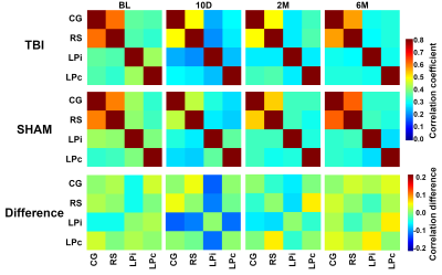 |
Association of thalamic neuroinflammation and the functional connectivity in a rat model of traumatic brain injury – a longitudinal study
Lenka Dvorakova1, Raimo A. Salo1, Petteri Stenroos1, Ekaterina Paasonen1, Kimmo Jokivarsi1, Jaakko Paasonen1, and Olli Gröhn1 1A. I. Virtanen Institute for Molecular Sciences, University of Eastern Finland, Kuopio, Finland Keywords: Brain Connectivity, fMRI (resting state), Light sedation Post traumatic brain injury (TBI) neuroinflammation has been linked to many long-term outcomes of TBI. To better understand the interrelationship of the neuroinflammation and changes of functional connectivity, we followed rats after TBI in a series of functional magnetic resonance imaging (fMRI) and positron emission tomography (PET) experiments. We observed hypoconnectivity in the corticothalamic connections, which was laterally altered at the acute time point and correlated with the observed level of neuroinflammation in the lateroposterior thalamic nuclei. This sheds light on the potential role of focal post-traumatic neuroinflammation shaping large scale functional connectivity in the post-traumatic brain. |
| 17:21 | 0661.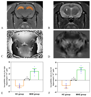 |
QSM in rats with minimal hepatic encephalopathy: does iron overload aggravate cognitive impairment by promoting neuroinflammation?
Xuhong Yang1, Pei Dang2, Jiarui Zheng1, Min Li3, Ruirui Lv1, and Xiaodong Wang2
1Ningxia Medical University, Yingchuan, China, 2Department of Radiology, General Hospital of Ningxia Medical University, Yingchuan, China, 3GE Healthcare, MR Enhancement Application, Beijing, China Keywords: Neurodegeneration, Quantitative Susceptibility mapping, Minimal hepatic encephalopathy; Iron In this study, we used the QSM technique to quantitatively evaluate the levels of brain iron in the hippocampus of MHE rats and by evaluating the neuroinflammatory response to explore the relationship between iron with neuroinflammation and cognition. It is expected to provide important insights into the occurrence of MHE disease symptoms and the related biological mechanisms behind them. Meanwhile, our findings also suggested that an abnormal susceptibility values in certain brain areas identified using the QSM may be a potential biomarker to reflect the severity of cognitive impairment and to monitor its progression in cirrhotic patients with MHE. |
| 17:29 | 0662.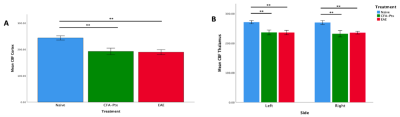 |
A Correlational Study of Changes in Behaviour and MRI Measures in the CNS in the EAE Mouse Model of Multiple Sclerosis
Rania Muhammed1, Rehman Tariq 2, Uche J Ohaezukosi2, Qandeel Shafqat2, Ying Wu2, and Jeff F. Dunn1
1University of Calgary, Calgary, AB, Canada, 2Department of Radiology, University of Calgary, Calgary, AB, Canada Keywords: Multiple Sclerosis, Brain Multiple sclerosis is an inflammatory disease of the central nervous system, with a posited hypoxia-inflammation cycle hypothesis. Hypoxia may increase disease severity in MS. We found R2* was increased (an MRI marker of hypoxia) in the cortex. We observed increased sickness and R2* in EAE mice compared to controls, but reduced CBF in both CFA-PTx and EAE. Inflammation is in both groups, while autoimmunity is only in EAE. Reduced CBF may relate to inflammation, whereas hypoxia may relate to autoimmune-mediated damage and increase disease severity. |
| 17:37 | 0663.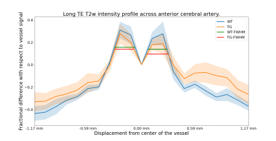 |
Imaging the perivascular and cerebrospinal fluid spaces in TgF344-AD rats using ultra-long echo time MRI
Martin Kozár1,2, Laura Parkes1,2, Hervé Boutin3, Igor Chernyavsky4, and Ben Dickie2,5
1Division of Psychology, Communication and Human Neuroscience, School of Health Sciences, Faculty of Biology, Medicine and Health, The University of Manchester, Manchester, United Kingdom, 2Geoffrey Jefferson Brain Research Centre, Manchester Academic Health Science Centre, Manchester, United Kingdom, 3Division of Neuroscience, School of Biological Sciences, Faculty of Biology, Medicine and Health, The University of Manchester, Manchester, United Kingdom, 4Department of Mathematics, The University of Manchester, Manchester, United Kingdom, 5Division of Informatics, Imaging and Data Sciences, Faculty of Biology, Medicine and Health, The University of Manchester, Manchester, United Kingdom Keywords: Neurofluids, Alzheimer's Disease, Perivascular space Enlargement of perivascular and ventricular spaces are associated with neuroinflammation in neurodegenerative diseases, including cerebral small vessel disease and Alzheimer’s disease. In this study we develop methodology for measuring the anatomy of the perivascular space around the anterior cerebral artery and partial-volume-free estimates of total CSF volume and apply this approach to TgF344-AD rats. Neither the area under the curve of perivascular space profiles, nor overall width of the perivascular space, or the total CSF volume differed between TgF344-AD rats and wild-types. |
 Back to Meeting Home
 Back to the Program-at-a-Glance
The International Society for Magnetic Resonance in Medicine is accredited by the Accreditation Council for Continuing Medical Education to provide continuing medical education for physicians.
 Back to Meeting Home
 Back to the Program-at-a-Glance
 View Presentation Video