Combined Educational & Scientific Session
Fetal MRI: Current Topics in Cardiovascular & Neuroimaging
ISMRM & ISMRT Annual Meeting & Exhibition • 03-08 June 2023 • Toronto, ON, Canada

Combined Educational & Scientific Session
Fetal MRI: Current Topics in Cardiovascular & Neuroimaging
| 13:45 | Fetal Cardiac MRI Mike Seed | |
| 14:05 |  |
Fetal Neuro MRI
Camilo Jaimes
Keywords: Cross-organ: Antenatal, Neuro: Brain, Neuro: Nervous system Fetal life is the most dynamic and crucial stage in human neurodevelopment. The exceptional tissue contrast offered by fetal MRI has revolutionized our ability to study brain development and identify emerging abnormalities. Processes like neuronal migration, parenchymal growth, and cortical folding are displayed in remarkable detail. The potential to detect abnormalities prior to birth opens the possibility for interventions earlier in the course of diseases, which could potentially improve the neurological outcomes of fetuses affected by congenital disorders. |
| 14:25 | 1243.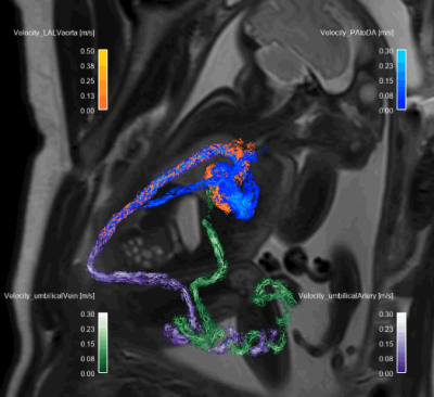 |
4D flow MRI for investigation of fetal cardiovascular hemodynamics in healthy development and ductal dependent lesions
Erin K Englund1, Takashi Fujiwara1, Sarah Smith2, Bettina Cuneo3, Mehdi Hedjazi Moghari1, Mariana L Meyers1, Richard M Friesen3, Lorna P Browne1, and Alex J Barker1 1Radiology, University of Colorado, Anschutz Medical Campus, Aurora, CO, United States, 2Radiology, Children's Hospital Colorado, Aurora, CO, United States, 3Pediatric Cardiology, University of Colorado, Anschutz Medical Campus, Aurora, CO, United States Keywords: Fetal, Velocity & Flow Fetal cardiovascular MRI has the potential to aid in the diagnosis and evaluation of congenital heart disease. Here, a fast, Doppler ultrasound- gated, 4D flow MRI acquisition with online reconstruction was achieved in twelve healthy fetuses and six patients with suspected congenital cardiovascular defects. Analysis of the 4D flow data revealed expected flow distributions across vascular territories in the healthy cohort. Using 4D flow MRI, we were also able to evaluate the presence and impact of ductal dependent lesions in patients with suspected congenital heart disease, finding confirmation of presence/absence of pathology on post-natal imaging. |
| 14:33 | 1244.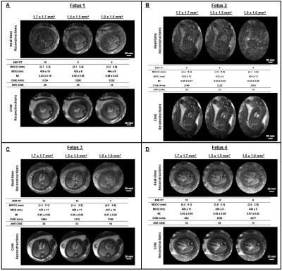 |
Multiresolution comparison of fetal CINE MRI at 0.55 T
Datta Singh Goolaub1, Ye Tian2, Joshua F.P. van Amerom1, John Wood3,4, Jon Detterich4, Krishna S. Nayak2, and Christopher K. Macgowan1,5
1Translational Medicine, The Hospital for Sick Children, Toronto, ON, Canada, 2Ming Hsieh Department of Electrical and Computer Engineering, Viterbi School of Engineering, University of Southern California, Los Angeles, CA, United States, 3Biomedical Engineering, University of Southern California, Los Angeles, CA, United States, 4Division of Cardiology, Department of Pediatrics and Radiology, Children's Hospital Los Angeles, Los Angeles, CA, United States, 5Medical Biophysics, University of Toronto, Toronto, ON, Canada Keywords: Prenatal, Fetus In this study, we demonstrate the feasibility of CINE fetal CMR at 0.55 T at multiple spatial resolutions. First, real-time images are reconstructed for motion-correction and cardiac gating. Fetal cardiac CINEs are then reconstructed using the corrected data. Retrospective CINEs have higher SNR relative to their corresponding real-time reconstructions. Feasibility of the pipeline is demonstrated for up to 1.0 mm in-plane resolution. Good cardiac structure conspicuity is observed at coarse spatial resolutions in real-times and at all spatial resolutions in CINEs. |
| 14:41 | 1245.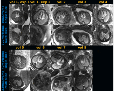 |
Real-time fetal cardiac MRI at 0.55T enables assessment of ventricular function and heart and great vessel anatomy
Ye Tian1, Jon Detterich2, Jay D. Pruetz2, Anand A. Joshi1, John Wood2, and Krishna S. Nayak1
1University of Southern California, Los Angeles, CA, United States, 2Children’s Hospital Los Angeles, Los Angeles, CA, United States Keywords: Fetal, Fetus We demonstrate a real-time spiral balanced steady-state free precession (bSSFP) pulse sequence for fetal cardiac examinations on a 0.55T scanner. The real-time sequence provides easy adjustments of scan plan, automatic volumetric sweeping covering the whole heart, and flexible choice of reconstruction temporal resolution, without relying on any maternal breath-hold or cardiac gating. In 9 experiments involving 8 volunteers, we demonstrated high-temporal resolution (40ms/frame) real-time videos that capture fetal cardiac dynamics, and low-temporal resolution (320ms/frame) volumetric images that capture cardiac anatomy. This approach provides both functional and structural evaluations of the fetal heart. |
| 14:49 | 1246.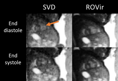 |
Fetal 3D cine cardiovascular MRI: Improved image quality with region-optimized virtual coils
Marjolein Piek1, Johannes Töger1, Erik Hedström1,2, and Anthony H. Aletras1,3
1Clinical Physiology, Department of Clinical Sciences Lund, Lund University, Skåne University Hospital, Lund, Sweden, 2Diagnostic Radiology, Department of Clinical Sciences Lund, Lund University, Skåne University Hospital, Lund, Sweden, 3Laboratory of Computing, Medical Informatics and Biomedical-Imaging Technologies, School of Medicine, Faculty of Health Sciences, Aristotle University of Thessaloniki, Thessaloniki, Greece Keywords: Fetal, Image Reconstruction Recently, the first 3D cine radial acquisition of the fetal heart with isotropic resolution was introduced. However, image quality suffers from radial streaking artifacts due to under-sampling. A new coil combination method, Region-Optimised Virtual coils (ROVir), was suggested to improve image quality by highlighting signal from the ROI while suppressing signal from unwanted regions. This study aimed to compare 3D fetal cardiac imaging ROVir to conventional SVD-based coil combination, in a parallel imaging based reconstruction. Streaking artifacts were reduced, and the cine could be reconstructed to more cardiac phases. This allowed for improved appreciation of fetal cardiac function. |
| 14:57 | 1247.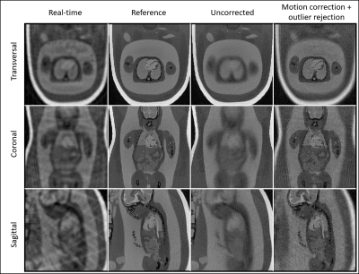 |
Motion-corrected free-running 4D MRI of the fetal heart - from in silico to in vivo
Robin Ferincz1, Mariana Baginha Da Lanca Falcão1, Aurelio Secinaro2, Guido Buonincontri3, Leonor Alamo4, Estelle Tenisch4, Milan Prša5, Davide Piccini1,6,7, Jérôme Yerly8, Matthias Stuber8, and Christopher William Roy1
1Department of Radiology, Lausanne University Hospital (CHUV) and University of Lausanne (UNIL), Lausanne, Switzerland, 2Advanced Cardiothoracic Imaging Unit, Department of Imaging, Bambino Gesù Children’s Hospital IRCCS, Rome, Italy, 3Siemens Healthcare srl, Milan, Italy, Milan, Italy, 4Department of Radiology and Interventional Radiology, Lausanne University Hospital (CHUV) and University of Lausanne (UNIL), Lausanne, Switzerland, 5Woman- Mother-Child Department, Lausanne University Hospital (CHUV) and University of Lausanne (UNIL), Lausanne, Switzerland, 6Advanced Clinical Imaging Technology (ACIT), Siemens Healthineers International AG, Lausanne, Switzerland, 7LTS5, École Polytechnique Fédérale de Lausanne (EPFL), Lausanne, Switzerland, 8Center for Biomedical Imaging (CIBM), Lausanne, Switzerland Keywords: Fetal, Cardiovascular MR imaging of the fetal heart is challenging due to resolution requirements and the impact of maternal respiration, fetal cardiac motion, and gross fetal movement. These factors have largely precluded the development of 3D acquisition techniques. In this work, a novel reconstruction algorithm is developed to estimate and correct for displacement of the fetal heart due to maternal respiration and gross fetal movement enabling the first-ever motion-corrected time-resolved 4D images of the fetal heart from 3D radial data. Proof-of-concept results are demonstrated using a comprehensive numerical simulation developed for this work and initial data acquired in utero. |
| 15:05 | 1248.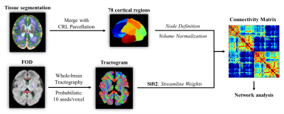 |
Development of the fetal brain structural connectivity during the second-to-third trimester based on diffusion MRI
Ruike Chen1, Ruoke Zhao1, Xinyi Xu1, Mingyang Li1, Cong Sun2, Guangbin Wang3, and Dan Wu1
1Key Laboratory for Biomedical Engineering of Ministry of Education, Department of Biomedical Engineering, College of Biomedical Engineering & Instrument Science, Zhejiang University, Hangzhou, China, 2Department of Radiology, Beijing Hospital, National Center of Gerontology, Institute of Geriatric Medicine, Chinese Academy of Medical Sciences, P.R. China., Beijing, China, 3Department of Radiology, Shandong Provincial Hospital Affiliated to Shandong First Medical University, Jinan, Shandong, China Keywords: Fetal, Brain Connectivity, Structural Connectivity Network Extensive cortico-cortical connections emerge in the fetal brain during the second-to-third trimester with the rapid development of white matter fiber pathways. However, the early establishment and prenatal development of the brain’s structural network are not yet understood. In this work, we built structural connectivity networks of the fetal brain using in-utero diffusion MRI data. Network analysis revealed the increasing overall efficiency of the fetal brain network. The strengthening of short-ranged cortico-cortical connections and the emerging hubs contributed to the reorganization of its sub-units. These findings provided valuable information on the early developmental patterns of brain cortico-cortical structural connectivity |
| 15:13 | 1249.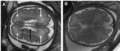 |
Can MRS detect metabolites differences in fetuses affected by CMV?
Or Rachel Sadan1,2, Netanell Avisdris2,3, Aviad Rabinowich2,4,5, Daphna Link-Sourani2, Liat Ben Sira1,4,5, and Dafna Ben Bashat1,2,5
1Sagol School of Neuroscience, Tel Aviv University, Tel Aviv, Israel, 2Sagol Brain Institute, Tel-Aviv Sourasky Medical Center, Tel Aviv, Israel, 3School of Computer Science and Engineering, The Hebrew University of Jerusalem, Jerusalem, Israel, 4Department of Radiology, Tel-Aviv Sourasky Medical Center, Tel Aviv, Israel, 5Sackler Faculty of Medicine, Tel-Aviv University, Tel Aviv, Israel Keywords: Fetal, Infection
We aimed to examine whether MR spectroscopy (MRS) within the deep grey matter can detect brain metabolic changes in fetal cytomegalovirus infection. Retrospective data from 47 fetuses with brain sonography, MRI, and MRS scans were used: 27 women had a positive PCR on amniocentesis. Seven (out of the 27) fetuses had brain MRI findings common to CMV. NAA+NAAG, and Cr were significantly lower in fetuses with PCR+ and MRI findings. No differences were detected in fetuses without imaging findings (PCR+ and PCR-). These metabolic changes could reflect brain impairments. |
| 15:21 | 1250.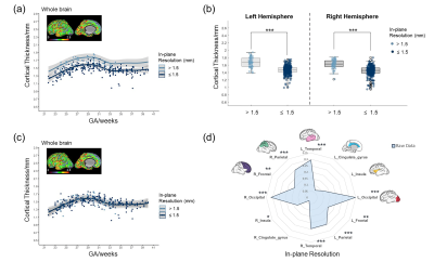 |
Site Effects in Multisite Fetal Brain MRI: a Morphological Study of Early Brain Development
Xinyi Xu1, Haoan Xu1, Tianshu Zheng1, Yutian Wang1, Chi Zhou1, Jiaxin Xiao1,2, Ruike Chen1, Mingyang Li1, Cong Sun3, Guangbin Wang4, Xianglei Kong5, Qingqing Zhu5, Jingshi Wang6, Hong Yu6, Yu Zou7, Guohui Yan7, and Dan Wu1
1Department of Biomedical Engineering, College of Biomedical Engineering & Instrument Science, Zhejiang University, Hangzhou, China, 2School of Biomedical Engineering & Imaging Sciences, Faculty of Life Sciences and Medicine, King’s College London, London, United Kingdom, 3Department of Radiology, Beijing Hospital, National Center of Gerontology, Institute of Geriatric Medicine, Chinese Academy of Medical Sciences, P.R. China., Beijing, China, 4Department of Radiology, Shandong Provincial Hospital Affiliated to Shandong First Medical University, Jinan, China, 5Department of Radiology, Sir Run Run Shaw Hospital, Zhejiang University School of Medicine, Hangzhou, China, 6Dalian Municipal Women and Children’s Medical Center (Group), Dalian, China, 7Department of Radiology, Women’s Hospital, Zhejiang University School of Medicine, Hangzhou, China Keywords: Fetal, Brain, Multisite; morphological development; harmonization; cortical thickness Studies have shown that the non-biological site-related effects may induce bias in multisite neuroimaging studies among adults and adolescents. It is unknown how site effects would affect the analysis of fetal brain MRI and which acquisition factors are critical in quantitative analysis. In this study, we identified site effects, including manufacture, field strength, in-plane resolution, and slice-thickness on volume and cortical thickness measurements in normal fetuses. We also showed these site effects could be effectively removed with ComBat-GAM while preserving developmental pattern indicating that the harmonization procedure is necessary when combing multisite imaging data to study fetal brain morphological development. |
| 15:29 | 1251.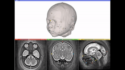 |
Automated atlas-based craniofacial biometry for 3D fetal MRI: multi-acquisition comparison of fetuses with Down syndrome and a control cohort.
Jacqueline Matthew1, Alena Uus1, Abi Fukami-Gartner1, Lucillio Cordero Grande1, Vanessa Kyriakopoulou1, Daniel Cromb1, Robert Wright1, Jonathan O'Muircheartaigh1, Ana Baburamani1, Christina Malamateniou2, Jana Hutter1, Joseph Hajnal1, Mary Rutherford1, and Maria Deprez1
1Centre for the Developing Brain, School of Biomedical Engineering and Imaging Sciences, King's College London, London, United Kingdom, 2City University London, London, United Kingdom Keywords: Prenatal, Fetus, Biometry We introduce the first automated atlas-based method for fetal craniofacial biometry. Using motion-corrected slice-to-volume reconstructions for 3D fetal head visualisation, an automated label propagation method extracted linear biometry across 12 measures. The optimisation process used retrospective data and no differences in automated biometry was seen between different MRI acquisition parameters. A comparison of measures made between a cohort of fetuses with Down syndrome and control fetuses, with normal development, found significant differences for the occipitofrontal skull, oral hard palate and anterior base of skull distances. This suggests a promising and meaningful method for large population-level investigation of MRI craniofacial morphology. |
| 15:37 | 1252.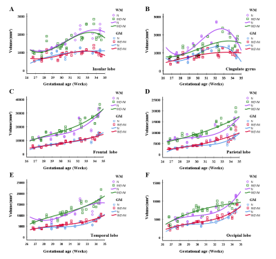 |
Evaluation of the brain volumes development of fetuses with isolated non-severe ventriculomegaly using MRI
Zhaoji Chen1, Fan Wu1, Xin Zhang1, Yuchao Li1, Zhenqing Liu1, Chenxin Xie1, and Hongsheng Liu1
1Department of Radiology, Guangzhou Women and Children's Medical Center, Guangzhou Medical University,Guangdong Provincial Clinical Research Center for Child Health, Guangzhou, 510623, China, Guangzhou of China, China Keywords: Prenatal, Brain The objective of our study to perform the development of brain volume in fetus with the isolated non-severe ventriculomegaly (INSVM). Then the MRI images of 36 INSVM fetuses and 22 normal fetuses were retrospectively collected, and were manually delineated to obtain quantitative data of brain regions. The gestational age ranged from 26 to 35 weeks. Our study found that the volume development of the basal ganglia, thalamus, and partial white matter in the INSVM fetuses were different from those in the normal fetuses. These differences in the volume development of brain regions need to be determined by expanding the sample. |
 Back to Meeting Home
 Back to the Program-at-a-Glance
The International Society for Magnetic Resonance in Medicine is accredited by the Accreditation Council for Continuing Medical Education to provide continuing medical education for physicians.
 Back to Meeting Home
 Back to the Program-at-a-Glance
 View Presentation Video