Combined Educational & Scientific Session
ets Novel Technology
ISMRM & ISMRT Annual Meeting & Exhibition • 03-08 June 2023 • Toronto, ON, Canada

Combined Educational & Scientific Session
ets Novel Technology
| 08:15 | Clinical Value of Quantitative MRI Christopher Hess | |
| 08:35 | New Technologies for Quantitative MRI
Mariya Doneva
Keywords: Image acquisition: Quantification This lecture will provide an overview of the latest advances in MRI techniques that enable quantification of tissue properties. Specific topics that will be covered include approaches for fast quantitative imaging and techniques that aim at improving the accuracy of QMRI. Remaining challenges in quantitative MRI will be briefly discussed as well as potential new research directions. |
|
| 08:55 | 1139.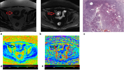 |
Quantitative diffusion and Kurtosis MRI in the evaluation of endometrial cancer: validation with histopathology
Alessandra Maiuro1,2, Francesca Di Stadio2, Serena Satta3, Giorgia Perniola4, Innocenza Palaia4, Angelina Pernazza3, Carlo Della Rocca3, Carlo Catalano3, Lucia Manganaro3, and Silvia Capuani1,2
1Physics, CNR Institute for Complex Systems (ISC), Rome, Italy, 2Physics, Sapienza University of Rome, Rome, Italy, 3Radiological, Oncological and Pathological Sciences, Umberto I Hospital, Sapienza University of Rome, Rome, Italy, 4Maternal and Child Health and Urological Sciences, Umberto I Hospital, Sapienza University of Rome, Rome, Italy Keywords: Pelvis, Cancer, Pelvis, Endometrium, Endometrial cancer, IVIM, Kurtosis To ameliorate the Endometrial cancer (EC) diagnosis and prognosis, IVIM and KURTOSIS models were used to elaborate DWIs obtained from 18 with EC and 20 healthy women. The b-values were 0,30,50,150,500,800,1000,1500,2000,2500s/mm2. DWIs were noise-corrected considering a homomorphic approach. For each EC subject, ROI in the tumor (T) and peritumoral (PT) area were analysed and endometrial area in healthy (H) subjects was also obtained. IVIM and Kurtosis parameters were quantified. K, which quantifies tissue’s complexity, is significantly higher in T and PT than in H. f is higher in PT compared to the other areas, highlighting the perfusive nature of EC. |
| 09:03 | 1140.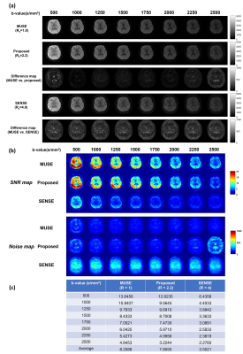 |
Feasibility of Highly-accelerated High b-value Multi-shot Diffusion Weighted Imaging by Using Parametric POCSMUSE and Kurtosis Model
Shihui Chen1 and Hing-Chiu Chang1,2
1The Department of Biomedical Engineering, The Chinese University of Hong Kong, Hong Kong, Hong Kong, 2Multi-scale Medical Robotics Center, Hong Kong, Hong Kong Keywords: Image Reconstruction, Diffusion/other diffusion imaging techniques, high b-value diffusion weighted imaging, multi-shot diffusion-weighted imaging High b-value DWI is promising in detection of white matter pathology and infarctions. However, the disadvantages of the acquired high b-value DWI, such as insufficient SNR and image distortions, prohibits its clinical application. Though the feasibility of computed high b-value has been estimated in prostate cancer, the parameters derived from low b-value images cannot be used for diffusion kurtosis model fitting and achieved inferior performance at high b-value. In this study, we proposed a framework based on parametric POCSMUSE and kurtosis model to generate multiple high b-value images with comparable image quality to MUSE from highly-accelerated high b-value DWI. |
09:11 |
1141.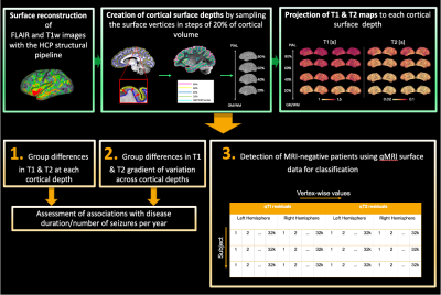 |
Widespread, depth-dependent microstructural damage in the cortex of children with focal epilepsy: A quantitative T1 and T2 mapping study
Chiara Casella1, Katy Vecchiato1,2, Daniel Cromb1, Yourong Guo1, Emer Hughes1, Louise Dillon1, Elaine Green1, Kathleen Colford1, Anthony Price1, Lucilio Cordero Grande3,4,5,6, Tobias C. Wood7, Shaihan Malik3, Rui Pedro A. G. Teixeira3, David W. Carmichael3, and Jonathan O'Muircheartaigh1,2,8
1Centre for the Developing Brain, School of Biomedical Engineering and Imaging Sciences, King's College London, London, United Kingdom, 2Department for Forensic and Neurodevelopmental Sciences, Institute of Psychiatry, Psychology and Neuroscience, King's College London, London, United Kingdom, 3Department of Biomedical Engineering, King's College London, London, United Kingdom, 4Biomedical Image Technologies, ETSI Telecomunicación, Madrid, Spain, 5Universidad Politécnica de Madrid, Madrid, Spain, 6Biomedical Research Networking Center in Bioengineering, Biomaterials and Nanomedicine (CIBER-BBN), Madrid, Spain, 7Department of Neuroimaging, King's College London, London, United Kingdom, 8MRC Centre for Neurodevelopmental Disorders, London, United Kingdom Keywords: Epilepsy, Relaxometry, Paediatric We assessed cortical microstructure in children with drug-resistant focal epilepsy using T1 and T2 relaxometry (qT1 and qT2). We show widespread, depth-mediated qT1 and qT2 increases, and alterations in intracortical organisation in patients. Changes did not correlate with clinical parameters, suggesting that they may be independent of disease severity. Using a random forest algorithm, we also show that qT1 and qT2 surface-features from patients with radiologically defined abnormalities (MRI-positive) and controls, can classify patients without reported radiological abnormalities (MRI-negative). This suggests a common imaging endophenotype of focal epilepsy irrespective of visible abnormalities that may be present at a pre-symptomatic disease-stage. |
| 09:19 |
1142. |
T1 and T2 Mapping Using Highly Sparse Unsuppressed Water Signals from MRSI Scans with Generalized Series-Assisted Low-Rank Tensor Modelling
Yudu Li1,2, Rong Guo1,3, Yibo Zhao1,4, Wen Jin1,4, Chao Ma5,6, Shirui Luo2, Georges El Fakhri5,6, Yao Li7, Maria Jaromin2, Volodymyr Kindratenko2,4, Brad Sutton1,2,8,9, and Zhi-Pei Liang1,2,4
1Beckman Institute for Advanced Science and Technology, University of Illinois at Urbana-Champaign, Urbana, IL, United States, 2National Center for Supercomputing Applications, University of Illinois at Urbana-Champaign, Urbana, IL, United States, 3Siemens Medical Solutions, Urbana, IL, United States, 4Department of Electrical and Computer Engineering, University of Illinois at Urbana-Champaign, Urbana, IL, United States, 5Radiology, Harvard Medical School, Boston, MA, United States, 6Radiology, Massachusetts General Hospital, Boston, MA, United States, 7School of Biomedical Engineering, Shanghai Jiao Tong University, Shanghai, China, 8Department of Bioengineering, University of Illinois at Urbana-Champaign, Urbana, IL, United States, 9Carle Illinois College of Medicine, University of Illinois at Urbana-Champaign, Urbana, IL, United States Keywords: Quantitative Imaging, Quantitative Imaging MR spectroscopic imaging (MRSI) without water suppression provides a unique opportunity to use the unsuppressed water spectroscopic signals for T1 and T2 mapping. This work presents a new image reconstruction method for reconstructing the T1/T2 maps from the highly sparse MRSI data. This method uses a novel generalized series-assisted low-rank tensor model to absorb the high-quality reference MRSI images to constrain the spatial-spectral-parametric variations. Experimental results demonstrated very encouraging reconstruction performance. |
| 09:27 | 1143.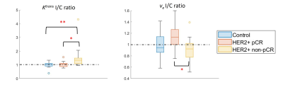 |
Bilateral asymmetry of parenchymal kinetics from ultrafast DCE-MRI predicts HER2+ breast cancer response to neoadjuvant chemotherapy
Zhen Ren1, Federico D. Pineda2, Frederick M. Howard3, Hiroyuki Abe1, Kirti Kulkarni1, Rita Nanda3, Nora T. Jaskowiak4, and Gregory S. Karczmar1
1Department of Radiology, The University of Chicago, Chicago, IL, United States, 2Department of Radiology, University of Pittsburgh, Pittsburgh, PA, United States, 3Department of Medicine - Section of Hematology/Oncology, The University of Chicago, Chicago, IL, United States, 4Department of Surgery, The University of Chicago, Chicago, IL, United States Keywords: Breast, Cancer, HER2-positive breast cancer We retrospectively reviewed data from 28 patients with HER2-positive breast cancer treated with neoadjuvant chemotherapy (NAC) who underwent a protocol that included ultrafast DCE-MRI (temporal resolution = 3 – 9 seconds) for the first minute after contrast administration prior to NAC. We measured quantitative kinetic background parenchymal enhancement parameters (kBPEs) from ipsi- and contra-lateral normal parenchyma separately to quantify bilateral parenchymal enhancement asymmetry. The results show that HER2-positive patients with similar pre-NAC $$$K^{trans}$$$ in ipsi-and contralateral normal parenchyma were more likely to achieve pathologic complete response post NAC. |
| 09:35 | 1144.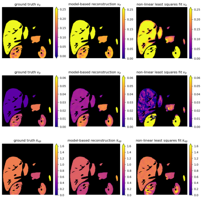 |
Direct estimation of pharmacokinetic parameters of DCE-MRI from raw k-space data with model-based reconstruction
Natalia V Korobova1, Susanne S Rauh2, Marian A Troelstra1, Matthew R Orton3, Eric M. Schrauben1, Oliver Maier4, Aart J Nederveen1, and Oliver J Gurney-Champion1
1Department of Radiology and Nuclear Medicine, Amsterdam University Medical Center, Amsterdam, Netherlands, 2Department of Biomedical Engineering and Physics, Amsterdam University Medical Center, Amsterdam, Netherlands, 3Division of Radiotherapy and Imaging, The Institute of Cancer Research, London, United Kingdom, 4Institute of Medical Engineering, Technical University Graz, Graz, Austria Keywords: Contrast Agent, Quantitative Imaging, Model-based reconstruction Dynamic contrast enhanced (DCE) MRI is a minimally invasive technique that is able to quantitatively investigate the tumor vasculature microenvironment. Such information shows great potential for treatment stratification and response monitoring. However, DCE typically suffers from low spatial resolution, Rician noise bias, and errors due to complex perfusion modeling. Model-based reconstruction, in which DCE parameters are estimated directly from k-space, may overcome these shortcomings. In this study, we implemented model-based reconstruction for DCE-MRI data, validated it in simulations, and showed its performance in-vivo. With model-based reconstruction the estimated parameter maps exhibited less noise and preserved more anatomical details. |
| 09:43 | 1145.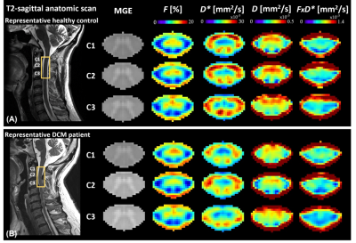 |
Investigating spinal cord perfusion impairment in degenerative cervical myelopathy (DCM) using Intravoxel Incoherent Motion (IVIM) MRI
Anna Lebret1, Simon Lévy2, Nikolai Pfender1, Mazda Farshad3, Virginie Callot4,5, Armin Curt1, Patrick Freund1,6, and Maryam Seif1,6
1Spinal Cord Injury Center, Balgrist University Hospital, University of Zurich, Zurich, Switzerland, 2MR Research Collaborations, Siemens Healthcare Pty Ltd, Melbourne, Australia, 3Department of Orthopedic Surgery, Balgrist University Hospital, University of Zurich, Zurich, Switzerland, 4Aix-Marseille Univ, CNRS, CRMBM, Marseille, France, 5APHM, Hôpital Universitaire Timone, CEMEREM, Marseille, France, 6Max Planck Institute for Human Cognitive and Brain Sciences, Leipzig, Germany Keywords: Spinal Cord, Neurodegeneration, Degenerative Cervical Myelopathy Degenerative cervical myelopathy (DCM) is the most common form of non-traumatic spinal cord injury, mainly caused by chronic cervical cord compression. Impaired spinal cord perfusion is a central pathophysiological tenet in DCM patients. This study aims at investigating non-invasively DCM-induced changes of blood perfusion in the spinal cord above a cervical myelopathy using quantitative MRI techniques. Cardiac-gated intravoxel incoherent motion (IVIM) 3T MRI, sensitive to perfusion, was applied to the cervical cord (C1-C3) in 25 DCM patients and 27 healthy controls. DCM showed tissue-specific perfusion impairment. IVIM maps suggested remote hemodynamic deficit induced by cervical cord compression in DCM. |
| 09:51 | 1146.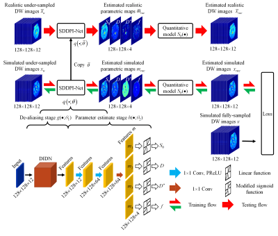 |
IVIM reconstruction from highly under-sampled DW-PROPELLER acquisition data via synthetic data-driven physics-informed deep learning
Jiechao Wang1, Wenhua Geng1, Jian Wu1, Taishan Kang2, Zhigang Wu3, Jianzhong Lin2, Congbo Cai1, and Shuhui Cai1
1Department of Electronic Science, Fujian Provincial Key Laboratory of Plasma and Magnetic Resonance, Xiamen University, Xiamen, China, 2Department of Radiology, Zhongshan Hospital of Xiamen University, School of Medicine, Xiamen University, Xiamen, China, 3Clinical & Technical Solutions, Philips Healthcare, Shenzhen, China Keywords: Quantitative Imaging, Diffusion/other diffusion imaging techniques A synthetic data-driven physics-informed network (SDDPI-Net) was proposed for intravoxel incoherent motion (IVIM) mapping based on highly under-sampled diffusion-weighted turbo spin echo PROPELLER (DW-TSE-PROPELLER) data. This reconstruction network directly estimated distortion-free and artifacts-free IVIM parameters by explored data redundancy in the k-b space and IVIM bi-exponential model with synthetic training data. The results of human brain experiments show that our method can significantly improve the accuracy of IVIM maps with 6´ under-sampled DW-TSE-PROPELLER than other methods. |
| 09:59 |
1147.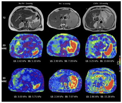 |
Noninvasive diagnosis of clinically significant portal hypertension with MR elastography, T1, and T1ρ mapping of the liver and spleen
Octavia Bane1,2, Efe Ozkaya1,2, Paul Kennedy1,2, Aaron Fischman1, Swan Thung3, and Bachir Taouli1,2
1Department of Diagnostic, Molecular and Interventional Radiology, Icahn School of Medicine at Mount Sinai, New York, NY, United States, 2BioMedical Engineering and Imaging Institute, Icahn School of Medicine at Mount Sinai, New York, NY, United States, 3Department of Pathology, Icahn School of Medicine at Mount Sinai, New York, NY, United States Keywords: Liver, Quantitative Imaging, Portal Hypertension, Clinically Significant Portal Hypertension In this prospective study, we explored the diagnostic value of MR elastography (MRE) as well as T1-pre and post-gadoxetate contrast at the hepatobiliary phase and T1ρ mapping of the liver and spleen for noninvasive diagnosis of clinically significant portal hypertension (CSPH) in patients with liver disease. 2D MRE liver (r=0.457, p<0.001) and spleen stiffness (r=0.438, p<0.001) showed a strong significant correlation with portal pressure measurement based on hepatic venous pressure gradient (HVPG). 2D MRE spleen stiffness outperformed other imaging parameters for prediction of CSPH [AUC = 0.867 (0.764-0.970)]. |
10:07 |
1148.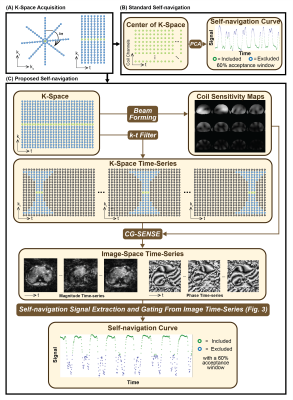 |
Image-Space Self-Navigation for Respiratory Motion Compensation in 2D Axial Radial Free-Breathing MRE of the Liver
Timoteo I. Delgado1,2, Sevgi Gokce Kafali1,3, Shu-Fu Shih1,3, Timothy R. Adamos4, Shahnaz Ghahremani1, Kara L. Calkins5, Xiaodong Zhong6, Vibhas Deshpande7, Bradley D. Bolster Jr.8, and Holden H. Wu1,2,3
1Radiological Sciences, University of California Los Angeles, Los Angeles, CA, United States, 2Physics and Biology in Medicine, University of California Los Angeles, Los Angeles, CA, United States, 3Bioengineering, University of California Los Angeles, Los Angeles, CA, United States, 4Gastroenterology, University of California Los Angeles, Los Angeles, CA, United States, 5Pediatrics, University of California Los Angeles, Los Angeles, CA, United States, 6Siemens Medical Solutions USA, Inc., Los Angeles, CA, United States, 7Siemens Medical Solutions USA, Inc., Austin, TX, United States, 8Siemens Medical Solutions USA, Inc., Salt Lake City, UT, United States Keywords: Motion Correction, Liver Previous work has proposed self-navigated golden-angle (GA) radial free-breathing (FB) MR elastography (MRE) of the liver. This work employed a standard DC-based motion compensation framework that proved suboptimal. Here we propose an image-space based motion-compensation framework for 2D free-breathing radial MRE. The proposed method is compared to the standard DC-based method by measuring the signal-to-noise (SNR) after self-navigated motion-compensation by each method (8 subjects). The median (interquartile range) SNR across subjects was 4.8 (3.6-5.7) and 6.8 (6.3-7.5) for the standard and proposed methods, respectively. Inclusion of image-space data may allow for more robust motion compensation for 2D radial axial acquisitions. |
 Back to Meeting Home
 Back to the Program-at-a-Glance
The International Society for Magnetic Resonance in Medicine is accredited by the Accreditation Council for Continuing Medical Education to provide continuing medical education for physicians.
 Back to Meeting Home
 Back to the Program-at-a-Glance
 View Presentation Video