Digital Poster
Radiomics
ISMRM & ISMRT Annual Meeting & Exhibition • 03-08 June 2023 • Toronto, ON, Canada

| Computer # | |||
|---|---|---|---|
2880.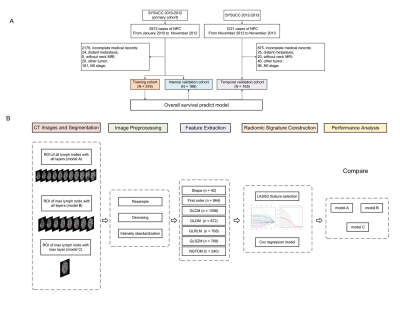 |
1 |
Three Delineating Radiomics Models Based on MR-determined
Metastatic Lymph Nodes for Prognostic Prediction in
Nasopharyngeal Carcinoma
Manqian Huang1,
Kan Deng2,
Hui Xie1,
Wenjie Huang1,
Xiaoyi Wang3,
Chao Luo1,
Shuqi Li1,
Chunyan Cui1,
Huali Ma1,
Lizhi Liu1,
and Haojiang Li1
1Sun Yat-sen University Cancer Center, Guangzhou, China, 2Philips Healthcare, Guangzhou, China, 3Hainan General Hospital (Hainan Affiliated Hospital of Hainan Medical University), Haikou, China Keywords: Radiomics, Cancer, Nasopharyngeal Carcinoma The purpose of this study was to assess the performance of radiomic models based on MR-determined metastatic lymph nodes phenotype in predicting the prognosis of nasopharyngeal carcinoma (NPC) with three different feature selection methods: all lymph nodes (ALN), the largest lymph node (LLN), and the largest slice of the largest lymph node (LSLN). The results showed that LSLN radiomic features showed better accuracy in predicting the overall survival (OS). |
|
2881. |
2 |
MRS combined with enhanced silhouette in the prediction of
malignant glioma radiomics classification
jing song1,
hui qian zong1,
ya zhang1,
jing wang1,
hong yang wei1,
and li zhi xie2
1The Second Hospital of Hebei Medical University, Shijiazhuang, China, 2GE Healthcare, Beijing, China Keywords: Radiomics, Brain, Magnetic resonance spectroscopy Question: Among the studies using radiomics methods to predict glioma grading, most of them are based on conventional magnetic resonance imaging sequences, and functional magnetic resonance imaging is less studied. Methods: This study predicted malignant glioma grading based on magnetic resonance structural images and magnetic resonance spectroscopy using an radiomics approach. Results: The test set AUC of the model constructed based on T1-enhanced images and the ratio of three metabolites of MRS was 0.95. Conclusion: Radiomics based on T1-CE and MRS has a good performance in identifying both grade III and grade IV gliomas. |
|
2882. |
3 |
Radiomic feature reliability of protein-based amide proton
transfer-weighted images of brain tumors acquired with
compressed sensing
Jingpu Wu1,2,
Yiqing Shen1,3,
Qianqi Huang3,
Pengfei Guo1,3,
Jinyuan Zhou1,
and Shanshan Jiang1
1Department of Radiology, School of Medicine, Johns Hopkins University, Baltimore, MD, United States, 2Department of Applied Mathematics and Statistics, Whiting School of Engineering, Johns Hopkins University, Baltimore, MD, United States, 3Department of Computer Science, Whiting School of Engineering, Johns Hopkins University, Baltimore, MD, United States Keywords: Radiomics, Radiomics Sensitivity encoding (SENSE) is a conventional practice for accelerating APTw image acquisition. To achieve an even higher acceleration, SENSE with compressed sensing (CS-SENSE) was introduced. However, its effect on the radiomic features extracted from the APTw images was yet studied. Here we extracted radiomic features from both SENSE- and CS-SENSE-APTw images and evaluated their reliability in different regions of interest (ROIs). Moreover, filters play an important part in emphasizing specific image characteristics in radiomics. The impact of filters on the radiomic features were also discussed. Our results provided a comprehensive reference for radiomic analyses where CS is implemented for acceleration. |
|
2883. |
4 |
Reproducibility of radiomics features between MRI-derived
synthetic-CT and true CT in prostate MR-guided radiotherapy
Yihang Zhou1,
Jing Yuan1,
Cindy Xue1,
Bin Yang2,
Kin Yin Cheung2,
and Siu Ki Yu2
1Research Department, Hong Kong Sanatorium and Hospital, Hong Kong, China, 2Medical Physics Department, Hong Kong Sanatorium and Hospital, Hong Kong, China Keywords: Radiomics, Prostate, MRCAT, synthetic-CT, reproducibility MR-guided radiotherapy and radiomics have gained considerable attention. Synthetic-CT (sCT) derived from MRI has been adopted to facilitate MRI-only radiotherapy planning. Researches have evaluated the sCT both qualitatively and quantitatively. However, few studies have looked into the potential that sCT offers at radiomics level. We hypothesize that sCT generated from MR can faithfully reproduce radiomics features compared to those extracted from true planning-CT. We aim to investigate the reproducibility of radiomics features derived from a commercially available sCT generation (MRCAT) acquired on a 1.5T MR-simulator to those obtained from a CT simulation scan in a cohort of prostate cancer patients. |
|
2884.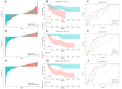 |
5 |
Prediction of overall survival for astrocytoma with whole-tumor
radiomics analysis based on diffusion kurtosis imaging
Yan Tan1,
Dawei Tian1,
Wenqiao Zheng1,
Xiaochun Wang1,
and Hui Zhang1
1Department of Radiology, First Hospital of Shanxi Medical University, Taiyuan, China Keywords: Radiomics, Diffusion/other diffusion imaging techniques For investigating the usefulness of radiomics signature based on diffusion kurtosis imaging in overall survival prediction for astrocytoma, radiomics features extracted from whole-tumor on MK and MD images were selected to construct radiomics signature. Then the radiomics signature was integrated with clinical-genetic risk factors to develop a combined model, which was represented as nomogram. The results showed that the radiomics signature have offered clinically relevant prognostic information for astrocytoma and further stratified patients into different risk groups. The combined model achieved the highest predictive performance and facilitated clinical decision-making by nomogram, demonstrating the incremental value of radiomics signature. |
|
2885.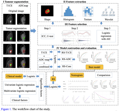 |
6 |
Potential anti-HER2 target therapy beneficiaries: can MRI
radiomics identify the status of HER2-low in breast cancer?
Xiaoqian Bian1,
Zhibin Yue2,
Siyao Du1,
Yan Xu3,
Yang Song3,
Min Zhao4,
and Lina Zhang1
1The First Hospital of China Medical University, Shenyang, China, 2Department of Biomedical Engineering, School of Intelligent Medicine, China Medical University, Shenyang, China, 3MR Scientific Marketing, Siemens Healthineers, Shanghai, China, 4Pharmaceutical Diagnostics, GE Healthcare, Beijing, China Keywords: Radiomics, Breast Recently, HER2-low expression tumor was proposed as a new entity in the field of breast cancer and radiomics studies related to HER2-low are rare. This study investigated whether multiparametric MRI-based radiomics can distinguish HER2- low from HER2-positive and HER2-zero in breast cancer. Results showed that clinico-radiomics nomograms including hormone receptor status and radiomics signatures combined contrast-enhanced T1-weighted and apparent diffusion coefficient map performed best in both prediction tasks: HER2-positive vs. HER2-negative and HER2-low vs. HER2-zero. This suggests that multiparametric MRI radiomics achieved effective prediction of HER2-low, which might be further guidance of the anti-HER2 targeted therapy in breast cancer. |
|
2886.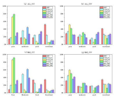 |
7 |
Effects of SMS accelerating factor on stability of radiomics
features from quantitative parametric maps of IVIM and DKI in
Cervical Cancer
Ai Shuangquan1,2,
Peng Wei1,
He Yaoyao1,
Zhang Huiting3,
Grimm Robert4,
Zhang Zhaoxi1,
Peng Lin5,
Liu Yulin1,
and Yuan Zilong1
1Department of Radiology, Hubei Cancer Hospital, Tongji Medical College, Huazhong University of Science and Technology, Wuhan, China, 2School of Biomedical Engineering, South-Central Minzu University, Wuhan, China, 3MR Scientific Marketing, Siemens Healthcare, Wuhan, China, Wuhan, China, 4MR Application Predevelopment, Siemens Healthcare GmbH, Erlangen, Germany, Erlangen, Germany, 5Department of Gynecology, Hubei Cancer Hospital, Tongji Medical College, Huazhong University of Science and Technology, Wuhan, China Keywords: Radiomics, Cancer, Cervical Cancer The aim of this study was to explore the effects of SMS on radiomics features in quantitative parametric maps based on IVIM and DKI models. Meanwhile, the effects of whole-tumor and maximal-tumor layer delineation on radiomics features were compared, whether stable features are associated with clinical staging of cervical cancer was analyzed. The results showed that SMS had the greatest effect on features extracted from D* and f map from IVIM model and the least effect on features extracted from ADC map; SMS had a greater effect on the radiomics features extracted by maximum-tumor layer delineation than by whole-tumor delineation. |
|
2887.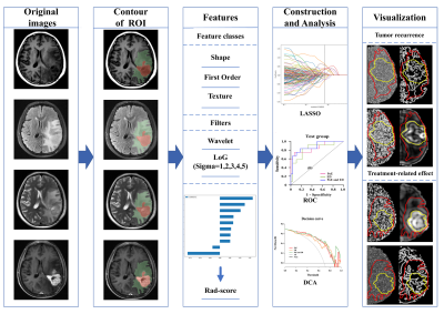 |
8 |
The value of regional radiomics score based on postoperative
conventional MRI in evaluation of glioma recurrence
Jinfa Ren1,
Xiaoyang Zhai1,
Dongming Han1,
Huijia Yin1,
Ruifang Yan1,
and Kaiyu Wang2
1Department of MR, The First Affiliated Hospital of Xinxiang Medical University, Weihui, China, 2GE Healthcare, MR Research China, Beijing, China Keywords: Radiomics, Radiomics Early diagnosis of postoperative glioma recurrence is difficult. But emerging measurements of radiomics could provide a powerful tool for this dilemma. We used the least absolute shrinkage and selection operator to select features and generate radiomics scores based on multiple modalities of conventional MRI to discriminate recurrence from treatment-related effects. We found that tumor recurrence could be independently identified by features from both the postoperative enhanced region and edematous region with a best performance of the combined one. |
|
2888.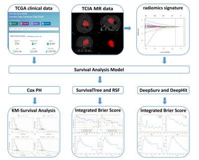 |
9 |
Comparison of radiomics-based machine learning survival models
in predicting prognosis of glioblastoma
Jixin Luan1,
Chuanchen Zhang2,
Bin Liu1,
Aocai Yang1,
Kuan Lv3,
Pianpian Hu3,
and Guolin Ma1
1China-Japan Friendship Hospital, Chinese Academy of Medical Sciences & Peking Union Medical College, Beijing, China, 2Department of Radiology, Liaocheng People's Hospital, Shandong First Medical University & Shandong Academy of Medical Sciences, Liaocheng, China, 3China-Japan Friendship Hospital, Beijing, China Keywords: Radiomics, Cancer In this study, we aimed to compare the performance of radiomics-based machine learning survival models in predicting the prognosis of glioblastoma multiforme (GBM) patients. The Cox proportional-hazards model (Cox-PH) and SurvivalTree, Random survival forest (RSF), DeepHit, DeepSurv four machine learning models were constructed, and the performance of the models was evaluated using C-index. We found that deep learning algorithms based on radiomics in predicting the overall survival of GBM patients, and the DeepSurv model showed the best predictive ability. |
|
2889.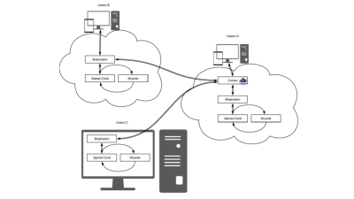 |
10 |
Rado – A Cloud-Based Toolbox for Radiomics Analysis
Eros Montin1,2 and
Riccardo Lattanzi1,2,3
1Center for Advanced Imaging Innovation and Research (CAI2R) Department of Radiology, Radiology Department, New York University Grossman School of Medicine, New York, New York, USA, New York, NY, United States, 2Bernard and Irene Schwartz Center for Biomedical Imaging, Department of Radiology, New York University Grossman School of Medicine, New York, New York, USA, New York, NY, United States, 3Vilcek Institute of Graduate Biomedical Sciences, New York University Grossman School of Medicine, New York, New York, USA, New York, NY, United States Keywords: Radiomics, Software Tools Rado is a web-based application designed to fully automatically perform a radiomic analysis. Rado allows users to extract features from medical images, apply feature selections, and mine the dataset using the most common classifiers and regressors. It also offers the option to augment the dataset by means of rigid transformations.The current version of Rado allows users to interact with the dataset by means of restful APIs or using a standardized web GUI. The application will be distributed via the Cloud MR portal, which allows running the feature extraction on sparse servers as well as on local computers. |
|
2890.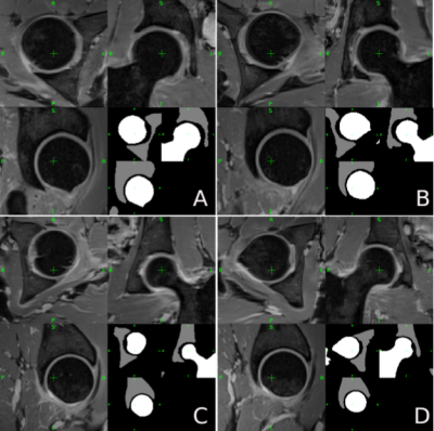 |
11 |
A radiomic approach to the diagnosis of femoroacetabular
impingement
Eros Montin1,2,
Richard Kijowski3,
and Riccardo Lattanzi1,2,4
1Center for Advanced Imaging Innovation and Research (CAI2R) Department of Radiology, Radiology Department, New York University Grossman School of Medicine, New York, New York, USA, New York, NY, United States, 2Bernard and Irene Schwartz Center for Biomedical Imaging, Department of Radiology, New York University Grossman School of Medicine, New York, New York, USA, New York, NY, United States, 3Department of radiology, New York University Grossman School of Medicine, New York, NY, United States, New York, NY, United States, 4Vilcek Institute of Graduate Biomedical Sciences, New York University Grossman School of Medicine, New York, New York, USA, New York, NY, United States Keywords: Radiomics, MSK The results of this study showed that radiomic can automatically distinguish a healthy joint from one with impingement using water-only Dixon MRI. To our knowledge, this is the first application of radiomic for FAI diagnosis. Our radiomic analysis achieved an accuracy greater than 97%, which is higher than the 90% accuracy for detecting FAI reported for standard diagnostic tests (90%). Combining our proposed radiomic analysis with methods for automated joint segmentation could be used to rapidly identify patients with FAI, avoiding time-consuming radiological measurements of bone morphology. |
|
2891.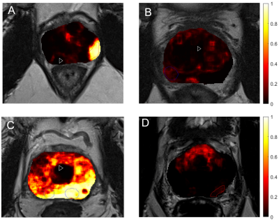 |
12 |
Radiomics-based Machine Learning for Predicting Clinically
Significant Cancer in Multicenter Cohort: Comparison to PI-RADS
Reading
Gabriel Addio Nketiah1,2,
Mohammed RS Sunoqrot 1,3,
Elise Sandsmark3,
Sverre Langørgen 3,
Kirsten M Selnæs 1,3,
Helena Bertilsson 1,4,
Mattijs Elschot 1,3,
and Tone F Bathen1,3
1Department of Circulation and Medical Imaging, Norwegian University of Science and Technology, Trondheim, Norway, 2Department of Radiology and Nuclear Medicine, St. Olavs Hospital, Trondheim University Hospital,, Trondheim, Norway, 3Department of Radiology and Nuclear Medicine, St. Olavs Hospital, Trondheim University Hospital, Trondheim, Norway, 4Department of Urology, St. Olavs Hospital, Trondheim University Hospital, Trondheim, Norway Keywords: Machine Learning/Artificial Intelligence, Prostate Synopsis: Recently, predictive machine learning models have shown promise for prostate cancer diagnosis. The utility of MRI radiomic features for prostate cancer detection and classification has been shown several studies, but mostly using relatively small and single centre cohort. In this study, we showed that radiomics-based machine learning can perform relatively well compared to clinical practice, especially in large multicentre settings. On the patient-level analysis, the areas under the receiver-operating curves for PI-RADS reading by a radiologist and machine learning model were 90% and 89%, respectively. |
|
2892.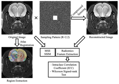 |
13 |
The Impact of Compressed Sensing on Radiomics of Fast Anatomical
MRI of Rat Brain at 7T
Arda Arpak1,
Esra Sümer1,
Asım Samlı1,
Pınar S. Özbay1,
Uluç Pamuk2,3,
Kübra Çağlar2,
Elif M. Demir2,
Mehmet Yumak2,
Can A. Yucesoy1,
and Esin Ozturk-Isik1
1Institute of Biomedical Engineering, Boğaziçi University, İstanbul, Turkey, 2Targeted Therapy Technologies Experimental Animal Imaging Laboratory, Boğaziçi University, İstanbul, Turkey, 3Department of Biomedical Engineering, İstinye University, İstanbul, Turkey Keywords: Image Reconstruction, Preclinical, Radiomics This study aims to investigate the effect of compressed sensing reconstruction on radiomics parameters of rat brain MRI at 7T. T2 weighted MRI of rat brain were randomly under-sampled with R=2.2 and reconstructed using compressed sensing. Brains were segmented after registration to a Wistar rat brain atlas. The radiomics of original and accelerated MRI data were compared in seven scans of rat brains using a Wilcoxon signed-rank test. Stable radiomics features were identified using intraclass correlation coefficients. The findings of this study indicated that not all radiomics features of rat brain were robust to compressed sensing acceleration. |
|
2893.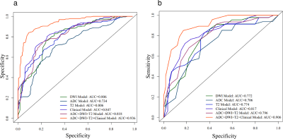 |
14 |
Preoperative MRI-based radiomic-clinical nomogram to predict
residual tumor for advanced high-grade serous ovarian carcinoma
Jingjing Lu1,
Songqi Cai1,
Fang Wang2,
Pu-Yeh Wu3,
Xianpan Pan2,
Jinwei Qiang4,
Haiming Li5,
and Mengsu Zeng1
1Department of Radiology, Zhongshan Hospital, Fudan University, Shanghai, China, 2Department of Research and Development, Shanghai United Imaging Intelligence Co., Ltd, Shanghai, China, 3GE Healthcare, Beijing, China, 4Department of Radiology, Jinshan Hospital, Fudan University, Shanghai, China, 5Department of Radiology, Fudan University Shanghai Cancer Center, Shanghai, China Keywords: Machine Learning/Artificial Intelligence, Radiomics, Ovarian carcinoma, Residual tumor prediction Residual tumor (RT) status is associated with the prognosis and survival rate of patients with high-grade serous ovarian carcinoma (HGSOC). However, current RT status prediction approach through laparoscopy has disadvantages of invasiveness, high cost and incidence of tumor metastases. In this study, we proposed a radiomic-clinical nomogram, based on multiple-sequence MRI combined with score of abdominal metastases and clinical markers, for preoperative prediction of RT status. We demonstrated that the radiomic-clinical nomogram had satisfactory prediction performance in all cohorts (AUC = 0.900-0.936). The clinical application value of the nomogram was further confirmed by decision curves. |
|
2894.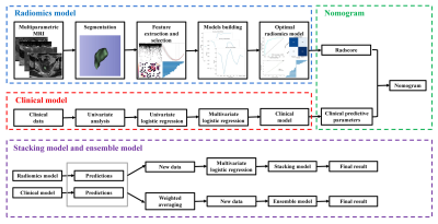 |
15 |
Different multiparametric MRI-based radiomics models for
differentiating stage IA endometrial cancer from benign
endometrial lesions: A multicenter study
Qiu Bi1,
Kunhua Wu1,
and Yunzhu Wu2
1the First People’s Hospital of Yunnan Province, Kunming, China, 2Siemens Healthcare, Shanghai, China Keywords: Artifacts, Uterus In the study, age and irregular vaginal bleeding were the valid predictive parameters in clinical model. On the basis of several common machine learning algorithms, the diverse multiparametric MRI-based radiomics models were developed to differentiate stage IA EC from benign endometrial lesions, and LR algorithm model were selected as the optimal radiomics model with the highest AUC and accuracy. Compared with clinical model and radiologist, the optimal radiomics model and the compositive models combining clinical parameters with radiomics features, like the nomogram, stacking model, and ensemble model showed better diagnostic performance and achieved good clinical net benefits. The nomogram had a higher AUC than that of the optimal radiomics model, and revealed more stable discrimination efficiency and better generalization ability than stacking and ensemble modals. |
|
2895.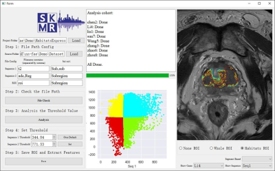 |
16 |
nnFAE: An Extended Module for FeAture Explorer (FAE) for
Radiomic Feature Processing
Yang Song1,
Chengxiu Zhang2,
Jing Zhang2,
Shaoyu Wang1,
Xu Yan1,
Yefeng Yao2,
and Guang Yang2
12. MR Scientific Marketing, Siemens Healthcare, Shanghai, China, 21. Shanghai Key Laboratory of Magnetic Resonance, East China Normal University, Shanghai, China Keywords: Machine Learning/Artificial Intelligence, Software Tools The biological meaning, model robustness and the harmonization of the features are the focuses in the current radiomics development. We designed a software named nnFAE which extends the open-source FeatureExplorer (FAE) to extract habitats features using multi-parameter MR images, to extract robust features recommended by IBSI, and to harmonize features from multi-vendors, etc. nnFAE has a graphic user interface to process the images and feature matrix in batch and can be used readily in radiomics studies. |
|
2896.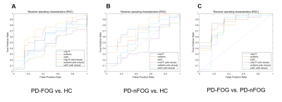 |
17 |
Application of radiomics approach on predicting freezing of gait
in Parkinson’s disease based on rs-fMRI indices
Miaoran Guo1,
Hu Liu1,
and Guoguang Fan1
1The First Hospital of China Medical University, Shenyang, China Keywords: Machine Learning/Artificial Intelligence, fMRI (resting state), Parkinson’s disease, freezing of gait, feedforward neural network, receiver operating characteristic.
|
|
2897. |
18 |
Feasibility of texture analysis of cocaine use disorder
patients’ MRI data to predict early brain changes
Pavan a Poojar1,
Priti Balchandani2,
Yasmin Hurd3,
Shilpa Taufique3,
and Sairam Geethanath1
1Accessible Magnetic Resonance Laboratory, Biomedical Imaging and Engineering Institute, Department of Diagnostic, Molecular and Interventional Radiology, Icahn School of Medicine at Mount Sinai, New York, NY, United States, 2Biomedical Imaging and Engineering Institute, Department of Diagnostic, Molecular and Interventional Radiology, Icahn School of Medicine at Mount Sinai, New York, NY, United States, 3Department of Psychiatry, Addiction Institute at Mount Sinai, New York, NY, United States Keywords: Data Analysis, Brain Substance use disorder (SUD) affects the structure, function, and metabolism of the brain, and MR imaging helps to track and manage the therapeutic efficacy. We analyzed cortical white matter and Amygdala regions for 5 patients with cannabis use disorder using changes in texture and volume. Each patient was scanned 4 times(at 0,2,12 and 24 weeks) with 3D fast spin echo on a 3T scanner. We found increased textural changes across intervals for cortical white matter. However, for Amygdala, volumetric changes were greater in weeks 2 and 12.Both these changes help in the early monitoring of the effectiveness of the therapy. |
|
2898. |
19 |
Prediction of Genomic Signature of Prostate Lesion
Radiosensitivity by mpMRI Radiomics and Machine Learning
Evangelia I Zacharaki1,
Mohammad Alhusseini 1,
Adrian L Breto1,
Isaac L Xu1,
Ahmad Algohary1,
Wendi Ma 1,
Sandra M Gaston 1,
Matthew C Abramowitz 1,
Alan Dal Pra 1,
Sanoj Punnen1,
Alan Pollack 1,
and Radka Stoyanova 1
1University of Miami, Miami, FL, United States Keywords: Radiomics, Prostate, multi-parametric MRI, prostate cancer radiosensitivity, genomic siganture, PORTOS Genomic classifiers, such as PORTOS, have shown great promise in the prediction of prostate cancer radiosensitivity. However, the spatial heterogeneity of prostate cancer may confound genomic assessment due to tumor sampling error. We aimed to develop a model predictive of PORTOS genomic signature using multiparametric MRI (mpMRI) radiomics features and machine learning. Lesions were localized based on Habitat Risk Score maps. Eight radiomic features were selected (out of 167) including T2, ADC, high B-value intensity and texture variables and used to build logistic regression models through cross-validation. Our analysis shows association between the radiomics profile and prostate lesion radiosensitivity phenotype. |
|
 Back to Meeting Home
 Back to the Program-at-a-Glance
The International Society for Magnetic Resonance in Medicine is accredited by the Accreditation Council for Continuing Medical Education to provide continuing medical education for physicians.
 Back to Meeting Home
 Back to the Program-at-a-Glance
 View Presentation Video