Digital Poster
Perfusion, Blood Flow & Blood Volume I
ISMRM & ISMRT Annual Meeting & Exhibition • 03-08 June 2023 • Toronto, ON, Canada

Digital Poster
Perfusion, Blood Flow & Blood Volume I
| Computer # | |||
|---|---|---|---|
2899.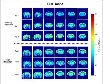 |
21 |
Six-fold enhancement in spatial-resolution of Pseudo-Continuous
Arterial Spin Labeling Perfusion Mapping using a Cryogenic Coil
at 9.4T
Sara Pires Monteiro1,2,
Lydiane Hirschler3,4,
Emmanuel L. Barbier4,
Patrícia Figueiredo2,
and Noam Shemesh1
1Champalimaud Research, Champalimaud Foundation, Lisbon, Portugal, 2Institute for Systems and Robotics - Lisboa and Department of Bioengineering, Instituto Superior Técnico, Universidade de Lisboa, Lisbon, Portugal, 3C.J. Gorter MRI Center for High Field MRI, Department of Radiology, Leiden University Medical Center, Leiden, Netherlands, 4Inserm, Grenoble Institut des Neurosciences, Université Grenoble Alpes, Grenoble, France Keywords: Data Acquisition, Arterial spin labelling, Preclinical High resolution CBF mapping using ASL can benefit multitude applications, yet its spatiotemporal resolution is limited in pre-clinical settings and at higher fields. To address this issue, we explored the possibility of improving the resolution of pCASL by using a cryogenic coil at 9.4T in the rat brain. Compared to the current state-of-the-art measurements, we managed to enhance the resolution of pCASL images by a factor of at least 6. The interpulse phase optimizations applied at the labelling plane are crucial for higher and stable inversion efficiency. |
|
2900.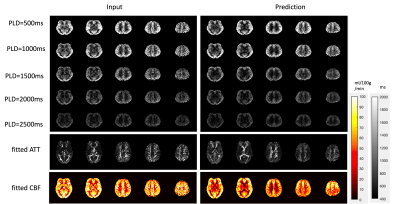 |
22 |
Denoising single and multi-delay 3D pCASL using SWIN Transformer
Qinyang Shou1,
Chenyang Zhao1,
Xingfeng Shao1,
and Danny JJ Wang1
1Laboratory of Functional MRI Technology (LOFT), Stevens Neuroimaging and Informatics Institute, University of Southern California, Los Angeles, CA, United States Keywords: Machine Learning/Artificial Intelligence, Machine Learning/Artificial Intelligence We developed a Transformer-based deep learning denoising model to improve the SNR for both single and multi-delay perfusion images acquired using 3D pseudo-continuous arterial spin labeling (pCASL). This method can significantly improve SNR (~2-fold) of the perfusion images without introducing bias for CBF and ATT quantification for both single-delay and multi-delay 3D pCASL. Further training and testing of this model on clinical datasets acquired on different vendor platforms is warranted. |
|
2901.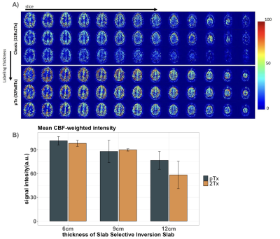 |
23 |
Improved perfusion-weighted images at 7T combining pTx and low
B1+ Adiabatic pulses
Ícaro Agenor Ferreira de Oliveira1,2,
Robin Schnabel1,
Matthias JP van Osch3,
Lydiane Hirschler3,
and Wietske van der Zwaag1,2
1Spinoza Centre for Neuroimaging, Amsterdam, Netherlands, 2Computational Cognitive Neuroscience and Neuroimaging, Netherlands Institute for Neuroscience, Amsterdam, Netherlands, 3Radiology, Leiden University Medical Center, Leiden, Netherlands Keywords: Data Acquisition, High-Field MRI A FAIR acquisition at 7T, using a TR-FOCI pulse for background suppression, was set up on a pTx (32Rx8Tx) system and on the standard (32Rx2Tx) system. CBF-imaging results were compared in terms of CBF-weighted signal intensities and drop-off with increasing thickness of the slab-selective inversion slab. The higher and more homogeneous B1 of the pTx system translated to higher CBF-weighted signal intensities. Both setups allowed functional mapping of the hand region in the motor cortex in approximately 5 minutes. |
|
2902.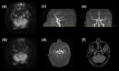 |
24 |
Subject-Specific Circular Artifacts in ASL Perfusion Imaging
with 3D Stack-of-Spirals FSE Readout
Yichen Hu1,
Junpu Hu2,
Zhongqi Zhang1,
Hui Liu1,
Qi Liu1,
Yongquan Ye1,
and Jian Xu1
1United Imaging, Houston, TX, United States, 2United Imaging, Shanghai, China Keywords: Artifacts, Arterial spin labelling In perfusion images of ASL scans of brain, artifacts originated from unlabeled blood inflow can be effectively suppressed by inferior saturation. In our study with spiral-based readout, artifacts in the patterns of bright spots and concentric rings were determined to arise from arterial blood inflow. We discovered that the emergence of the artifacts is directly related to the carotid artery anatomies of human subjects. The artifacts appeared persistently for just one out of four participating volunteers. The application of inferior saturation should be favored to avoid such artifacts, which had been a fact not fully recognized from recent ASL studies. |
|
2903.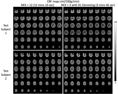 |
25 |
Deep Learning-denoised Isotropic 2mm Whole Brain
pseudo-Continuous Arterial Spin Labeling at 7T
Chenyang Zhao1,
Qinyang Shou1,
Xingfeng Shao1,
and Danny JJ Wang1,2
1Mark & Mary Stevens Neuroimaging and Informatics Institute, Keck School of Medicine, University of Southern California, Los Angeles, CA, United States, 2Department of Neurology, Keck School of Medicine, University of Southern California, Los Angeles, CA, United States Keywords: Machine Learning/Artificial Intelligence, Arterial spin labelling, Noise reduction Optimized pseudo-Continuous Arterial Spin Labeling has been implemented at 7T. To achieve whole brain high-resolution (2mm isotropic) perfusion imaging at 7T, however, requires prolonged scan time with an increased number of segments. A deep learning (DL) model was trained to boost the signal-to-noise ratio (SNR) for a scan with fewer repetitions and thus a shorter scan time. The analysis of SNR and temporal SNR suggests that at least 3 repetitions are needed to make a high-SNR prediction comparable to the full scan without compromising quantification accuracy. With DL denoising, the original 12 mins scan can be finished in 4 min. |
|
2904.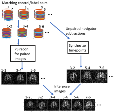 |
26 |
Increasing temporal SNR, sharpness, specificity, and sensitivity
of ASLfMRI using the partial separability model
Charles John Marchini1 and
Brad Sutton 1
1Bioengineering, University of Illinois Urbana-Champaign, Urbana, IL, United States Keywords: Sparse & Low-Rank Models, Arterial spin labelling ASLfMRI temporal SNR (tSNR) and sharpness in the inferior-superior direction was improved by using partial separability, a low rank model. The method requires an additional novelty to the partial separable model which allows time points with no corresponding imaging data, only temporal navigator data, to be reconstructed. A finger tapping task was used to demonstrate the detection of cerebral blood flow to the motor cortex. Mean squared error, structural similarity index, and the area under the curve of a receiver operating characteristic curve was also improved as shown by using a simulation of ASLfMRI data. |
|
2905.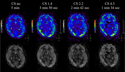 |
27 |
Accelerated Brain ASL using 3D Gradient and Spin-Echo
pseudo-Continuous ASL (GraSE-pCASL) with Compressed SENSE.
Yutaka Hamatani1,
Kayoko Abe2,
Masami Yoneyama3,
Johannes M Peeters4,
Kim van de Ven4,
Michinobu Nagao2,
Yasuhiro Goto1,
Isao Shiina1,
Kazuo Kodaira1,
Takumi Ogawa1,
Mana Kato1,
and Shuji Sakai2
1Department of Radiological Services, Tokyo Women's Medical University Hospital, Tokyo, Japan, 2Department of Diagnostic imaging & Nuclear Medicine, Tokyo Women's Medical University Hospital, Tokyo, Japan, 3Philips Japan, Tokyo, Japan, 4Philips Healthcare, Best, Netherlands Keywords: Data Acquisition, Arterial spin labelling In this study, 3D GraSE-pCASL was combined with Compressed SENSE (CS) to accelerate the acquisition time of perfusion images. The results showed that accelerated 3D CS-GraSE-pCASL could provide sufficient perfusion information in half the acquisition time compared to the conventional method. This technique may be useful in the diagnosis of cerebral blood flow disorders especially for pediatric patients and/or patients with cognitive impairments. |
|
2906.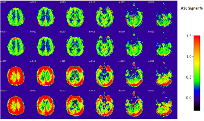 |
28 |
Comparison of velocity-selective-inversion arterial spin
labeling schemes
Ke Zhang1,
Simon M.F. Triphan1,
Oliver Sedlaczek1,2,
Christian Ziener2,
Hans-Ulrich Kauczor1,
Heinz-Peter Schlemmer2,
and Felix T. Kurz2
1Department of Diagnostic and Interventional Radiology, Heidelberg University Hospital, Heidelberg, Germany, 2Department of Radiology, German Cancer Research Center, Heidelberg, Germany Keywords: Pulse Sequence Design, Arterial spin labelling Velocity-selective pulses include VS saturation pulses (VSS) and VS Inversion (VSI) pulses. Previous study (1) concluded that both dual-sBIR8-VSS and sinc-VSI achieved the highest SNR efficiency among the VS labeling schemes. Overall, the dual-sBIR8-VSS pulse was the most robust against field imperfections, whereas sinc-modulated VSI pulse showed greater tSNR and was the best among the VSI methods. In this study, VSI sequence with rectangular small flip-angle RF pulses (rect-VSI), sinc-VSI with and without VS gradients during the control condition are compared. Bloch simulation and in vivo experiments for their robustness against B1, B0 variation and eddy current (EC) are investigated. |
|
2907.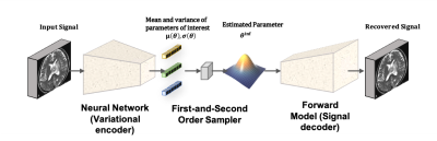 |
29 |
Rapid Bayesian inference for perfusion quantification using
ASL-MRI with a VAE-based neural network structure
Yechuan Zhang1,
Michael Chappell2,3,4,5,6,
and Jian-Qing Zheng1
1University of Oxford, Oxford, United Kingdom, 2Institute of Biomedical Engineering, Department of Engineering Science, University of Oxford, Oxford, United Kingdom, 3Wellcome Centre for Integrative Neuroimaging, FMRIB, Nuffield Department of Clinical Neuroscience, University of Oxford, Oxford, United Kingdom, 4Mental Health and Clinical Neuroscience, School of Medicine, University of Nottingham, Nottingham, United Kingdom, 5Sir Peter Mansfield Imaging Centre, School of Medicine, University of Nottingham, Nottingham, United Kingdom, 6Nottingham Biomedical Research Centre, Queen’s Medical Centre, University of Nottingham, Nottingham, United Kingdom Keywords: Data Analysis, Arterial spin labelling A Variational Autoencoder (VAE) based framework was created to solve perfusion parameter estimation problem for ASL non-linear forward models. The ultimate goal was to build up an efficient and uncertainty-aware framework for parameter estimation problem in medical imaging, using the concept from Variaitonal Bayes (VB) which was already applied to ASL. Evaluation was performed using simulation and real data experiments with a bi-exponential model and two ASL-MRI forward models with different complexity. Compared with Markov Chain Monte Carlo (MCMC) and analytical VB (aVB), our VAE-based model achieved comparable accuracy, and hundredfold improvement in computational time. |
|
2908.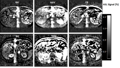 |
30 |
Navigator-based slice tracking for liver pCASL using spin-echo
EPI acquisition
Ke Zhang1,
Simon M.F. Triphan1,
Oliver Sedlaczek1,2,
Christian Ziener2,
Hans-Ulrich Kauczor1,
Heinz-Peter Schlemmer2,
and Felix T. Kurz2
1Department of Diagnostic and Interventional Radiology, Heidelberg University Hospital, Heidelberg, Germany, 2Department of Radiology, German Cancer Research Center, Heidelberg, Germany Keywords: Pulse Sequence Design, Arterial spin labelling Liver perfusion is an important physiological parameter in health and disease (1,2). In the measurement of liver perfusion using arterial spin labelling (ASL), respiratory motion is a major challenge. In this study, respiratory motion information is acquired from a projection signal and used to adjust the position of the excited slice in real time. The feasibility of free-breathing multi-slice liver perfusion imaging using spin-echo EPI based pseudo-continuous ASL (pCASL) with navigator-based slice tracking method is investigated. |
|
2909.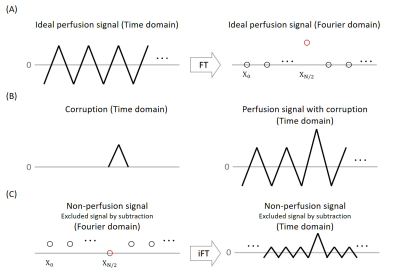 |
31 |
A novel Fourier-based perspective to analyze and compensate for
corruption in arterial spin labeling MRI
Seon-Ha Hwang1 and
Sung-Hong Park1
1Department of Bio and Brain Engineering, Korea Advanced Institute of Science and Technology, Daejeon, Korea, Republic of Keywords: Data Processing, Arterial spin labelling A new perspective in Fourier domain to understand the ASL signal and compensate for corruption artifacts is proposed. The averaged perfusion-subtraction image corresponds to a single frequency component and the other frequency components could be interpreted as non-perfusion signals. In this viewpoint, the multiple corruptions could be compensated by finding the optimal perfusion frequency component to minimize the zigzag amplitude in the non-perfusion signals. The proposed compensation reduced the corruption without eliminating any measured data, preserving SNR. This new perspective would open up the possibility of processing the ASL signal in the Fourier domain rather than the time domain. |
|
2910.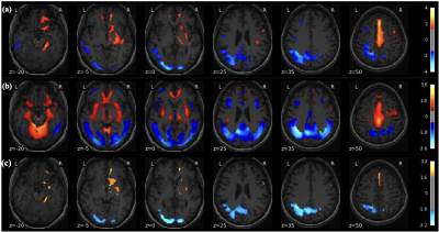 |
32 |
Combined Parkinson’s Disease Related Patterns using ASL MRI and
FDG PET
Yu Zeng1,
Zizhao Ju2,
Weiying Dai3,
Yong Zhang4,
David Alsop5,
Chuantao Zuo2,
and Li Zhao1
1College of Biomedical Engineering & Instrument Science, Zhejiang University, Zhejiang, China, 2PET Center & National Center for Neurological Disorders & National Clinical Research Center for Aging and Medicine, Huashan Hospital, Fudan University, Shanghai, China, 3Department of Computer Science, State University of New York at Binghamton, Binghamton, NY, United States, 4GE Healthcare, Shanghai, China, 5Radiology, Beth Israel Deaconess Medical Center and Harvard Medical school,, Bostom, MA, United States Keywords: Data Analysis, Parkinson's Disease, Parkinson's disease-related pattern (PDRP), multimodality Parkinson's disease-related patterns (PDRPs) of metabolism and perfusion have been reported to reflect brain abnormalities in Parkinson’s disease (PD). However, differences between the glucose metabolism PDRP derived using FDG-PET and the perfusion PDRP derived using ASL have not been compared directly in the same patient cohort. In this work, PDRPs were compared using PET and ASL images of 43 PD patients and 28 health controls using Scaled Subprofile Model analysis. In addition, a new method was proposed to build a multi-modality pattern. Our primary results showed that combined metabolism-perfusion PDRP resulted in superior accuracy than ASL- and PET-derived PDRP alone. |
|
2911.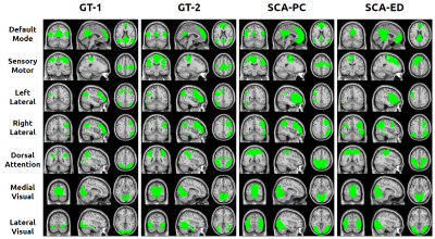 |
33 |
Identifying Brain Resting-state Networks from Arterial Spin
Labeling with Spectral Clustering
Jason Barrett1,
Haomiao Meng2,
Zongpai Zhang1,
Song Chen1,
Li Zhao3,
David Alsop3,
Xingye Qiao2,
and Weiying Dai1
1Computer Science, Binghamton University, Vestal, NY, United States, 2Mathematical Sciences, Binghamton University, Vestal, NY, United States, 3Department of Radiology, Beth Israel Deaconess Medical Center and Harvard Medical School, Boston, MA, United States Keywords: Data Analysis, fMRI (resting state), network detection We propose a spectral clustering algorithm (SCA) based on the Pearson correlation metric (SCA-PC) to identify large-scale brain networks in arterial spin labeling (ASL) images. It was shown to be more robust to Gaussian distributed noise sources based on simulations. We studied the robustness of SCA-PC vs. the traditional SCA method based on a Euclidean distance metric (SCA-ED) for deriving resting-state networks from real human fMRI data. Our results indicate that SCA-PC can derive better brain networks from ASL data than traditional SCA-ED. |
|
2912.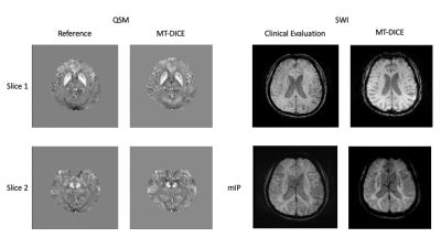 |
34 |
MT-DICE for Simultaneous Quantification of Perfusion,
Permeability, and Susceptibility
Yang Chen1,2,
Zhehao Hu1,
Jiayu Xiao1,
Mark Shiroishi1,
Wensha Yang1,
Debiao Li3,
Anthony G. Christodoulou3,
and Zhaoyang Fan1
1Department of Radiology, Keck School of Medicine, University of Southern California, Los Angeles, CA, United States, 2Department of Biomedical Engineering, University of Southern California, Los Angeles, CA, United States, 3Cedars-Sinai Medical Center, Los Angeles, CA, United States Keywords: Quantitative Imaging, Permeability, perfusion, susceptibility We have recently developed an MR MultiTasking based Dynamic Imaging for Cerebrovascular Evaluation (MT-DICE) technique that provides DCE- and leakage-corrected DSC-MRI parameters simultaneously with one scan and a single-dose contrast injection. This work further expanded the technique by incorporating quantitative susceptibility mapping. The refined technique was validated in healthy volunteers and patients, demonstrating the good agreement with reference methods. |
|
2913.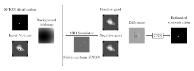 |
35 |
Intensity-based Deep Learning for SPION concentration estimation
in MR imaging
Alberto Di Biase1,2,
Shuang Liu3,
Masaki Sekino3,
and Pablo Irarrázabal1,2
1Department of Electrical Engineering, Pontificia Universidad Católica de Chile, Santiago, Chile, 2Biomedical Imaging Center, Pontificia Universidad Católica de Chile, Santiago, Chile, 3Department of Bioengineering, School of Engineering, University of Tokyo, Tokyo, Japan Keywords: Machine Learning/Artificial Intelligence, Quantitative Imaging, SPION SPION is a contrast agent with a wide range of biomedical applications. A new Deep Learning based method is presented for the quantification of SPION from intensity images. This contrast agent cause off-resonance artifacts, distorting the image. The field map is encoded in the difference of two images taken alternating the direction of the slice selection gradient. The network was trained on simulated data. The network is based on U-net and uses only 2D convolution to process the whole 3D volume, interpreting the last dimension as filters. Results are shown in simulations and on phantoms acquired on a 7T scanner. |
|
2914.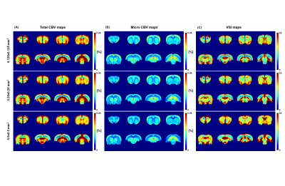 |
36 |
Resolution Dependency Study of Cerebral Blood Volume and Vessel
Size Index
Yelim Gong1,
DongKyu Lee1,
and HyungJoon Cho1
1BME, UNIST, Ulsan, Korea, Republic of Keywords: Data Analysis, Blood vessels To obtain cerebral vasculature related information, various kinds of cerebrovascular magnetic resonance imaging (MRI) technique are being applied. However, it turned out that cerebral blood volume (CBV) and vessel size index (VSI) values differ depending on their spatial resolutions. To find an adjustment solution for this phenomenon, the resolution dependency must be confirmed in advance. Therefore, this study aims to investigate the resolution dependency of CBV and VSI through whole brain and region of interest based analysis. The results show that micro CBV and VSI values are resolution-dependent, while total CBV did not show any distinct patterns. |
|
2915.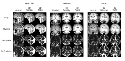 |
37 |
3D Artificial Cerebral Blood Volume Generation from T1W
Structural MRI
Vishwanatha Mitnala Rao1,
Scott A Small2,
and Jia Guo3
1Biomedical Engineering, Columbia University, Acton, MA, United States, MA, United States, 2Department of Neurology, Columbia University Medical Center, New York, NY, United States, 3Department of Psychiatry, Columbia University, New York, NY, United States Keywords: Machine Learning/Artificial Intelligence, Brain While gadolinium-based contrast agents are necessary to generate a quantitative mapping of brain metabolism, they are invasive with unclear long-term side-effects. As such, convolutional neural networks (CNNs) have been explored as a method to generate artificial cerebral blood volume (aCBV) maps from T1W structural MRI scans. However, prior implementations process MRI in 2D slices, severely limiting output resolution, production time, and utility. In this study, we propose a 3D CNN-Transformer hybrid aCBV generation tool that outperforms both 2D and 3D implementations of the prior state-of-the-art model (PSNR: 29.46, P.R.: 0.836, SSIM: 0.875, S.R.: 0.681). |
|
2916.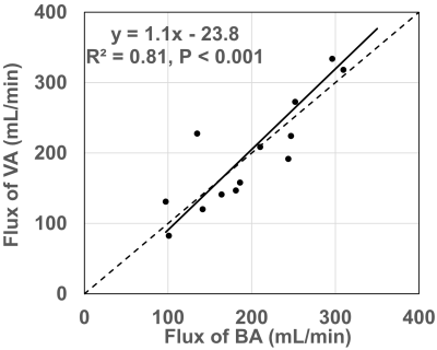 |
38 |
Comparison of Blood Flow Measurement of Posterior Cerebral
Circulation at Vertebral and Basilar Arteries Using Phase
Contrast MRI
Feng Xu1,2 and
Qin Qin1,2
1The Russell H. Morgan Department of Radiology and Radiological Science, Johns Hopkins University, Baltimore, MD, United States, 2F.M. Kirby Research Center for Functional Brain Imaging, Kennedy Krieger Institute, Baltimore, MD, United States Keywords: Data Acquisition, Velocity & Flow Phase contrast (PC) MRI provides a quantitative measurement of the total flux of blood flow. As it is not easy to find a perpendicular plane to the vertebral arteries (VA) at the base of the brain, especially in the elderly and people who developed tortuous arteries, we explored the alternative imaging location at the basilar artery (BA) where two VA merged in front of the pon. The flux of BA shows an excellent correlation (ρ=0.9, P<0.01) with that of VA, suggesting that BA is a practical choice for measuring the global blood flow of posterior cerebral circulation using PC MRI. |
|
2917.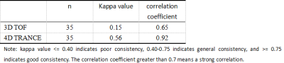 |
39 |
Visualization of Collateral Vessels in 4D TRANCE MR Angiography
Compaired with 3D TOF in Moyamoya Disease
Zhihua Chen1,
Kaiyin Liang1,
Yongkun Lan1,
and Wen Zhou1
1PEKING UNIVESITY SHENZHENG HOSPITAL, Shen Zheng, China Keywords: Visualization, Blood vessels Compared with 3D-CE-MRA, TOF technology isn't conducive to display slow blood flow. Moreover, TOF angiography is a static approach that lacks flow dynamic information. In our study, the 4D TRANCE MRA depicted the proximal arteries better than 3D TOF and may replace TOF angiography. Our study also indicated that LMA collaterals were better visualized in 4D TRANCE angiography and visualization of distal cerebral arteries and collateral vessels with Moyamoya disease with 4D TRANCE MR angiography was good to excellent. Besides conventional MRA to show the shape and distribution of blood vessels, it can also provide hemodynamic information of vascular diseases. |
|
2918.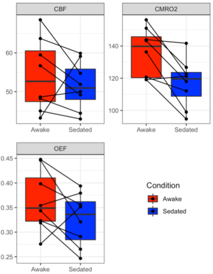 |
40 |
MRI evidence for reduced human brain oxygen metabolism during
midazolam sedation
Hannah L Chandler1,
Ian Driver2,
Sharmila Khot1,
Zoltán Auer3,
Murthy Varanasi3,
Neeraj Saxena1,3,
Michael Germuska2,
and Richard G Wise1,4,5
1Cardiff University Brain Research Imaging Centre (CUBRIC), Department of Psychology, Cardiff University, Cardiff, United Kingdom, 2Cardiff University Brain Research Imaging Centre (CUBRIC), School of Physics and Astronomy, Cardiff University, Cardiff, United Kingdom, 3Department of Anaesthetics, ICU and Pain medicine, Cwm Taf Morgannwg University Health Board, Merthyr Tydfil, United Kingdom, 4Neurosciences, Imaging and Clinical Sciences, University “G. d'Annunzio” of Chieti-Pescara, Chieti, Italy, 5Institute for Advanced Biomedical Technologies (ITAB), G. d’Annunzio University’ of Chieti-Pescara, Chieti, Italy Keywords: Data Analysis, Arterial spin labelling We investigated the effects of mild sedation with the type A GABA receptor positive allosteric modulator (GABAA-R PAM) midazolam, on cerebral blood flow (CBF), oxygen extraction fraction (OEF) and the rate of cerebral metabolic oxygen consumption (CMRO2) in the healthy human brain using ASL and TRUST MRI. CMRO2 was significantly reduced during sedation compared to wakefulness but no statistically significant change in CBF or OEF was detected. Our data are consistent with prior imaging evidence of reduced brain energy consumption during sedation with midazolam. |
|
 Back to Meeting Home
 Back to the Program-at-a-Glance
The International Society for Magnetic Resonance in Medicine is accredited by the Accreditation Council for Continuing Medical Education to provide continuing medical education for physicians.
 Back to Meeting Home
 Back to the Program-at-a-Glance
 View Presentation Video