Digital Poster
Motion Correction II
ISMRM & ISMRT Annual Meeting & Exhibition • 03-08 June 2023 • Toronto, ON, Canada

| Computer # | |||
|---|---|---|---|
1997.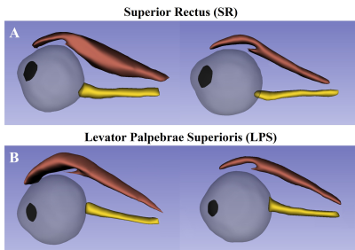 |
1 | Changes in the position of the eyeball in TAO patients with unilateral upper eyelid retraction by T2-weighted SPIR imaging
Xinyi Gou1, Yi Wang2, Lingli Zhou3, Jianxiu Lian4, Zilong Chen4, Xiuying Zhang3, Jingyi Cheng1, Lei Chen1, Nan Hong1, and Jin Cheng1
1Department of Radiology, Peking University People’s Hospital, Beijing, China, 2Department of Ophthalmology, Peking University Third Hospital, Beijing, China, 3Department of Endocrinology, Peking University People’s Hospital, Beijing, China, 4Philips Healthcare, Beijing, China Keywords: Image Reconstruction, Software Tools For patients with thyroid-associated orbitopathy (TAO) presenting unilateral upper eyelid retraction, the phenomenon that the impaired eye is lower than the healthy eye (“eyeball descending”) has been noticed. This study investigated the eyeballs' position changes by 3D reconstitution of magnetic resonance (MR) images. With reference to the central plane of the healthy side, 70.37% impaired eyeballs (19/27) were found in significantly lower positions, and with significant positive correlations of increased thicknesses of levator palpebrae superioris (LPS), superior rectus (SR), and LPS-SR complex volume. This study provided objective evidence of “eyeball descending” in unilateral upper eyelid retraction TAO patients. |
|
1998.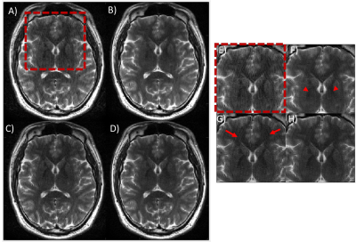 |
2 | A combined model-based and ML-based approach performs better than model-based or ML-based applied individually
Srikant Kamesh Iyer1, Hassan Haji-Valizadeh1, and Samir Sharma1
1Canon Medical Research USA, Inc., Mayfield, OH, United States Keywords: Motion Correction, Motion Correction A motion correction framework was developed to suppress artifacts from rigid and non-rigid motion using a combination of model-based and ML-based approaches. The performance of this framework was compared with model-based only and ML-based only motion correction approaches on motion-simulated data and motion-corrupted in-vivo data using visual inspection and image quality metrics. The combined approach showed superior image quality than model-based and ML-based approaches applied individually. |
|
1999.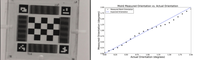 |
3 | Optical position tracking fiducial marker for rigid body motion parameter estimation with high performance
Marina Silic1,2, Aravinthan Jegatheesan1,2, Fred Tam2, and Simon J Graham1,2
1Department of Medical Biophysics, University of Toronto, Toronto, ON, Canada, 2Sunnybrook Research Institute, Toronto, ON, Canada Keywords: Data Acquisition, Motion Correction, Optical Position Tracking Optical position tracking (OPT) using fiducial markers is advantageous for data acquisition of rigid body head motion parameters and motion correction in magnetic resonance imaging (MRI). Many opportunities still remain to improve OPT through the development of new devices. A promising prototype OPT marker and analysis pathway are described. The marker through-plane degree of freedom (DOF) precision was enhanced via moiré patterns and stereovision. Precision was assessed using positional and rotational stages in all 6DOF. Initial results strongly suggest that with minimal additional work, the OPT marker will provide excellent performance in a 3 T MRI system. |
|
2000.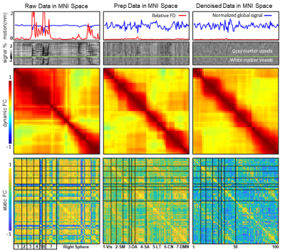 |
4 | The noise suppression of resting fMRI data based on eigenvector correction
Wei Zhao1, Huanjie Li 1, Yunge Zhang1, Dongyue Zhou1, and Fengyu Cong1,2
1Dalian University of Technology, Dalian, China, 2University of Jyvaskyla, Jyvaskyla, Finland Keywords: Motion Correction, Artifacts The fMRI signal has been very noisy for artifacts induced various reasons, and yet the head motion and non-neuronal contributions are always the most tickle one. And too severe of motion corruption usually will lead to abandonment of the participant’s data. In this study, proposed method manages to effectively control these noises meanwhile without losing valuable signals. Proposed method exceeds standard pipeline in both quantitative and qualitative metrics. |
|
2001.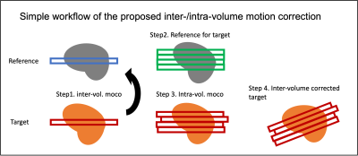 |
5 | The development of Inter-/intra-volume motion correction algorithm for fMRI using a custom MRI acquisition with prospectively injected motion.
Wanyong Shin1 and Mark J Lowe1
1Radiology, Cleveland Clinic, Cleveland, OH, United States Keywords: Motion Correction, fMRI In this study, we propose the new inter-/intra-volume motion correction algorithm. To test the proposed method, we modify simulated Prospective Acquisition CorrEcted (SIMPACE) EPI sequence and inject both inter- and intra-volume motion during actual EPI acquisition. Using ex-vivo brain phantom, we synthesize inter-/intra volume motion corrupted MR data. We evaluate the proposed method, compared to volume motion correction method. We find that the proposed inter-/intra-volume method outperforms volume motion correction method. |
|
2002.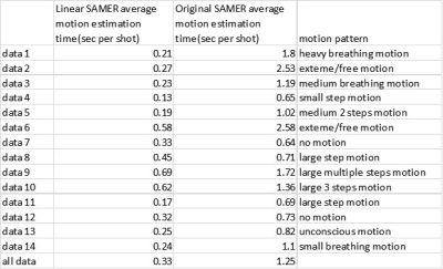 |
6 | Linear SAMER: linear motion optimization for scout accelerated retrospective motion estimation and reduction with linear+ reordering in MPRAGE
Yantu Huang1, Daniel Nicolas Splitthoff2, Bryan Clifford3, Daniel Polak2,4, Stephen Cauley4,5, Wei-Ching Lo3, Nan Xiao1, and Huixin Tan1
1Siemens Shenzhen Magnetic Resonance Ltd., Shenzhen, China, 2Siemens Healthcare GmbH, Erlangen, Germany, 3Siemens Medical Solutions, Boston, MA, United States, 4Massachusetts General Hospital, Charlestown, MA, United States, 5Harvard Medical School, Boston, MA, United States Keywords: Motion Correction, Head & Neck/ENT SAMER uses a fast reference scan (“scout”) and short additional guidance lines in each shot of an MPRAGE sequence (“linear+ reordering”) to calculate motion parameters. Optimization of rigid body motion parameters are typically nonlinear. In this work we exploit local linearity with SAMER to improve the performance. During motion estimation, the initial guess of motion parameters for a given shot is taken from the previous most similar shot. Similarity is determined by correlations of the linear+ guidance lines. Retrospective reconstructions of volunteer data show the proposed method has very good computation performance and can handle large motion well. |
|
2003.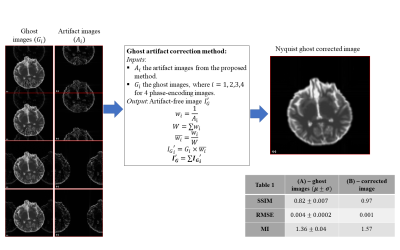 |
7 | Detection and remediation of ghost artifacts in low b-value diffusion MRI with ghost re-synthetization and 4-way phase-encoded data
Anh S Thai1,2, Carlo Pierpaoli2, Lin-Ching Chang1, and M. Okan Irfanoglu2
1Catholic University of America, Washington, DC, United States, 2QMI, NIBIB/National Institutes of Health, Bethesda, MD, United States Keywords: Artifacts, Artifacts, diffusion mri We propose a novel post-processing method for ghost artifact correction in low-b diffusion weighted images (DWIs) using ghost synthetization and four different phase-encoding direction (PED) images. The results from both simulations and real data show that our method can robustly detect and correct ghost artifacts on b=0 s/mm2 images for artifact-free image. The proposed method can be used further to generate ground truth training images for a machine-leaning method to remedy ghost artifacts in single PED acquisition. |
|
2004.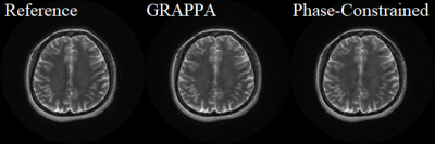 |
8 | Phase-Constrained Reconstruction for Enhancing PROPELLER SNR
Yuchou Chang1, Gulfam Ahmed Saju1, Jasina Yu1, Reza Abiri2, Tianming Liu3, and Zhiqiang Li4
1Computer and Information Science, University of Massachusetts Dartmouth, North Dartmouth, MA, United States, 2Electrical, Computer and Biomedical Engineering, University of Rhode Island, Kingston, RI, United States, 3Computer Science, University of Georgia, Athens, GA, United States, 4Neuroradiology, Barrow Neurological Institute, Phoenix, AZ, United States Keywords: Motion Correction, Parallel Imaging PROPELLER blade acquisition has been accelerated by undersampling blade k-space data. Missing data on each blade can be reconstructed by parallel MRI reconstruction methods. However, noise deteriorates the blade and the overall image quality. To enhance the signal-to-noise ratio (SNR), phase-constrained reconstruction is studied for improving the SNR of PROPELLER imaging. Phase-constrained reconstruction can improve PROPELLER imaging SNR when the acceleration of data acquisition is used. Optimal selection of the ACS lines and the outer reduction factors is expected to achieve a better SNR and accelerate the PROPELLER imaging speed simultaneously. |
|
2005.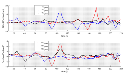 |
9 | Fast Prospective Motion Correction using Directional Couplers as Motion Sensors
Jason Daniel van Schoor1,2, Mark Gosselink3, Dennis Klomp3, Giel Mens3, Hans Hoogduin3, Wim Prins4, and Tijl van der Velden3
1High Field MRI, UMC Utrecht, Utrecht, Netherlands, 2Utrecht University, Utrecht, Netherlands, 3UMC Utrecht, Utrecht, Netherlands, 4Philips, Best, Netherlands Keywords: Motion Correction, Motion Correction, Directional coupler Motion is a prevalent issue in MRI. Here, we present a fast prospective motion correction (PMC) algorithm using directional couplers as head-motion sensors. The mean reflectances (per volume) of an 8 channel pTx head coil are modelled to head-position recorded by the systems built-in PMC during a calibration scan. Thereafter, the model is used for motion correction at a per RF pulse temporal resolution. Results indicate partial motion correction with error in correcting fast motions due to the necessary presence of filters to remove unidentified low-frequency noise. Mitigating low-frequency noise contributions would allow for less filtering thereby improving dynamic performance. |
|
2006.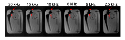 |
10 | Spectrally-encoded multi-spectral imaging (SEMSI) at 0.55T provides improved imaging adjacent to metallic implants
Bochao Li1, Kübra Keskin2, Daehyun Yoon3, Nam Gyun Lee1, Brian Hargreaves3, and Krishna Shrinivas Nayak2
1Department of Biomedical Engineering, University of Southern California, Los Angeles, CA, United States, 2Department of Electrical and Computer Engineering, University of Southern California, Los Angeles, CA, United States, 3Stanford University, Stanford, CA, United States Keywords: Artifacts, Low-Field MRI, Metal Spectrally-encoded multi-spectral imaging (SEMSI) enables imaging adjacent to metal implants without blurring due to off-resonance, and has been demonstrated at 3T. Here, we demonstrate application of this technique at 0.55T with appropriate adjustments to the spectral resolution and RF pulse bandwidth. A total hip arthroplasty phantom was imaged with SEMSI, turbo spin echo (TSE), and Slice Encoding for Metal Artifact Correction (SEMAC). Experiments demonstrate the feasibility of SEMSI at 0.55T, and examine ΔTE to support multi-echo acquisitions in a single TR. |
|
2007.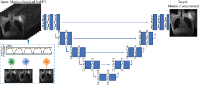 |
11 | Iterative Motion-Compensated Reconstruction with Convolutional Neural Network (iMoCo-Net) for Ultrashort Echo Time (UTE) Proton Lung MRI
Fei Tan1, Ke Wang2, Michael Lustig1,2, and Peder E. Z. Larson1,3
1UC Berkeley-UCSF Graduate Program in Bioengineering, University of California Berkeley and University of California, San Francisco, San Francisco, CA, United States, 2Electrical Engineering and Computer Sciences, University of California, Berkeley, Berkeley, CA, United States, 3Department of Radiology and Biomedical Imaging, University of California, San Francisco, San Francisco, CA, United States Keywords: Motion Correction, Motion Correction This abstract explores the feasibility of machine learning-based motion-compensated reconstruction for free-breathing UTE lung MRI. Specifically, we used respiratory motion-resolved Non-uniform Fourier Transform (NuFFT) reconstruction as input, iterative motion-compensated (iMoCo) reconstruction as target, and a 2D U-Net convolutional neural network. Test results demonstrate a sharper diaphragm and a higher apparent SNR compared to the averaged input. In conclusion, iMoCo-Net accelerates the reconstruction of 3D radial UTE data substantially, shortening the required time from hours to minutes. |
|
2008.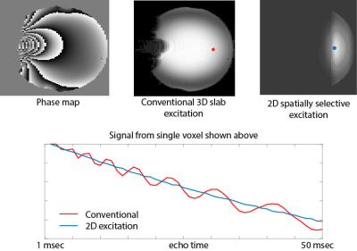 |
12 | In-plane B0 inhomogeneity effects in T2* mapping
Glen Morrell1
1Radiology and Imaging Sciences, University of Utah, Salt Lake City, UT, United States Keywords: Artifacts, Relaxometry Main field B0 inhomogeneity may adversely affect the accuracy of T2* mapping in the abdomen even at locations spatially distant from the B0 variation. We show this effect in both simulation and phantom studies, and show that it can be ameliorated with the use of two-dimensional spatially selective excitation. These findings have important ramifications for applications of BOLD imaging in the abdomen such as renal BOLD or liver T2* mapping, where large B0 inhomogeneity is often present in areas of the image volume spatially remote from the organ of interest. |
|
2009.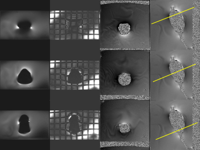 |
13 | SPAMM Tagged EPI Acquisition for Assessment of Geometric Distortion Caused by an Endorectal Coil
Ken-Pin Hwang1, R. Jason Stafford1, and Tharakeswara K. Bathala2
1Department of Imaging Physics, The University of Texas M.D. Anderson Cancer Center, Houston, TX, United States, 2Department of Abdominal Imaging, The University of Texas M.D. Anderson Cancer Center, Houston, TX, United States Keywords: Artifacts, System Imperfections: Measurement & Correction Diffusion weighted imaging is an essential sequence for diagnosis of prostate cancer but is often impacted by distortion. Current existing phantoms designed for assessing geometric accuracy do not sufficiently simulate the environmental factors that affect distortion in diffusion weighted EPI. In this work we apply a rectilinear tagging technique to visualize and understand patterns of distortion caused by an endorectal coil immersed in water when imaged with a PI-RADS compliant protocol. Maximum spatial distortion including regions of spatial collapse was observed near the coil element. Observed distortion pattern supports proper centering of the coil to avoid distortion. |
|
2010.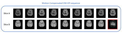 |
14 | Reduction of motion artifacts in diffusion weighted brain images using first order motion-compensated diffusion gradients
Danielle Kara1, Mark J. Lowe2, Christopher T Nguyen1, and Ken E. Sakaie2
1Cardiovascular Innovation Research Center, Cleveland Clinic, Cleveland, OH, United States, 2Imaging Institute, Cleveland Clinic, Cleveland, OH, United States Keywords: Motion Correction, Diffusion/other diffusion imaging techniques, Diffusion Tensor Imaging, Motion Correction, Neuro Patient motion during acquisition of diffusion weighted (DW) images causes significant signal dropout. To perform motion correction, DW images of the brain were acquired during gross head motion using a first order motion-compensated diffusion gradient scheme and compared to a traditional DW-EPI sequence. Of the images acquired with the motion-compensated sequence, only 1.5% contained significant signal dropout compared to 36% of the images acquired with the traditional sequence. Overall, the motion-compensated sequence reduced signal dropout by 96% compared to the traditional sequence without increasing scan time or introducing new post-processing protocols. |
|
2011.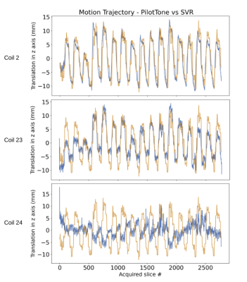 |
15 | Respiratory binning with PilotTone Navigator For Motion Compensated Liver DW-MRI
SERGE DIDENKO VASYLECHKO1, CEMRE ARIYUREK2, ONUR AFACAN2, and SILA KURUGOL2
1HARVARD MEDICAL SCHOOL, BOSTON, MA, United States, 2RADIOLOGY, BOSTON CHILDREN'S HOSPITAL, HARVARD MEDICAL SCHOOL, BOSTON, MA, United States Keywords: Motion Correction, Body Respiratory motion substantially affects the accuracy of quantitative DWI-MR techniques in the upper abdomen. A conventional approach for motion correction in abdominal MRI uses a respiratory belt or other navigators for prospective triggering. This study explores use of a new approach - PilotTone (PT) navigator for binning. |
|
2012.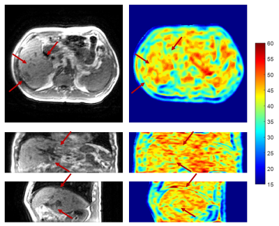 |
16 | Artifacts Map-guided Nonlocal Mean denoising for abdominal 4D-MR images acquired by 3D view-sharing sequence
Yat Lam Wong1, Hing Chiu Charles Chang2, Weiwei Liu3, Weihu Wang3, Yibao Zhang3, Hao Wu3, Victor Ho Fun LEE4, Lai Yin Andy Cheung5, Tian Li1, and Jing Cai1
1Department of Health Technology and Informatics, The Hong Kong Polytechnic University, Hong Kong, Hong Kong, 2Department of Biomedical Engineering, The Chinese University of Hong Kong, Hong Kong, Hong Kong, 3Peking University Cancer Hospital & Institute, Beijing, China, 4Department of Clinical Oncology, The University of Hong Kong, Hong Kong, Hong Kong, 5Department of Clinical Oncology, Queen Mary Hospital, Hong Kong, Hong Kong Keywords: Data Processing, Data Processing, 4D-MRI 3D view-sharing sequences have been demonstrated its great promise for abdominal 4D-MRI due to its high temporal resolution and availability in clinical scanners. However, the sub-optimal sampling methods in these sequences under free-breathing condition deteriorates the image quality by ghost artifacts. Here, we propose a novel technique, Artifacts Map-guided Nonlocal Mean (AM-NLM), to suppress motion artifacts and to increase image quality of the 4D-MR images of liver cancer patients acquired by a 3D view-sharing sequence, TRICKS, on a 1.5T scanner. |
|
2013.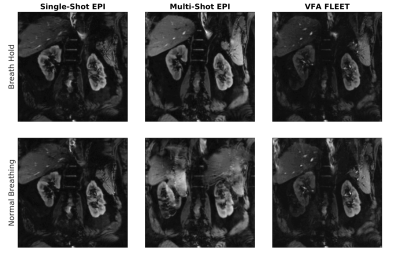 |
17 | Motion robust multi-shot EPI of the abdomen using VFA-FLEET
Mustafa Utkur1,2, Tess E Wallace1,2, Sila Kurugol1,2, and Onur Afacan1,2
1Radiology, Harvard Medical School, Boston, MA, United States, 2Computational Radiology Laboratory, Boston Children's Hospital, Boston, MA, United States Keywords: Motion Correction, Body Multi-shot EPI can be a promising technique to improve the resolution limits of the single shot EPI. However, its application has been so far limited in the abdominal MRI, as phase errors between shots due to unavoidable breathing motion creates artifacts in the images. Here, we tested a Variable-Flip-Angle Fast Low-angle Excitation Echo planar Technique (VFA-FLEET) pulse sequence and showed that it can be used to create high-resolution motion-robust images with high geometric fidelity without breath holding. |
|
2014.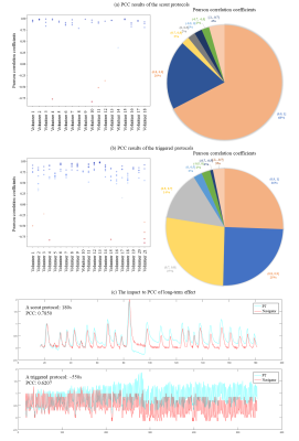 |
18 | Pilot Tone respiratory signal processing with RF interference suppression and validation against image navigator
Yantu Huang1, Huixin Tan1, Qiuyi Zhang1, and Peter Speier2
1Siemens Shenzhen Magnetic Resonance Ltd., Shenzhen, China, 2Siemens Healthcare GmbH, Erlangen, Germany Keywords: Data Processing, Data Processing, physiology signal Pilot Tone (PT) has been demonstrated to extract respiratory signal successfully. However, RF interferences are not considered. We propose a method to create a high-quality PT respiratory signal, while suppressing RF interference. At beginning of a measurement, initial RF suppression matrix and respiratory combination vector are learned. Then a PT respiratory signal can be generated to trigger measurement. Afterwards, RF suppression matrix is updated during each RF train. We validate our method in a moving phantom and in 39 volunteers against image navigator. Results show that our method can suppress RF interference effectively- highly correlated with image navigator. |
|
2015.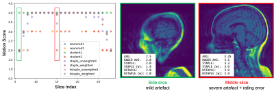 |
19 | Consolidation of expert ratings of motion artifacts using hierarchical label fusion
Yael Balbastre1,2, Robert Frost1,2, Khushi Morparia1,3, Richard L Carrington III1, Yuh-Shin Chang1,4,5, Brooks P Applewhite1,4, Marcio Aloisio Bezerra Cavalcanti Rockenbach6, and Bruce Fischl1,2,7,8
1Athinoula A. Martinos Center for Biomedical Imaging, Massachusetts General Hospital, Boston, MA, United States, 2Department of Radiology, Harvard Medical School, Boston, MA, United States, 3Northeastern University, Boston, MA, United States, 4Department of Radiology, Massachusetts General Hospital, Boston, MA, United States, 5Department of Radiology, Massachusetts Eye and Ear Infirmary, Boston, MA, United States, 6Data Science Office, Mass General Brigham, Somerville, MA, United States, 7Computer Science and Artificial Intelligence Laboratory, Massachusetts Institute of Technology,, Cambridge, MA, United States, 8Harvard-MIT Divison of Health Sciences and Technology, Massachusetts Institute of Technology, Cambridge, MA, United States Keywords: Data Analysis, Motion Correction Intra-scan motion costs tens of thousands of dollars per scanner annually due to the need to repeat non-diagnostic scans1. When assessing the scale of the problem and potential solutions, radiologists’ ratings of artifacts are considered the gold standard. However, inconsistent and conflicting ratings must be consolidated into a single gold-standard. We introduce a hierarchical label fusion algorithm that infers each rater's performance and promotes consistency across slices from a volume. This algorithm reduces label noise compared to majority votes, and allows non-expert ratings to be calibrated and included as additional silver-standards. |
|
2016.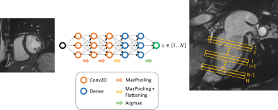 |
20 | Recovering slice location for unconventional acquisition plane with deep learning algorithm: cardiac magnetic resonance case
Habib Rebbah1 and Timothé Boutelier1
1Research & Innovation, Olea Medical, La Ciotat, France Keywords: Machine Learning/Artificial Intelligence, Data Analysis In cardiac MR field, the slice location along the long axis is a key parameter for the standardization proposed by the American heart association. We explore here the ability of a CNN to estimate it. |
|
 Back to Meeting Home
 Back to the Program-at-a-Glance
The International Society for Magnetic Resonance in Medicine is accredited by the Accreditation Council for Continuing Medical Education to provide continuing medical education for physicians.
 Back to Meeting Home
 Back to the Program-at-a-Glance
 View Presentation Video