Digital Poster
Segmentation I
ISMRM & ISMRT Annual Meeting & Exhibition • 03-08 June 2023 • Toronto, ON, Canada

| Computer # | |||
|---|---|---|---|
3585.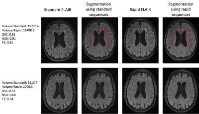 |
1 |
Ultra rapid volumetric imaging for diagnosis in memory clinic:
assessment of white matter hyperintensities quantification
Carole Helene Sudre1,2,
Haroon R Chughtai2,
David L Thomas3,4,
David Cash4,
Miguel Rosa-Grilo4,
Millie Beament4,
Frederik Barkhof2,4,5,
Daniel Alexander2,
Cath Mummery4,
Nick Fox4,
and Geoff JM Parker2
1MRC Unit for Lifelong Health and Ageing at UCL, University College London, London, United Kingdom, 2Centre for Medical Image Computing, University College London, London, United Kingdom, 3Department of Brain Repair and Rehabilitation, UCL Queen Square Institute of Neurology, University College London, London, United Kingdom, 4Dementia Research Centre, UCL Queen Square Institute of Neurology, University College London, London, United Kingdom, 5Department of Radiology & Nuclear Medicine, Amsterdam UMC, Amsterdam, Netherlands Keywords: Segmentation, Dementia Acquisition time is a crucial factor in the tolerability and availability of MRI examinations for patients attending memory clinics. The suitability of using prototype ultra-rapid sequences for the quantification of white matter hyperintensities was assessed by comparing segmentation outputs from standard clinical and ultra-rapid sequences. Despite a slightly lower sensitivity to smaller lesions, quantification of white matter hyperintensities when using ultra-rapid sequences was very highly correlated with lesion volumes obtained from standard sequences. |
|
3586.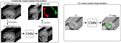 |
2 |
Fully Automated Hippocampus Segmentation Pipeline using Deep
Convolutional Neural Networks
Maximilian Sackl1,
Christian Tinauer1,
Christian Enzinger1,
Reinhold Schmidt1,
and Stefan Ropele1
1Department of Neurology, Medical University Graz, Graz, Austria Keywords: Segmentation, Alzheimer's Disease, Multi-Contrast, AI Segmentation of the hippocampus on T1-weighted structural MR images is required to quantify the neurodegenerative effects in Alzheimer’s disease studies. In this work, we propose an automated artificial intelligence-based pipeline for hippocampus segmentation combined with manual ground truth (GT) data that originates from high-resolution T2-weighted MR images. Results are evaluated against the manual GT-labels and compared to the segmentation results from FreeSurfer v732. Our deep learning-based segmentation outperforms FreeSurfer in terms of accuracy and speed, while reference experiments using the T2-based GT-labels yield the best results. Thus, using T2-weighted images for ground truth generation can improve automated HC segmentation. |
|
3587.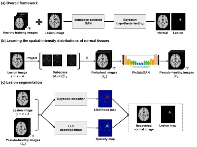 |
3 |
Improved Brain Lesion Detection by Integrating Subspace-based
Deep Learning of Normal Intensity Distributions and Bayesian
Hypothesis Testing
Huixiang Zhuang1,
Yue Guan1,
Yi Ding1,
Yuhao Ma1,
Yunpeng Zhang1,
Ziyu Meng1,
Ruihao Liu1,2,
Zhi-Pei Liang2,3,
and Yao Li1
1School of Biomedical Engineering, Shanghai Jiao Tong University, Shanghai, China, 2Beckman Institute for Advanced Science and Technology, University of Illinois at Urbana-Champaign, Urbana, IL, United States, 3Department of Electrical and Computer Engineering, University of Illinois at Urbana-Champaign, Urbana, IL, United States Keywords: Segmentation, Machine Learning/Artificial Intelligence, Unsupervised learning; Lesion segmentation Unsupervised segmentation of brain lesions is desirable in many applications and has been investigated extensively. In this work, we proposed a new method for brain lesion segmentation, which effectively learns the spatial-intensity distribution of normal brain tissues and then treats lesion segmentation as an anomaly detection problem. We overcame the high-dimensional distribution learning problem using a subspace-assisted generative network. With the learned distribution, the anomaly detection problem was solved using Bayesian hypothesis testing. Our method has been validated using simulated and real brain MR images with stroke and tumor lesions, and produced significantly improved results than several state-of-the-art methods. |
|
3588.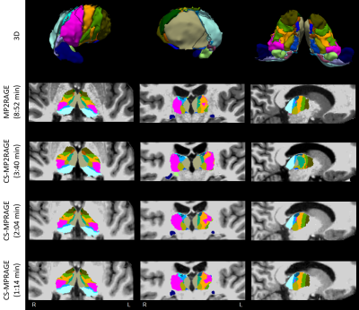 |
4 |
Human brain thalamic nuclei segmentation: compressed-sensing
effects on 3D MPRAGE variants
Sebastian Hübner1,
Stefano Tambalo1,
Tobias Kober2,3,4,
and Jorge Jovicich1
1Center for Mind/Brain Sciences, University of Trento, Rovereto (Trento), Italy, 2Advanced Clinical Imaging Technology, Siemens Healthineers International AG, Lausanne, Switzerland, 3Department of Radiology, Lausanne University Hospital and University of Lausanne, Lausanne, Switzerland, 4LTS5, École Polytechnique Fédérale de Lausanne (EPFL), Lausanne, Switzerland Keywords: Segmentation, Brain, Thalamus Accurate segmentation of thalamic nuclei can significantly support clinical decisions, especially in diseases such as Parkinson’s. At the same time, highly accelerated T1w acquisition techniques find wider clinical acceptance as they not only increase radiological efficiency and patient comfort, but also reduce motion artefacts. Here, we study how compressed sensing affects thalamic nuclei volumetry based on MPRAGE or MP2RAGE in healthy subjects with respect to clinical baseline. Segmentation results are promising and agree with expectations: acceleration to about 1 min is possible maintaining overall good image quality, contrast and thalamic segmentations, with acceleration-related biases. |
|
3589.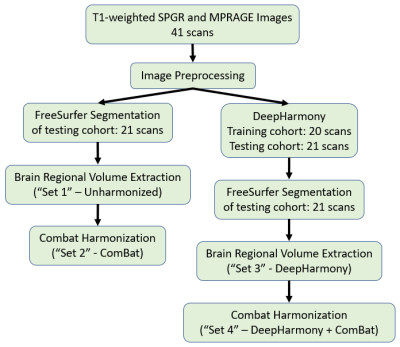 |
5 |
Comparison of image-based and statistical approaches for brain
volumetry harmonization
Yuan-Chiao Lu1,2,
Blake E Dewey3,
Yi-Yu Chou1,2,
Danielle Greenman1,2,
Russell T Shinohara4,
Daniel S Reich5,
Jerry L Prince6,
John A Butman2,
and Dzung L Pham2,7
1Center for Neuroscience and Regenerative Medicine, Henry M Jackson Foundation for the Advancement of Military Medicine, Bethesda, MD, United States, 2Radiology and Imaging Sciences, National Institutes of Health, Bethesda, MD, United States, 3Neurology, Johns Hopkins University, Baltimore, MD, United States, 4Department of Biostatistics, Epidemiology and Informatics, University of Pennsylvania, Philadelphia, PA, United States, 5Translational Neuroradiology Section, National Institute of Neurological Disorders and Stroke, Bethesda, MD, United States, 6Electrical and Computer Engineering, Johns Hopkins University, Baltimore, MD, United States, 7Radiology and Radiological Sciences, Uniformed Services University, Bethesda, MD, United States Keywords: Segmentation, Brain, Harmonization Image harmonization approaches have been proposed for reducing the variation of brain image measurements in studies involving acquisitions from multiple scanner protocols and hardware. This study compares two harmonization methods, DeepHarmony, a deep learning-based image synthesis approach, and ComBat, a statistical batch correction tool, based on their ability to yield consistent brain volume measurements from two different T1-weighted acquisitions. Our study showed that DeepHarmony outperformed the ComBat approach, although both approaches significantly improved consistency when compared to unharmonized images. |
|
3590.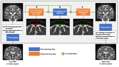 |
6 |
A new cascaded fully convolutional neural network for the
simultaneous segmentation of parasagittal dural space and
arachnoid granulations
Kilian Hett1,
Colin D. McKnight2,
Jennifer S. Lindsey2,
Melanie Leguizamon1,
Jarrod Eisma1,
Alexander K. Song1,
Jason Elenberger1,
Ciaran M. Considine1,
Daniel O. Claassen1,
and Manus J. Donahue1
1Department of Neurology, Vanderbilt University Medical Center, Nashville, TN, United States, 2Department of Radiology, Vanderbilt University Medical Center, Nashville, TN, United States Keywords: Segmentation, Neurofluids The overarching goal of this work is to develop and validate novel deep learning algorithms for segmenting the peri-sinus space, including parasagittal dural (PSD) space, which has been hypothesized to harbor cerebral lymphatic channels, and intra-veinous arachnoid granulations, which has been long-hypothesized as a site a CSF egress, from standard non-contrast anatomical imaging. The new segmentation method is based on cascaded neural networks using non-contrasted 3D T2-weighted MRI; the method is method in a mixed cohort of adults with and without neurodegeneration. |
|
3591.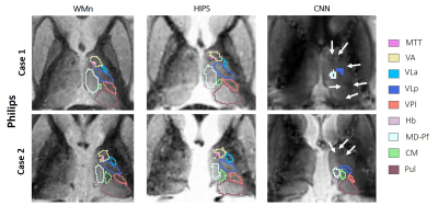 |
7 |
Robust thalamic nuclei segmentation from T1 MRI data
Julie P. Vidal1,2,
Lola Danet2,3,
Patrice Péran2,
Jérémie Pariente2,3,
Dolf Pfefferbaum4,
Edith Sullivan4,
Emmanuel J. Barbeau1,
and Manojkumar Saranathan5
1CNRS, CerCo (Brain and Cognition Research Center) - Université Paul Sabatier, Toulouse, France, 2INSERM, ToNiC (Toulouse NeuroImaging Center) - Université Paul Sabatier, Toulouse, France, 3Hôpital Purpan, Centre Hospitalier Universitaire de Toulouse, Département de Neurologie, Toulouse, France, 4Stanford University School of Medicine, Department of Psychiatry & Behavioral Sciences, Stanford, CA, United States, 5UMass Chan Medical School, Department of Radiology, Worcester, MA, United States Keywords: Segmentation, Data Processing, WMn synthesis, HIPS This work presents a methodology for thalamic nuclei segmentation from T1w MRI. Thalamus Optimized Multi-Atlas Segmentation (THOMAS), which was originally developed for white-matter nulled (WMn) MPRAGE, is adapted for standard T1 MRI by employing synthesis techniques to make the T1 images closer to WMn contrast. Robustness is tested across image contrast, MRI manufacturer, and field strength. |
|
3592.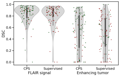 |
8 |
Improving Early Post-Operative Glioblastoma Segmentation With
Semi-Supervised Deep Learning
Lidia Luque1,2,3,
Karoline Skogen4,
Bradley J MacIntosh3,5,6,
Kyrre Eeg Emblem1,
Christopher Larsson3,7,
Einar O Vik-Mo7,
and Atle Bjørnerud1,3
1Department of Physics and Computational Radiology, Oslo University Hospital, Oslo, Norway, 2Department of Physics, University of Oslo, Oslo, Norway, 3Computational Radiology and Artificial Intelligence (CRAI), Oslo University Hospital, Oslo, Norway, 4Department of Radiology and Nuclear Medicine, Oslo University Hospital, Oslo, Norway, 5Department of Medical Biophysics, University of Toronto, Toronto, ON, Canada, 6Sandra E Black Centre for Brain Resilience and Recovery, Sunnybrook Research Institute, Toronto, ON, Canada, 7Department of Neurosurgery, Oslo University Hospital, Oslo, Norway Keywords: Segmentation, Machine Learning/Artificial Intelligence, Semi-supervision Improving automatic segmentation of glioblastoma on early post-operative MRI is key to study the effect of resection volumes on patient outcomes. We curate a dataset of over 700 MRI examinations, of which 87 include annotations, and train a supervised and a semi-supervised deep-learning model. Semi-supervision improves the segmentation of the high-intensity FLAIR signal with 3% to a Dice score of 0.83 (p=0.031), while the segmentation of the enhancing tumor increases with 9% to 0.55 (p=0.056). However, enhancing tumor segmentations show high variability, possibly due to imperfect annotations. Segmentation of enhancing tumor on early post-operative MRI remains a challenging task. |
|
3593.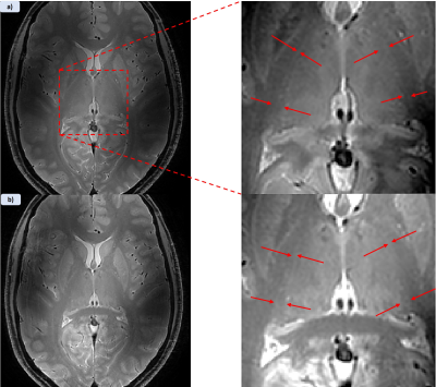 |
9 |
Imaging The Thalamic Reticular Nucleus at 7T
ross shaw1,
Penny Gowland1,
and Richard Bowtell2
1University of Nottingham, Nottingham, United Kingdom, 2Physics, University of Nottingham, Nottingham, United Kingdom Keywords: Segmentation, Contrast Mechanisms, Thalamus The Thalamic Reticular Nucleus is a micro-structure surrounding the dorsal edge of the thalamus that is of fundamental significance to understanding many neurological disorders in the brain. Here we present methods to visualise the TRN using various imaging modalities to explore different sources of contrast at the edge of the thalamus. |
|
3594.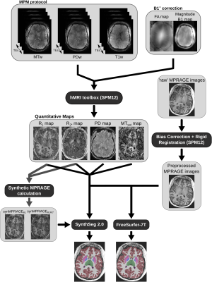 |
10 |
High quality brain segmentation from multi-parameter mapping at
Ultra-High Field MRI
Marc-Antoine Fortin1,
Yannik Völzke2,
Rüdiger Stirnberg2,
Siya Sherif3,
Laurent Lamalle3,
Tony Stöcker2,4,
and Pål Erik Goa1
1Department of Physics, Norwegian University of Science and Technology (NTNU), Trondheim, Norway, 2German Center for Neurodegenerative Diseases (DZNE), Bonn, Germany, 3GIGA Cyclotron Research Centre (GIGA-CRC) in vivo imaging, University of Liège, Liège, Belgium, 4Department of Physics and Astronomy, University of Bonn, Bonn, Germany Keywords: Segmentation, High-Field MRI, Multi-Parameter Mapping, Quantitative MRI, 7T Very few segmentation techniques are optimized for 7T images. Due to the lack of approaches and the high level of inhomogeneity observed with Ultra-High Field images, neuroscientists typically limit the analysis to the upper half of the brain. A novel resolution- and contrast-agnostic technique, called SynthSeg, may remedy this limitation. A multi-parameter mapping protocol and MPRAGE images were acquired to test SynthSeg against FreeSurfer on 7T sub-millimeter brain images. Our results showed that SynthSeg can surpass FreeSurfer, especially for the cerebellum and temporal lobes. Moreover, we showed that quantitative maps can be great surrogates to high-resolution T1w images like MPRAGE. |
|
3595.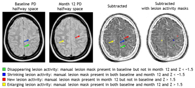 |
11 |
Validation of a Semi-Automated Method to Quantify Lesion Volume
Changes in Multiple Sclerosis Using Subtraction Images
Rozemarijn M. Mattiesing1,
Serena Stel1,
Alysha S. Mangroe1,
Iman Brouwer1,
Adriaan Versteeg1,
Ronald A. van Schijndel1,
Bernard M.J. Uitdehaag2,
Frederik Barkhof1,3,
Hugo Vrenken1,
and Joost P.A. Kuijer1
1MS Center Amsterdam, Radiology and Nuclear Medicine, Amsterdam Neuroscience, Amsterdam UMC location VUmc, Amsterdam, Netherlands, 2MS Center Amsterdam, Neurology, Amsterdam Neuroscience, Amsterdam UMC location VUmc, Amsterdam, Netherlands, 3UCL London, Institutes of Neurology and Healthcare Engineering, London, United Kingdom Keywords: Segmentation, Multiple Sclerosis Monitoring changes in white matter lesions with MRI is important to evaluate the effects of treatment in multiple sclerosis. In this study a validation of a semi-automated method to quantify lesion volume changes based on 2D proton-density-weighted images and image subtraction was performed. With this method new and enlarging but also disappearing and shrinking lesion activity can be quantified. As assessed with the intraclass correlation coefficient for absolute agreement, we found that the reproducibility was excellent and the accuracy was good overall. This semi-automated subtraction method can reliably quantify lesion volume changes in patients with (early) multiple sclerosis. |
|
3596.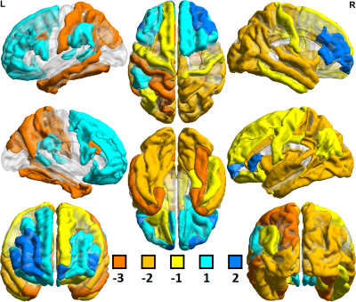 |
12 |
Differential patterns of automatic segmentation of 3T and 7T
MRI: Implications in neuropsychiatric disease
Gaurav Verma1,
Ki-Sueng Choi1,
Helen Mayberg2,
and Priti Balchandani1
1Radiology, Icahn School of Medicine at Mount Sinai, New York, NY, United States, 2Neurology, Icahn School of Medicine at Mount Sinai, New York, NY, United States Keywords: Segmentation, High-Field MRI, FreeSurfer, MDD Automatic segmentation was performed on T1-MPRAGE structural MRI data acquired at 3T and 7T from 37 and 69 distinct healthy controls, respectively. Additionally, segmentation was performed on imaging acquired from 215 major depressive disorder (MDD) patients at 3T and 40 MDD patients at 7T. Of 259 segmentation-derived imaging features evaluated, 120 showed significant 3T vs. 7T differences among controls, and 153 among patients. 7T imaging metrics showed consistently lower cortical thickness and cortical gray/white matter ratios. Subcortical and cortical volumes measured at 7T were more mixed, with 7T images showing greater frontal lobe volume, but lower cortical volumes elsewhere. |
|
3597.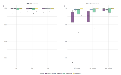 |
13 |
Reliability of brain volume measurements on 3D-FLAIR is similar
to 3D-T1 in multiple sclerosis
David Rudolf van Nederpelt1,
Samantha Noteboom2,
Eva M.M. Strijbis3,
Iman Brouwer1,
Bastiaan Moraal1,
Bas M.S. Jasperse1,
Henk-Jan M.M. Mutsaerts1,
Menno M. Schoonheim2,
Frederik Barkhof1,4,
Hugo Vrenken1,
and Joost P.A. Kuijer1
1Radiology and nuclear medicine, MS Center Amsterdam, Radiology and Nuclear Medicine, Vrije Universiteit Amsterdam, Amsterdam Neuroscience, Amsterdam UMC location VUmc, Amsterdam, The Netherlands, Amsterdam, Netherlands, 2Anatomy and Neurosciences, MS Center Amsterdam, Anatomy and Neurosciences, Vrije Universiteit Amsterdam, Amsterdam Neuroscience, Amsterdam UMC location VUmc, Amsterdam, The Netherlands, Amsterdam, Netherlands, 3MS Center Amsterdam, Neurology, Vrije Universiteit Amsterdam, Amsterdam Neuroscience, Amsterdam UMC location VUmc, Amsterdam, The Netherlands, Amsterdam, Netherlands, 4Queen Square Institute of Neurology and Centre for Medical Image Computing, University College London, UK, London, United Kingdom Keywords: Segmentation, Multiple Sclerosis New methods have enabled segmentation on sequences other than T1 for brain volume measurements in multiple sclerosis (MS), such as those more readily available in clinic as FLAIR and PD. However, reliability studies have not been performed yet in MS, limiting their use. Here, we assessed within-scanner and between-scanner reliability of FLAIR segmentations in 30 people with MS on three different MR scanners. We found similar reliability compared to T1w scans, with high intra-scanner reliability but systematic differences between scanners. This suggests that within-scanner volume measurements of FLAIR are possible, but standardization is needed for between-scanner single patient measurements. |
|
3598.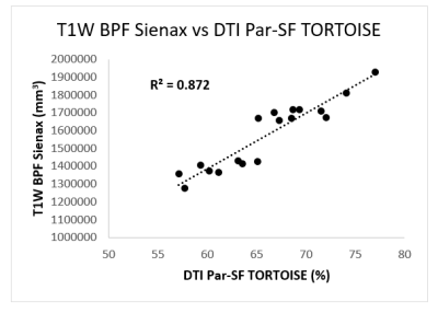 |
14 |
Comparison of whole brain parenchyma fraction (BPF) from T1W and
diffusion MRI in the assessment of CLN3, a neurodegenerative
disease
Amritha Nayak1,2,
An N Dang Do3,
Audrey E Thurm4,
Ariane Soldatos5,
Forbes D Porter3,
and Carlo Pierpaoli1
1Laboratory on Quantitative Medical Imaging, NIBIB, NIH, Bethesda, MD, United States, 2Henry Jackson Foundation for Advancement of Military Medicine, Bethesda, MD, United States, 3Division of Translational Medicine, NICHD,NIH, Bethesda, MD, United States, 4Office of Clinical Director, NIMH,NIH, Bethesda, MD, United States, 5Office of Clinical Director, NINDS,NIH, Bethesda, MD, United States Keywords: Segmentation, Quantitative Imaging, Neurodegeneration, Neurodegeneration diseases, aging, T1-weighted segmentation, T1W, DTI, Diffusion Tensor Imaging, brain parenchyma volume, brain atrophy In this work, we have compared if whole brain parenchyma fraction (BPF) measured from conventionally used T1-weighted (T1W) based brain segmentation method is comparable to signal fraction attributable to parenchymal water (Par-SF) measured from method using diffusion MRI, in assessing the overall disease state of participants with CLN3, a pediatric neurodegenerative disease. |
|
3599.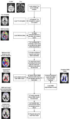 |
15 |
Tract-specific Evaluation of White Matter Hyperintensity
Segmentation
Aaron Sinclair1,
Ross Callaghan2,
and Hui Zhang1
1Centre for Medical Image Computing and Department of Computer Science, University College London, London, United Kingdom, 2AINOSTICS Ltd., Manchester, United Kingdom Keywords: Segmentation, Neuroinflammation, White Matter Hyperintensity Segmentation We propose a tract-specific white matter hyperintensity (WMH) segmentation evaluation as a new way to assess WMH segmentation techniques. Currently, WMH segmentation is assessed globally or between periventricular and deep white matter (WM) regions, which has no obvious functional relevance. WM tracts have known functional relevance and associations to neurological conditions. We demonstrate that this new approach allows for functionally-relevant assessment of techniques. Two WMH segmentation techniques are compared to highlight the performance differences across a key tract associated with processing speed. We also found significant performance differences across this tract, which would be missed by standard evaluation methods. |
|
3600.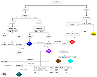 |
16 |
Intensity-Based Segmentation of Tumor on Multiparametric MRI to
Aid Response Assessment of High-Grade Gliomas Treated with
Immunoradiotherapy
Harshan Ravi1,
Samuel H. Hawkins1,2,
Olya Stringfield3,
Malesa Pereira1,4,5,
Heiko Enderling6,7,
H-H Michael Yu7,8,
John A. Arrington5,7,
Solmaz Sahebjam7,9,10,
and Natarajan Raghunand1,7
1Moffitt Cancer Center and Research Center, Tampa, FL, United States, 2Department of Computer Science, Bradley University, Peoria, IL, United States, 3Quantitative Imaging Core, Moffitt Cancer Center and Research Institute, Tampa,, FL, United States, 4Behavioral and Community Health Sciences,, LSU Health School of Public Health, New Orleans, LA, United States, 5Department of Radiology, Moffitt Cancer Center and Research Center, Tampa,, FL, United States, 6Department of Integrated Mathematical Oncology, Moffitt Cancer Center and Research Institute, Tampa,, FL, United States, 7Department of Oncologic Sciences, University of South Florida, Tampa, FL, United States, 8Department of Radiation Oncology, Moffitt Cancer Center and Research Center, Tampa,, FL, United States, 9National Cancer Institute, National Institutes of Health, Bethesda, MD, United States, 10Department of Neuro-Oncology, Moffitt Cancer Center and Research Center, Tampa,, FL, United States Keywords: Segmentation, Machine Learning/Artificial Intelligence, Glioblastoma, immunotherapy, radiotherapy Confounding appearance of radiographic changes in recurrent high grade glioma (HGG) patients treated with multimodality immunotherapy presents a challenge to the neuro-radiologist. A clinical need exists to improve upon conventional criteria for assessment of GBM on standard-of-care (SOC; T1w, T2w, FLAIR, and T1w-enhanced) MRI to help distinguish treatment-related effects from true disease progression. We have investigated the feasibility of intensity-based segmentation of tumor tissue types on multiparametric MRI (mpMRI) to inform response assessment in HGG patients treated with bevacizumab, hypofractionated stereotactic radiotherapy, and pembrolizumab. |
|
3601.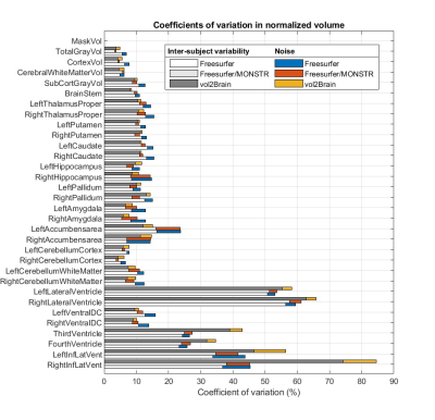 |
17 |
A general framework for analyzing the various contributions to
reproducibility in brain morphometry
Ruifeng Dong1,
Amritha Nayak1,2,
Leighton Chan3,
and Carlo Pierpaoli1
1Laboratory on Quantitative Medical Imaging, National Institute of Biomedical Imaging and Bioengineering, National Institutes of Health, Bethesda, MD, United States, 2Henry Jackson Foundation for Advancement of Military Medicine, Bethesda, MD, United States, 3Rehabilitation Medicine Department, National Institutes of Health, Bethesda, MD, United States Keywords: Segmentation, Software Tools We propose a framework for differentiating the contributions to the reproducibility in brain morphometry from the true inter-individual differences, experimental procedures, and data-processing methods. As an application, we build a linear mixed-effect model to evaluate and compare two segmentation software tools, Freesurfer and vol2Brain, in their reproducibility in measuring volumes of 32 regions of interest. For both software and for most structures under study, our approach successfully reveals the dominance of inter-subject variability over noise. Vol2Brain introduces less noise than Freesurfer for all subcortical nuclei while Freesurfer shows better performance for gray matter, cortex, cerebral white matter, and cerebellum cortex. |
|
3602.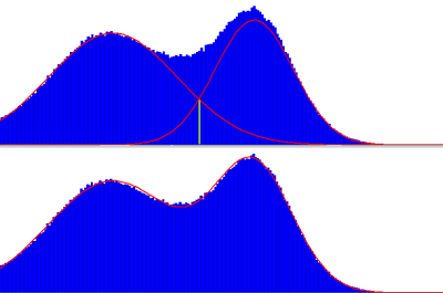 |
18 |
Globally optimal region thresholding based on bimodal Gaussian
distribution with physiological constraints : head MR example.
Artem Mikheev1 and
Henry Rusinek1
1Radiology, NYU Grossman School of Medicine, New York, NY, United States Keywords: Segmentation, Segmentation, Thresholding In medical image processing thresholding of the image region is one of the most basic operations and preprocessing. While there are well-proven histogram-based partitioning methods including Otsu and Gaussian Mixture Model ) it is challenging to combine those methods with physiological constraints such as tissue volume ratio or average signal ratio to avoid anatomically invalid segmentation. We propose a new method GOTC when the distribution of the signal over the ROI is described as a bimodal Gaussian of two tissues. We describe the algorithm and validate it on brain MRI segmentation into the White Matter and Gray Matter. |
|
3603.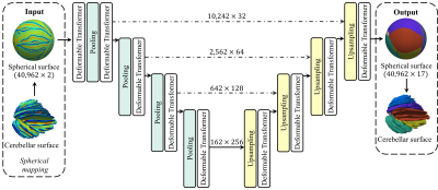 |
19 |
CEREBELLAR SURFACE PARCELLATION BASED ON DEFORMABLE SPHERICAL
TRANSFORMER
Jiale Cheng1,2,
Fenqiang Zhao2,
Zhengwang Wu2,
Ya Wang2,
Xinrui Yuan2,
Weili Lin2,
Li Wang2,
Xin Zhang1,
and Gang Li2
1School of Electronic and Information Engineering, South China University of Technology, Guangzhou, China, 2Department of Radiology and BRIC, University of North Carolina at Chapel Hill, Chapel Hill, NC, United States Keywords: Segmentation, Brain Accurate parcellation of the extremely folded cerebellar cortex is of immense importance for both brain structural and functional studies. Manual parcellation is time-consuming and expertise dependent, which motivates us to propose a novel end-to-end deep learning-based method for cerebellar cortical surface parcellation. Leveraging the spherical topology of the cerebellar surface, we propose the Deformable Spherical Transformer, which combines the advantages of the Spherical Transformer to extract the long-range dependency and the deformable attention mechanism to adaptively focus on the critical regions. Its superior performance has been validated by comparing with advanced algorithms with an average Dice ratio of 86.40%. |
|
3604.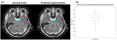 |
20 |
Deep-learning auto-segmentation and subtype classification of
pituitary adenoma based on MRI radiomics
Bing-Fong Lin1,
Dao-Chen Lin2,3,4,
and Chia-Feng Lu1
1Department of Biomedical Imaging and Radiological Sciences, National Yang Ming Chiao Tung University, Taipei, Taiwan, 2Department of Radiology, Taipei Veterans General Hospital, Taipei, Taiwan, Taipei, Taiwan, 3Division of Endocrine and Metabolism, Department of Medicine, Taipei Veterans General Hospital, Taipei, Taiwan, 4School of Medicine, National Yang Ming Chiao Tung University, Taipei, Taiwan Keywords: Radiomics, Quantitative Imaging, Pituitary adenoma Pituitary adenoma (PA) accounts for approximately 15% in intracranial neoplasms. The classification of PA generally based on the hormone level of blood as the gold standard test, while the analysis of hormone condition using neuroimaging biomarkers was less explored. Accordingly, our study developed a model to automatically segment PA and further used the quantitative and non-invasive MRI technique as image biomarkers to classify the three types of hormone pattern, focusing on corticortroph, gonadotroph, and plurihormonal type. We aimed to provide a feasible classification model based MRI to benefit the clinical management of patients with PA. |
|
 Back to Meeting Home
 Back to the Program-at-a-Glance
The International Society for Magnetic Resonance in Medicine is accredited by the Accreditation Council for Continuing Medical Education to provide continuing medical education for physicians.
 Back to Meeting Home
 Back to the Program-at-a-Glance
 View Presentation Video