Digital Poster
Image Reconstruction: UTE & ZTE
ISMRM & ISMRT Annual Meeting & Exhibition • 03-08 June 2023 • Toronto, ON, Canada

| Computer # | |||
|---|---|---|---|
5103.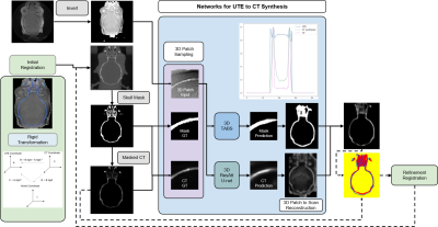 |
1 |
Synthesizing CT Image from Single-echo UTE-MRI using Multi-Task
Framework
Zhuoyao Xin1,
Vishwanatha Mitnala Rao2,
Dong Liu3,
Yanting Yang2,
Ye Tian2,
Chenghao Zhang2,
Andrew F. Laine2,
and Jia Guo2
1Biomedical Engineering, Columbia University, New York City, NY, United States, 2Columbia University, New York City, NY, United States, 3Neuroscience, Columbia University, New York City, NY, United States Keywords: Image Reconstruction, Multimodal This abstract proposed a cross-modality conversion method from UTE MRI to CT images. Using the TABS and ResidualAttentionU-net model in a processing framework combining image segmentation and prediction, the skull structure can be extracted from UTE MRI with high similarity of CT based skull. Five UTE-CT image pairs of mouse brains were used in the study. And a 3D-patch based training strategy was adopted, which took the advantage of structural continuity between slices in very limited datasets. The results show that the proposed combined image segmentation and prediction framework can achieve higher accuracy in medical image synthesizing for cross-modality conversion. |
|
5104. |
2 |
Direct Imaging and T1 and T2* Quantification of Semi-Solid Red
Blood Cell Membrane Lipid via Ultrashort Echo Time (UTE)
Sequences
Soo Hyun Shin1,
Dina Moazamian1,
Arya Suprana1,2,
Xiaojun Chen1,
Jiyo S Athertya1,
Chun Zeng1,
Michael Carl3,
Yajun Ma1,
Hyungseok Jang1,
and Jiang Du1,2,4
1Department of Radiology, University of California, San Diego, La Jolla, CA, United States, 2Department of Bioengineering, University of California, San Diego, La Jolla, CA, United States, 3GE Helathcare, San Diego, CA, United States, 4Radiology Service, Veterans Affairs San Diego Healthcare System, La Jolla, CA, United States Keywords: Relaxometry, Relaxometry Direct imaging of semi-solid lipids, such as cell membranes and myelin, is of great interest. Yet, the short T2 of semi-solid lipid protons hampers direct detection through conventional pulse sequences. In this study, we examined whether an ultrashort echo time (UTE) sequence can directly acquire signals from membrane lipids. Cell membranes from red blood cells were subject to D2O exchange and freeze-drying for complete water removal. High MR signals were detected with the UTE sequence, which showed an ultrashort T2* of ~77-252 µs and a short T1 of 189 ms for semi-solid membrane lipids. |
|
5105.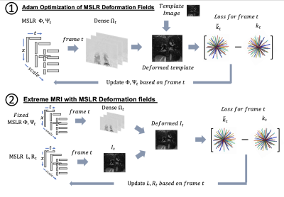 |
3 |
Motion Corrected Multi-scale Low Rank Reconstructions for Highly
Accelerated 3D Dynamic Acquisitions
Zachary Miller1,
Luis A Torres1,
Sean Fain2,
and Kevin M Johnson1
1University of Wisconsin-Madison, Madison, WI, United States, 2University of Iowa, Iowa City, IA, United States Keywords: Image Reconstruction, Motion Correction This work develops a method for large-scale motion compensated 4D reconstructions of fully 3D radial acquisitions at near 1mm isotropic spatial resolution and sub-second temporal resolution. We combine and extend multi-scale image reconstruction (Extreme MRI) with k-space based motion field estimation (MOTUS) to solve for a multi-scale low rank representations of both images and motion fields. This combined approach outperforms the multi-scale low rank reconstruction without motion fields. |
|
5106.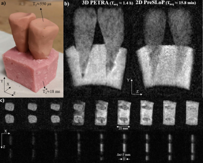 |
4 |
Slice-selective Zero Echo Time imaging of ultra-short 𝑇2
tissues
Jose Borreguero1,
Fernando Galve2,
Jose Miguel Algarín2,
Jose María Benlloch2,
and Joseba Alonso2
1Tesoro Imaging S.L., Valencia, Spain, 2Institute for Molecular Imaging and Instrumentation, Spanish National Research Council & Universitat Politècnica de València, Valencia, Spain Keywords: New Trajectories & Spatial Encoding Methods, New Signal Preparation Schemes Here we provide an MRI sequence which allows slice selection and 2D-imaging of hard tissues with T2 as short as 275 μs within clinically acceptable scan times even at fields as low as 260 mT. Our proposed sequence combines slice selection through spin-locking, which suffers a much more benign decay (T1ρ>>T2), and the fastest imaging sequence (ZTE), providing a new and robust tool for slice selection of the shortest-lived tissue signals in the body. |
|
5107.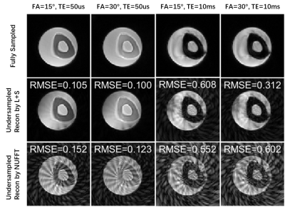 |
5 |
Low-Rank Plus Sparse Accelerated Proton Resonance Frequency
Shift and T1-mapping with a Dual-Echo 3D Spiral Ultra-Short Echo
Time Sequence
Sheng Chen1,
Zhixing Wang1,
Yekaterina K. Gilbo2,
Helen L. Sporkin1,
Samuel W. Fielden3,
Steven P. Allen4,
John P. Mugler III5,
G. Wilson Miller5,
and Craig H. Meyer1,5
1Biomedical Engineering, University of Virginia, Charlottesville, VA, United States, 2Netflix, Los Gatos, CA, United States, 3U.S. Food and Drug Administration, Silver Spring, MD, United States, 4Brigham Young University, Provo, UT, United States, 5Radiology and Medical Imaging, University of Virginia, Charlottesville, VA, United States Keywords: Sparse & Low-Rank Models, Image Reconstruction, UTE, Spiral, Focused Ultrasound Unintended heating of the skull and nearby brain tissue during MR-guided Focused Ultrasound (MRgFUS) treatment is not standardly monitored. A method for addressing this issue, which combines variable flip angle (VFA) T1 mapping and proton resonance frequency (PRF) shift thermometry based on a dual-echo 3D spiral ultra-short echo time (UTE) acquisition, was accelerated with a low-rank plus sparse model in image reconstruction and validated using retrospectively undersampled data in vitro. |
|
5108. |
6 |
DUDE: Diffusion tensor imaging with Ultra-short echo time Double
Echo steady state – a proof of concept
Stefan Sommer1,2,3,
Constantin von Deuster1,2,3,
Tom Hilbert3,4,5,
and Daniel Nanz2
1Siemens Healthineers International AG, Zurich, Switzerland, 2Swiss Center for Musculoskeletal Imaging (SCMI), Balgrist Campus, Zurich, Switzerland, 3Advanced Clinical Imaging Technology (ACIT), Siemens Healthineers International AG, Lausanne, Switzerland, 4Department of Radiology, Lausanne University Hospital and University of Lausanne, Lausanne, Switzerland, 5LTS5, École Polytechnique Fédérale de Lausanne (EPFL), Lausanne, Switzerland Keywords: Data Acquisition, Pulse Sequence Design, DESS, UTE, UTE-DESS, DW-DESS, DW-UTE-DESS, ZTE The quality of echo-planar diffusion images can suffer from severe distortions and signal dropouts, especially in regions with high susceptibility gradients and rapidly decaying (short T2*) signal. To overcome these problems, we implemented a diffusion sensitive UTE-DESS sequence. The acquisition along six distinct diffusion directions allows for fitting of a diffusion tensor. We show the feasibility of Diffusion tensor imaging with Ultra-short echo time Double Echo steady state (DUDE) at 3T and present preliminary results in the brain of a healthy volunteer. |
|
5109. |
7 |
Adapted rosette trajectory for functional 2D lung imaging
Hanna Frantz1,
Patrick Metze1,
and Volker Rasche1
1Ulm University Hospital, Ulm, Germany Keywords: New Trajectories & Spatial Encoding Methods, Lung, Function Lung MRI is a steadily evolving field of research, especially concerning the evaluation of chronic lung diseases such as cystic fibrosis or COPD. The major limitation of lung MRI is the ultrashort T2* relaxation time of lung parenchyma, which demands ultrashort echo time sequences. Sufficient SNR values in the parenchyma are crucial for clinical evaluations and the assessment of physiological parameters. This abstract presents an adapted rosette trajectory for UTE imaging, yielding higher SNR per unit time in comparison to radial UTE sampling approaches. |
|
5110. |
8 |
Multi-contrast multi-resolution UTE for simultaneous
quantitative and high-resolution MRI
Serhat Ilbey1,
Antonia Susnjar2,
Uzay Emir2,3,
Michael Bock1,
and Ali Caglar Özen1
1Division of Medical Physics, Department of Radiology, University Medical Center Freiburg, Freiburg, Germany, 2Weldon School of Biomedical Engineering, Purdue University, West Lafayette, IN, United States, 3School of Health Science Department, Purdue University, West Lafayette, IN, United States Keywords: New Trajectories & Spatial Encoding Methods, New Trajectories & Spatial Encoding Methods A multi-contrast multi-resolution UTE sequence was developed with a dual-echo acquisition between 1-1 binomial water excitation pulses and a conventional UTE readout. This acquisition scheme provides 3 images with different echoes, flip angles, spatial resolution, and contrast (water excitation). T2* was quantified in the whole brain in TA<15min, and UTE images of the brain were reconstructed with 0.5mm isotropic resolution without additional scan time. |
|
5111. |
9 |
On the elimination of the fat saturation effect in ultrashort
echo-time imaging of Achilles tendons.
Peter Latta1,
Vladimir Juras2,
Zenon Starcuk3,
Martin Kojan1,
Ivan Rektor1,
Pavol Szomolanyi2,
and Siegfried Trattnig2
1Masaryk University, Brno, Czech Republic, 2Medical University of Vienna, Vienna, Austria, 3Institute of Scientific Instruments, Czech Academy of Sciences, Brno, Czech Republic Keywords: Tendon/Ligament, Skeletal, Achilles tendon, Bi-exponential . T2* We investigated tri‑component analysis of 2D-UTE in-vivo measurements on a clinical 3T scanner and tested its ability to eliminate the effect of fat suppression on short T2* values in Achilles tendons. We found that fat-suppressed acquisition with two-component analysis provides ratios of short-T2* to long-T2* component intensities that are substantially lower (by a factor 0.95 to 0.55) than those resulting from fat-unsuppressed acquisition and tri-component fitting. Hence it seems that the combination of 2D-UTE acquisition with tri-component analysis might contribute to further improvement of T2* estimation accuracy. |
|
5112.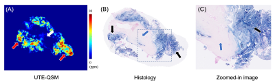 |
10 |
Quantification of Hemosiderin Deposition in Hemophilic
Arthropathy Using Ultrashort Echo Time Quantitative
Susceptibility Mapping (UTE-QSM)
Sam Sedaghat1,
James V Luck2,
Annette von Drygalski3,
Scott T Ball4,
Soo Hyun Shin1,
Eddie Fu1,
Eric Y Chang1,5,
Jiang Du1,5,6,
and Hyungseok Jang1
1Department of Radiology, University of California San Diego, San Diego, CA, United States, 2Department of Orthopaedic Surgery, University of California Los Angeles, Los Angeles, CA, United States, 3Department of Medicine, University of California San Diego, San Diego, CA, United States, 4Department of Orthopaedic Surgery, University of California San Diego, San Diego, CA, United States, 5Radiology Service, Veterans Affairs San Diego Healthcare System, San Diego, CA, United States, 6Department of Bioengineering, University of California San Diego, San Diego, CA, United States Keywords: MSK, Hematologic We investigated the quantitative capability of the UTE-QSM technique in the assessment of hemosiderin compared to histology. UTE-QSM and histology were performed in knee synovium tissues from hemophilia patients to visualize hemosiderin. The susceptibility maps from UTE-QSM showed a visually high similarity to the corresponding histological findings for all tissues. There was a significant difference in tissues with low and high iron load (p<0.001) in UTE-QSM. The susceptibility values on UTE-QSM showed a significant strong positive correlation to the blue signal in histology (R=0.908; p<0.001). We showed that UTE-QSM has excellent performance for detecting and quantifying hemosiderin in HA. |
|
5113.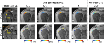 |
11 |
Quantitative Ultrashort Echo-Time MRI to Assess In Vivo Rotator
Cuff Tendon Degeneration
Misung Han1,
Peder EZ Larson1,2,
Thomas M Link1,
Drew A Lansdown3,
Brian T Feeley3,
and Sharmila Majumdar1,2
1Radiology and Biomedical Imaging, University of California, San Francisco, San Francisco, CA, United States, 2UCSF-UC Berkely Graduate Program in Bioengineering, University of California, San Francisco, San Francisco, CA, United States, 3Department of Orthopaedic Surgery, San Francisco, CA, United States Keywords: Tendon/Ligament, Quantitative Imaging Rotator cuff tears are a common source of pain and disability. Surgical treatment is normally considered for patients who fail in non-operative management, but a failure of tendon healing after surgery can be dependent on tendon quality. In this work, we applied two ultrashort-echo time (UTE) quantification imaging methods, UTE T2* quantification as well as UTE magnetization transfer (MT) quantification, on healthy subjects and patients with rotator cuff tears, and evaluated their ability to assess tendon degeneration. Significant difference was observed for quantified T2* and MT parameters over segmented supraspinatus tendon between the healthy control and patient groups. |
|
5114.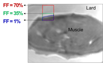 |
12 |
Robust assessment of macromolecular fraction in muscle with
differing fat fraction using ultrashort echo time magnetization
transfer modeling
Saeed Jerban1,
Yajun Ma1,
Qingbo Tang2,
Eddie Fu2,
Nikolaus Szeverenyi1,
Hyungseok Jang1,
Christine B Chung1,
Jiang Du1,
and Eric Y Chang1,2
1Radiology, University of California, San Diego, San Diego, CA, United States, 2Radiology Service, Veterans Affairs San Diego Healthcare System, San Diego, La Jolla, CA, USA, San Diego, CA, United States Keywords: Muscle, Magnetization transfer, Collagen Content; Fat Content Magnetization transfer (MT) MR imaging can indirectly characterize the spatial distribution of the relative contents of the macromolecular and water proton pools (MMF) of skeletal muscles. Fat presence in muscle has always been a source of concern in MMF calculation where some studies have reported significant underestimation of MMF for muscles with a considerable fat fraction (FF). We investigated the impact of FF on MMF in muscle/fat phantoms using an ultrashort echo time (UTE) MT model after T1 compensation. MMF demonstrated a relatively robust value with under 5% and 20% changes for FF increases up to 30 and 45%, respectively. |
|
5115.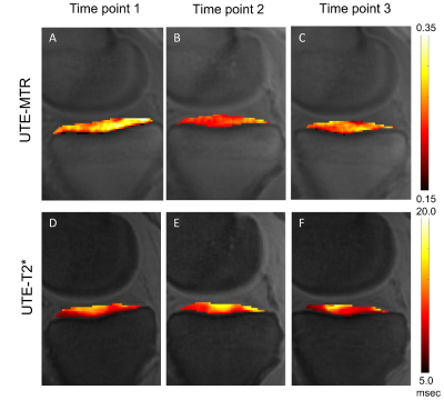 |
13 |
Ultrashort Echo Time Magnetization Transfer Imaging of Knee
Cartilage After Long-Distance Running
Dantian Zhu1,
Yijie Fang1,
Wenhao Wu1,
Shaolin Li1,
Long Qian2,
and Yajun Ma3
1Department of Radiology,Fifth Affiliated Hospital, Sun Yat-Sen University, Zhuhai, China, 2MR Research, GE Healthcare, Beijing, China, 3University of California, San Diego, Department of Radiology, San Diego, CA, United States Keywords: Cartilage, Cartilage To assess the detection of changes in the knee cartilage of amateur marathon runners before and after long-distance running. We recruited 23 amateur marathon runners prospectively. MRI scans using UTE-MT and UTE-T2* sequences. UTE-MTR and UTE-T2* were measured for knee cartilage. The UTE-MTR values in lateral tibial plateau, central medial femoral condyle, and medial tibial plateau showed a significant decrease at 2 days post-race compared to the other two time points (P < 0.05). But no significant UTE-T2* changes were found for any cartilage subregions. UTE-MTR is a promising method for the detection of dynamic changes in knee cartilage. |
|
5116.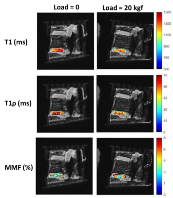 |
14 |
Feasibility of detecting subvoxel deformation in intervertebral
disc using quantitative ultrashort echo time (UTE) techniques
Saeed Jerban1,2,3,
Dina Moazamian1,
Sheronda Statum1,
Hyungseok Jang1,2,
Eric Y Chang1,2,
Jiang Du1,2,
Christine B Chung1,2,
and Yajun Ma1
1Department of Radiology, University of California, San Diego, La Jolla, CA, USA, San Diego, CA, United States, 2Radiology Service, Veterans Affairs San Diego Healthcare System, San Diego, La Jolla, CA, USA, San Diego, CA, United States, 3Department of Orthopedic Surgery, University of California, San Diego, La Jolla, CA, USA, San Diego, CA, United States Keywords: Skeletal, Magnetization transfer, Intervertebral disc Quantitative ultrashort echo time (UTE) MRI can be used for quantitative assessment of intervertebral discs (IVDs). It is hypothesized that the investigation of quantitative UTE MRI properties of IVD under mechanical loading may highlight the affected regions of IVD by diseases and injuries. We investigated the feasibility of using UTE-T1, UTE-Adiab-T1ρ, and UTE-MT measures for detecting the IVD deformation under loading. T1 and T1 ρ decreased in IVDs under loading while MMF from UTE-MT modeling as an index for collagen content increased by loading. This study highlights the potential of UTE-MRI to detect subvoxel deformations in IVDs caused by loading. |
|
5117.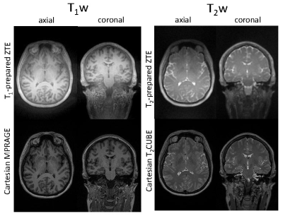 |
15 |
Progress toward a ZTE-based silent and motion-robust protocol
for pediatric neuroimaging
James H Holmes1,
Vincent A Magnota1,
Mathews Jacob2,
Yan Chen2,
Joshua Hanson3,
Paul A DiCamillo1,
and Curtis A Corum3
1Radiology, University of Iowa, Iowa City, IA, United States, 2Electrical and Computer Engineering, University of Iowa, Iowa City, IA, United States, 3Champaign Imaging, LLC, Shoreview, MN, United States Keywords: Neuro, Pulse Sequence Design, silent imaging In this work, we present progress in developing a ZTE-based silent and motion-robust neuroimaging protocol using intermittent magnetization preparation for generation of standard imaging contrasts including T1w and T2w. |
|
5118. |
16 |
Density-Controllable Spiral Trajectory Design Combined with
CS-UTE Improves High-Resolution Single Breath-Hold Lung Imaging
Yupeng Cao1,
Jun Zhao1,
Weinan Tang2,
Wentao Liu1,
and Dong Han1
1National Center for Nanoscience and Technology, Beijing, China, 2Wandong Medical Inc, Beijing, China, Beijing, China Keywords: New Trajectories & Spatial Encoding Methods, Lung Single breath-hold high-resolution lung imaging is challenging due to the short T2* and scan efficiency. The stack-of-spirals UTE enables fast lung imaging in a single breath hold. However, fast scanning of stack-of-spirals results in a long readout time, introducing adverse effects of the short T2* of the lung. Herein, we proposed a density-controllable spiral trajectory (DCST) design method to design a short readout time trajectory concurrently satisfying the criteria of optimal compressed sensing (CS), reducing the adverse effect of the short T2*. The short readout time trajectory combined with CS-UTE improves the single breath-hold high-resolution lung imaging. |
|
5119. |
17 |
Multi-Echo FLORET UTE MRI
Matthew Willmering1,
Guruprasad Krishnamoorthy2,3,
Joseph Plummer1,4,
Abdullah Bdaiwi1,
Laura Walkup1,4,5,
Zackary Cleveland1,4,5,
James Pipe3,
and Jason Woods1,4,5
1Cincinnati Children’s Hospital Medical Center, Cincinnati, OH, United States, 2Philips Healthcare, Rochester, MN, United States, 3Mayo Clinic, Rochester, MN, United States, 4University of Cincinnati, Cincinnati, OH, United States, 5University of Cincinnati Medical Center, Cincinnati, OH, United States Keywords: Data Acquisition, Data Acquisition FLORET UTE MRI has been used for many applications including lung, brain, musculoskeletal, and multinuclear imaging. The appeal of the pulse sequence comes from its high SNR and sampling efficiencies. With recent improvements minimizing artifacts, high-quality FLORET images are obtained routinely. However, acquisition of multiple echoes has not been investigated. Here, we determined the feasibility and optimal pulse-sequence design of multi-echo FLORET. Collecting echoes independently, but interleaved, produces the highest quality images, especially for ≥3 echoes. Furthermore, additional echoes provide improvements to image quality and add clinically useful contrast like T2*. |
|
5120.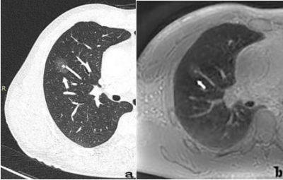 |
18 |
Assessing the clinical value of PETRA sequence for detection of
subsolid pulmonary nodules:comparison with CT
Hui Feng1,
Li Yang1,
Chen Zhang2,
Hui Liu1,
Ning Zhang1,
and Gaofeng Shi1
1The Fourth Hospital of Hebei Medical University, Shijiazhuang, China, 2Siemens Healthineers, Beijing, China Keywords: Visualization, Lung The PETRA technique had high sensitivity for the detection of subsolid pulmonary nodules and can accurately assess their diameter and morphologic characteristics. |
|
5121. |
19 |
Myelin Content Imaging Using Ultra-Short TE MRI with Variable
Flip Angles
Kwan-Jin Jung1,
Ryan Larsen2,
Laurie Rund2,
and Andrew Steelman3,4,5
1Biomedical Imaging Center, Beckman Institute, University of Illinois at Urbana-Champaign, Urbana, IL, United States, 2Department of Animal Sciences, University of Illinois at Urbana-Champaign, Urbana, IL, United States, 3Neuroscience Program, 2325/21, University of Illinois at Urbana-Champaign, Urbana, IL, United States, 4Carl R. Woese Institute for Genomic Biology, University of Illinois at Urbana-Champaign, Urbana, IL, United States, 5Division of Nutritional Sciences, University of Illinois at Urbana-Champaign, Urbana, IL, United States Keywords: Relaxometry, Neuro, Myelin, Piglet Myelin content was measured from the fast T1 relaxation component using bi-exponential T1 relaxation regression. The data was collected using UTE with variable flip angles to detect short T2 signal of myelin and to avoid magnetic susceptibility corruption by T2*-based myelin contrast methods. The estimated myelin content was influenced by CSF, which was suppressed by use of the slow T1 relaxation time. The estimated myelin content was higher in white matter than other brain regions. However, the myelin content was increased in the anterior pole and low in motor areas in this in-vivo piglet data. |
|
5122.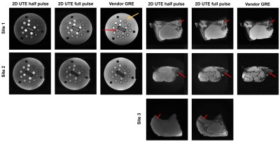 |
20 |
Validation of open-source minimal TE sequences across three
independent sites
Gehua Tong1,
Andreia S. Gaspar2,
Rita G. Nunes2,
Jon-Fredrik Nielsen3,
John Thomas Vaughan, Jr.1,4,
and Sairam Geethanath4,5
1Biomedical Engineering, Columbia University, New York, NY, United States, 2Institute for Systems and Robotics (ISR-Lisboa)/LaRSyS, Department of Bioengineering, Instituto Superior Técnico, Universidade de Lisboa, Lisbon, Portugal, 3Biomedical Engineering, University of Michigan, Ann Arbor, MI, United States, 4Columbia Magnetic Resonance Research Center, Columbia University, New York, NY, United States, 5Accessible MR Laboratory, BioMedical Engineering and Imaging Institute, Dept. of Diagnostic, Molecular and Interventional Radiology, Icahn School of Medicine at Mt. Sinai, New York, NY, United States Keywords: Validation, Pulse Sequence Design We validated five open-source PyPulseq minimal TE sequences (half/full pulse 2D/3D UTEs and COncurrent Dephasing and Excitation (CODE)) at three sites with different scanner models. The ISMRM/NIST T2 array and a porcine muscle and bone sample were imaged. SNR, contrast-to-noise ratio (CNR), and T2 contrast curves were calculated and compared. We found that the sequences were able to recover more SNR from bone tissue compared to vendor GRE (an additional 50% - 1060% for 3T and 34% - 474% for 1.5T) and higher signal for short-T2 spheres but intersite differences existed that were consistent with field strengths. |
|
 Back to Meeting Home
 Back to the Program-at-a-Glance
The International Society for Magnetic Resonance in Medicine is accredited by the Accreditation Council for Continuing Medical Education to provide continuing medical education for physicians.
 Back to Meeting Home
 Back to the Program-at-a-Glance
 View Presentation Video