Digital Poster
Quantitative Muscle Imaging
ISMRM & ISMRT Annual Meeting & Exhibition • 03-08 June 2023 • Toronto, ON, Canada

| Computer # | |||
|---|---|---|---|
4206.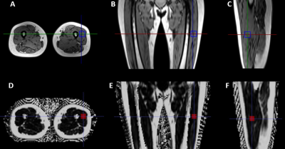 |
121 |
Q-Dixon MRI and High-Speed T2-Corrected Multiecho MR
Spectroscopy to Quantify Thigh Muscle Fat Content in Dialysis
Patients
Wenshuang Zhang1,
Ling Wang1,
Dong Yan1,
Yanglei Wu2,
Yi Yuan1,
and Xiaoguang Cheng1
1Department of Radiology, Peking University Fourth School of Clinical Medicine, Beijing Jishuitan Hospital, Beijing, China, 2MR collaborations, Siemens Healthineers Ltd., Beijing, China Keywords: Muscle, Fat This study evaluated the correlation and consistency of proton density fat fraction (PDFF) measured by Q-Dixon magnetic resonance imaging (MRI) and high-speed T2-corrected multiecho (HISTO) magnetic resonance spectroscopy (MRS) for quantifying thigh muscle fat content in patients undergoing dialysis. Q-Dixon MRI exhibited good correlation and consistency with the HISTO MRS for thigh muscle fat quantitative measurements indicating that Q-Dixon MRI may be a reliable alternative to HISTO MRS in clinical assessment and radiological quantitative evaluation of thigh muscle fat infiltration. |
|
4207. |
122 |
Elevated Muscle Water T2 in Dystrophinopathy
Glenn Walter1,
Alison Barnard2,
Sean Forbes2,
Rebecca Willcocks2,
Eric Baetscer3,
William Triplett2,
William Rooney 3,
and Krista Vandenborne2
1Physiology, University of Florida, Gainesville, FL, United States, 2Physical Therapy, University of Florida, Gainesville, FL, United States, 3Advanced Imaging Research Center, Oregon Health & Science University, Portland, OR, United States Keywords: Muscle, Muscle, muscular dystrophy Both MRI and MRS measures reveal that water T2 can be elevated in individuals living with dystrophin deficits, which includes patients with DMD, BMD and manifesting carriers. This has important implications when evaluating therapeutics aimed at decreasing inflammation (e.g., corticosteroids or NFkb inhibitors) or disease modifying interventions, such as gene therapies. |
|
4208.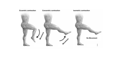 |
123 |
Diffusion Tensor Imaging parameters predict quadriceps
strength in younger and older healthy adults
Yael Vainberg1,
Jessica Asay1,
Andrew Schmidt1,
Julie Muccini1,
Garry Gold1,
Feliks Kogan1,
and Valentina Mazzoli1
1Radiology, Stanford University, Stanford, CA, United States Keywords: Muscle, Diffusion Tensor Imaging, Sarcopenia The loss of muscle strength was previously thought to be primarily impacted by the loss of muscle volume due to sarcopenia during aging, however, this did not explain why strength declined more rapidly than muscle volume. To better understand this, our study aimed to explore Diffusion Tensor Imaging parameters and their correlations to muscle strength in addition to volume. As expected, volume had a strong positive correlation with all functional strength tests. However, we also observed that radial diffusivity had a significant negative correlation with eccentric functional strength tests, but no correlation was found with the concentric tests. |
|
4209.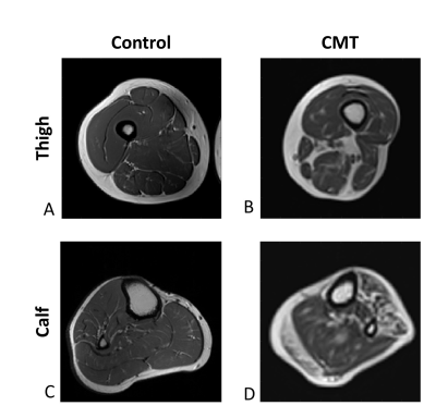 |
124 |
Assessment of Charcot-Marie-Tooth disease severity using
quantitative T2 and fat fraction biomarkers
Maria Holodov1,
Shimon Shahar2,
Tamar Blumenfeld-Katzir1,
David Bendahan3,
and Noam Ben-Eliezer1,4,5
1Department of Biomedical Engineering, Tel Aviv University, Tel Aviv, Israel, 2Center of AI and Data Science (TAD), Tel Aviv University, Tel Aviv, Israel, 3CNRS, CRMBM, Aix Marseille University, Marseille, France, 4Sagol School of Neuroscience, Tel Aviv University, Tel Aviv, Israel, 5Center for Advanced Imaging Innovation and Research (CAI2R), New-York University Langone Medical Center, New York, NY, United States Keywords: Muscle, Muscle, Muscular disorder; Neuropathy; Fat water separation Charcot-Marie-Tooth (CMT) is the most prevalent inherited neurological disease, commonly presenting in the first or second decade of life. Notwithstanding the ongoing advancement of non-invasive imaging, effective and sensitive tools for monitoring CMT and other neuromuscular conditions are lacking. Here we investigated the utility of the extended echo-modulation-curve (EMC) algorithm, for precise mapping of T2 relaxation times and fat fraction (FF) in CMT patients. Results indicate that quantitative T2 and FF biomarkers correlate with clinical scores. These can be useful for precise and objective monitoring of microstructural processes occurring in CMT. |
|
4210.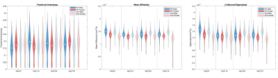 |
125 |
Diffusion Tensor Imaging in the Lower Leg of a Preclinical Model
of Amyotrophic Lateral Sclerosis
Ethan Mathew1,
Natenael Semmineh2,
Alberto Fuentes1,
Deborah Healey2,
Richard Dortch1,
Bruce Damon3,
and Chad Quarles2
1Barrow Neuroimaging Innovation Center, Barrow Neurological Institute, Phoenix, AZ, United States, 2Cancer Systems Imaging, MD Anderson Cancer Center, Houston, TX, United States, 3University of Illinois Urbana-Champaign, Urbana, IL, United States Keywords: Muscle, Diffusion Tensor Imaging This study examined changes in diffusion tensor parameters during amyotrophic lateral sclerosis progression using a transgenic rodent model. DTI was performed at four timepoints with an even amount of male and female rats to probe sex differences. Leg muscles were excised following scanning for histological analysis. Sex had a statistically significant effect on all parameters. Mean, radial, and axial diffusivity as well as the second eigenvalue were the most sensitive markers of disease progression. |
|
4211.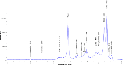 |
126 |
1H-MRS human skeletal muscle quantification: a repeatability
analysis
Alejandro Amador1,2 and
Michael D. Noseworthy1,2,3,4
1School of Biomedical Engineering, McMaster University, Hamilton, ON, Canada, 2Imaging Research Centre, St. Joseph's Healthcare, Hamilton, ON, Canada, 3Electrical and Computer Engineering, McMaster University, Hamilton, ON, Canada, 4Department of Radiology, McMaster University, Hamilton, ON, Canada Keywords: Muscle, Spectroscopy 1H-MRS of skeletal muscle presents challenges due to the highly structured arrangement of muscle fibres and their orientation to B0, complicating metabolite visibility and quantification. This study aimed to assess the repeatability of 1H-MRS in the anterior tibialis muscle in healthy volunteers by acquiring multiple spectra during and within different scanning sessions. A robust method for positioning of the voxel-of-interest was used to ensure reproducible acquisition. Data analysis was done using LCModel. Statistical analysis showed no significant difference in metabolite ratios against water across sessions, suggesting good repeatability of 1H-MRS of skeletal muscle. |
|
4212. |
127 |
Quantification of lumbar paraspinal muscle injury in patients
with low back pain using intravoxel incoherent motion diffusion
weighted imaging
Yinqi Liu1,
Weiyin Vivian Liu2,
and Kun Zhang1,3
1Department of Radiology, First Affiliated Hospital of Hunan University of Chinese Medicine, Changsha, China, 2MR Research, GE Healthcare, Beijing, China, 3College of Integrated Traditional Chinese and Western Medicine, Hunan University of Chinese Medicine, Changsha, China Keywords: Muscle, Diffusion/other diffusion imaging techniques Chronic low back pain (CLBP) has been reported to be associated with lumbar paraspinal muscles injury. Lumbar paraspinal muscles injury may result from oxidative stress and inflammation. In our study, D* at the middle of L4 and L5 levels of the paraspinal muscles was significantly higher in LBP group than in HCs. The fluid oozing from the inflammation can lead to greater unrestricted molecular movement. Therefore, non-invasive and contrast-agent-free IVIM could be a good perfusion predictor of lumbar paraspinal muscle injury. |
|
4213.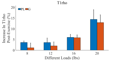 |
128 |
Exercise-induced changes of macromolecular content in calf
muscles quantified with T1ρ magnetic resonance imaging
Yanbin Li1,2,
Chang Ni2,
Jialin Wang1,
Zhenfeng Lv3,4,
Xin Mu2,
Mingyan Wu1,
Haikun Qi3,4,
and Jeff L. Zhang2
1Central Research Institute, Shanghai United Imaging Healthcare Co., Ltd, Shanghai, China, 2Vascular and Physiologic Imaging Research (VPIR) Lab, School of Biomedical Engineering, ShanghaiTech University, Shanghai, China, 3School of Biomedical Engineering, ShanghaiTech University, Shanghai, China, 4Shanghai Clinical Research and Trial Center, Shanghai, China Keywords: Muscle, Quantitative Imaging Exercise could induce changes in skeletal muscle’s macromolecular content. In this study, we used an exercise-then-imaging protocol to verify and characterize such phenomenon for calf muscles of healthy subject. A T1ρ mapping technique was used for imaging calf muscles before and after planter flexion of different loads. It was found that post-exercise increases in T1ρ values of gastrocnemius and peroneus longus at exercise load of 16 and of 20 lbs were significantly higher than that of 8 lbs. In conclusion, T1ρ is sensitive to changes of macromolecular content in calf muscles induced by plantar flexion of moderate or high intensity. |
|
4214.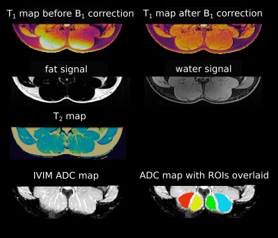 |
129 |
A multiparametric protocol to investigate the effects of
physical activity on lumbar spine muscles
Marta B. Maggioni1,
Renat Sibgatulin1,
Martin Krämer1,2,
Daniel Güllmar1,
and Jürgen R. Reichenbach1,2,3,4,5
1Medical Physics Group, Institute of Diagnostic and Interventional Radiology, Jena University Hospital, Jena, Germany, 2Institute of Diagnostic and Interventional Radiology, Jena University Hospital - Friedrich Schiller University, Jena, Germany, 3Michael Stifel Center for Data-driven and Simulation Science, Friedrich Schiller University, Jena, Germany, 4Abbe School of Photonics, Friedrich Schiller University, Jena, Germany, 5Center of Medical Optics and Photonics, Friedrich Schiller University, Jena, Germany Keywords: Muscle, Quantitative Imaging It is well established in literature that MRI quantitative parameters such as T2 or diffusion coefficients change significantly immediately after a strenuous exercise session. In this work, we instead focus on the long-term effects of regular repeated physical activity on three cohorts characterized by different training regimes. A multiparametric protocol was developed to assess T1, T2, fat fraction, and IVIM diffusion values to examine differences in lumbar spine muscle physiology between the different cohorts. |
|
4215.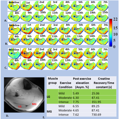 |
130 |
Enhanced Precision of the OXPHOS Measurement with integrated
CrCEST MRI and Down Field Proton MR Spectroscopy
Dushyant Kumar1,
Ryan Armbruster1,
Blake Benyard1,
Ravi Prakash Reddy Nanga1,
Neil Wilson1,
and Ravinder Reddy1
1Radiology, University of Pennsylvania, Philadelphia, PA, United States Keywords: Muscle, CEST & MT, creatine kinase; OXPHOS; Oxidative Phosphorylation Creatine recovery time constant (τCr) of exercised muscle is strongly coupled to net mitochondrial oxidative phosphorylation. However, local in vivo pH drop plays the role of a potential confounder, leading to significantly slower creatine recovery. Even under the same prescribed exercised regime, the extent of acidification and hence its impact on τCr would be variable across participant. To the best of our knowledge, this is the first study where downfield 1H-MR spectroscopy and 3D creatine chemical exchange saturation transfer are extensively used to prescribe mild exercise regime in terms of maximal voluntary contraction of participants while avoiding any acidification. |
|
4216.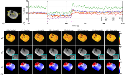 |
131 |
Monitoring of T2 changes in skeletal muscles using multiple
overlapping-echo detachment imaging
Qizhi Yang1,
Jiechao Wang1,
Hongjian He2,
Jianhui Zhong2,3,
Congbo Cai1,
Zhong Chen1,
and Shuhui Cai1
1Xiamen University, Xiamen, China, 2Zhejiang University, Hangzhou, China, 3University of Rochester, Rochester, NY, United States Keywords: Muscle, Muscle, multiple overlapping-echo detachment imaging Quantitative magnetic resonance imaging (qMRI), a noninvasive imaging tool without ionizing radiation, provides quantitative physiological information, whilst its lengthy scan time hinders its application in dynamic or real-time scenarios. Herein, the MOLED, a single-shot qMRI method we proposed previously, was applied in an ischemia and post-occlusive reactive hyperemia experiment to measure the T2 variation of the musculoskeletal system. The results disclose regular T2 fluctuations of different muscles during different periods. Inter-day repeatability experiments demonstrate that our method is accuracy and reliable. |
|
4217.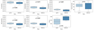 |
132 |
Diffusion Tensor Imaging of Swimmers’ Supraspinati: Fiber
Distinctions Between Sprint and Distance Freestyle
Grant M Shoults1,
Evan Gold1,
Feliks Kogan1,
and Valentina Mazzoli1
1Radiology, Stanford University, Stanford, CA, United States Keywords: Muscle, Diffusion Tensor Imaging Knowledge surrounding post-injury supraspinati in swimmers is vast, however a deep, preventative understanding at the cellular and fiber level is lacking. Through DTI, sixteen elite college swimmers were analyzed to evaluate mechanically stress induced differences between sprint and distance freestylers. Significant results (p-value<0.05) were found between groups for λ1, λ2, λ3, RD, MD, and volume. Findings indicated sprinters having a significantly larger MD and RD which was not compatible with the heightened levels microtrauma associated with aerobic distance training. DTI’s ability to inspect microstructural characteristics may pose it as an addition tool to conventional imaging techniques for athletes. |
|
4218.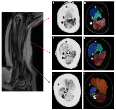 |
133 |
Quantitative MRI of the upper arm in Duchenne muscular
dystrophy: Consistent slice positioning is crucial due to
fat-fraction variability
Michel Cakici1,
Donnie Cameron2,
Karin J Naarding1,3,
Erik H Niks1,
and Hermien E Kan2
1Neurology, Leiden University Medical Center, Leiden, Netherlands, 2C J Gorter MRI Center, Leiden University Medical Center, Leiden, Netherlands, 3Rehabilitation, Amsterdam University Medical Center, Amsterdam, Netherlands Keywords: Muscle, Fat, Neuromuscular Skeletal muscle fat fraction determined using quantitative MRI is a promising outcome measure in Duchenne muscular dystrophy (DMD). However, fat fraction varies along the length of muscles in the lower limb, hampering consistency of longitudinal assessments. Our results show that fat fraction also varies over muscles in the upper limb. This variability is reduced when assessments are standardized at 40% of the humerus length. This is important for reproducibly measuring the fat fraction in the muscles of the upper limb in DMD in clinical trials and follow-up. |
|
4219.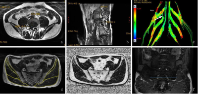 |
134 |
Quantitative evaluation of gluteal and multifidus muscle atrophy
and fatty infiltration in patients with lumbosacral nerve root
compression
Shi Yin1 and
Dou Weiqiang2
1The First Affiliated Hospital of Nanjing Medical University, Nanjing, China, 2GE Healthcare, MR Research, Beijing, P.R. China, Beijing, China Keywords: Muscle, Quantitative Imaging This study aims to investigate if MR technology can be applied to quantitatively evaluate the atrophy and fatty infiltration of gluteal and multifidus muscles in patients with lumbosacral nerve root compression. By quantitative evaluating 88 patients, we found the the change of multifidus muscle is related to the level of compressed nerve root and duration. As an indirect manifestation of nerve root compression, muscle change can help diagnosis in conventional lumbar MR images. Muscle content, especially that of multifidus is suggested to be a good indicator to complete the postoperative evaluation. |
|
4220.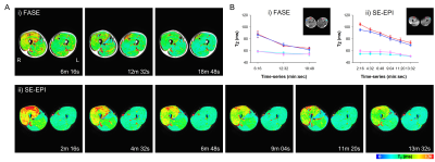 |
135 |
Dynamic T2 mapping following muscle exercise measured by fast
spin-echo and ultrafast spin-echo EPI
Jaekyun Ryu1,
Won Beom Jung1,
Chuluunbaatar Otgonbaatar2,
Jinil Park3,
Juho Kim1,
Jeonghak Song4,
and Hackjoon Shim1,4
1Medical Imaging AI Research Center, Canon Medical Systems Korea, Seoul, Korea, Republic of, 2College of Medicine, Seoul National University, Seoul, Korea, Republic of, 3Department of Intelligent Precision Healthcare, Sungkyunkwan University, Suwon, Korea, Republic of, 4Magnetic Resonance Business Unit, Canon Medical Systems Korea, Seoul, Korea, Republic of Keywords: Muscle, Muscle The changes in T2 driven by water content, oxygenation, pH, and blood volume in tissue altered during intense physical activity provides chance to measure the muscle performance. In this study, we investigated feasibility of dynamic T2 measurement for exercise-induced thigh muscle activity using conventional fast spin-echo (FASE, Fast Advanced Spin Echo) and ultrafast spin-echo EPI (SE-EPI) with 3T MRI. |
|
4221.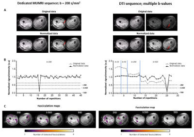 |
136 |
Longitudinal evaluation of fasciculation in a healthy population
using muscle DTI
Linda Heskamp1,
Lara Schlaffke2,
and Martijn Froeling1
1Radiology, University Medical Centre Utrecht, Utrecht, Netherlands, 2Neurology, BG-University Hospital Bergmannsheil gGmbH, Bochum, Germany Keywords: Muscle, Muscle Spontaneous motor unit (MU) contractions (fasciculation) are a hallmark of motor neuron diseases and can be measured with motor unit MRI. Previous work used a dedicated sequence with one optimized b-value. Here, we demonstrated that muscle DTI with variable b-values can also detect fasciculation. This allows simultaneous assessment of muscle architecture and fasciculation. Furthermore, we presented a retrospective analysis of fasciculation in healthy cohort muscle DTI data. This reveals higher and more variable fasciculation in lower legs compared to upper legs, and stable fasciculation distribution in repeated measures. Fasciculation detection in patients should therefore preferably focus on the upper legs. |
|
4222.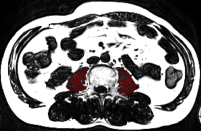 |
137 |
Fat Free Muscle Area measurement based on clinical liver MRI
workflow to assess sarcopenia
Xin Shi1,
Yueluan Jiang2,
Lei Zhang1,
Jiping Wang1,
Dongdong Qiu1,
and Shiyu Guo1
1the First Hospital of Jilin University, Changchun, China, 2MR Scientific Marketing, Siemens Healthineers, Beijing, China Keywords: Muscle, Fat Fat free muscle area (FFMA) is an important measurement to evaluate sarcopenia and myosteatosis, and it is associated with poor prognosis in patients with chronic liver and oncologic diseases. FFMA was measured on T2WI-TSE sequence in previous study, which is not in routine MRI workflow, and the scanning time of T2WI-TSE sequence is obviously longer than T1WI edixon sequence, so we develop a method to measure FFMA by T1WI edixon sequence in clinical liver MRI sequence and explore a clinical friendly sarcopenia measurement. |
|
4223.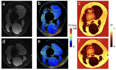 |
138 |
Longitudinal quantitative imaging of skeletal muscles and
peripheral nerves in CMT1A patients using Double-Echo in
Steady-State at 7 Tesla
Bragi Sveinsson1,2,
Robert L Barry1,2,3,
Matthew S Rosen1,2,4,
and Reza Sadjadi5
1Radiology, Massachusetts General Hospital, Boston, MA, United States, 2Radiology, Harvard Medical School, Boston, MA, United States, 3Harvard-MIT Health Sciences and Technology,, Cambridge, MA, United States, 4Physics, Harvard University, Cambridge, MA, United States, 5Neurology, Massachusetts General Hospital, Boston, MA, United States Keywords: Nerves, Quantitative Imaging Charcot-Marie-Tooth (CMT) is a neuromuscular disease causing progressive loss of muscle mass and function as well as nerve damage. Quantitative MRI offers the ability to investigate the condition of nerves and skeletal muscle non-invasively. In this work, we report findings from measuring various quantitative neuromuscular metrics in four CMT1A patients in high-resolution images obtained at 7T, measured at two time points. |
|
4224.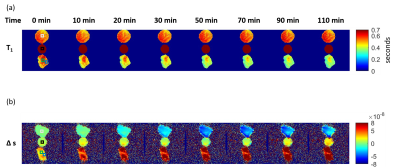 |
139 |
Magnetic resonance fingerprinting thermometry (MRFT) versus PRFS
thermometry: comparative temperature prediction in ex vivo
bovine muscle
Enlin Qian1,
Pavan Poojar2,
Maggie Fung3,
Zhezhen Jin4,
John Thomas Vaughan1,
Devashish Shrivastava1,
David Gultekin1,
and Sairam Geethanath2
1Columbia Magnetic Resonance Research Center, Columbia University, New York, NY, United States, 2Accessible MR Laboratory, Biomedical Engineering and Imaging Institute, Dept. of Diagnostic, Molecular and Interventional Radiology, Icahn School of Medicine at Mt. Sinai, New York, NY, United States, 3GE Healthcare, New York, NY, United States, 4Department of Biostatistics, Columbia University, New York, NY, United States Keywords: Safety, Safety We explored T1-based magnetic resonance fingerprinting thermometry (MRFT) and compared it with proton resonance frequency shift (PRFS) thermometry in ex vivo bovine muscle data. We conducted temperature sensitivity calibration experiments for both methods. We compared MRFT and PRFS thermometry predicted temperatures in bovine muscles, validated by fluoroptic probe measurements. We observed strong correlation in temperature sensitivity calibration experiments (R2>0.958). Both methods predict temperature accurately (RMSE<1.37 ℃ and RMSE<0.93 ℃ for MRFT and PRFS, over a range of 25 ℃). We observed strong correlation between PRFS and MRFT predicted temperature in heated bovine muscle (R2>0.93). |
|
4225.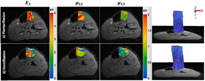 |
140 |
Estimation of isotropic and anisotropic biomechanical properties
of the lower leg muscle during muscle contraction using DTI and
MR elastography
Mahsa Salimi Majd1,
Yang Yang1,
Heiko Tzschätzsch1,
Steffen Görner1,
Jürgen Braun1,
Ingolf Sack1,
and Jing Guo1
1Charité - Universitätsmedizin Berlin, Berlin, Germany Keywords: Elastography, Elastography In this study, DTI and multifrequency MR elastography (MRE) were applied to 6 subjects to investigate the changes of fiber orientation and the biomechanical properties of the lower leg muscle during passive plantarflexion and dorsiflexion. Using the pentation angle estimated from DTI, MRE data were processed to obtain both isotropic shear wave speed (c) and anisotropic shear moduli (μ12, μ13) as well as Young’s modulus (E3). Preliminary findings showed significant changes of the biophysical properties upon lower leg muscle contraction. |
|
 Back to Meeting Home
 Back to the Program-at-a-Glance
The International Society for Magnetic Resonance in Medicine is accredited by the Accreditation Council for Continuing Medical Education to provide continuing medical education for physicians.
 Back to Meeting Home
 Back to the Program-at-a-Glance
 View Presentation Video