Digital Poster
Perfusion, Inflammation & Malignancy
ISMRM & ISMRT Annual Meeting & Exhibition • 03-08 June 2023 • Toronto, ON, Canada

Digital Poster
Perfusion, Inflammation & Malignancy
| Computer # | |||
|---|---|---|---|
2254.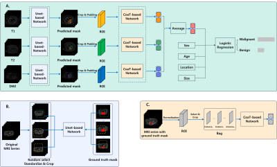 |
81 | Attention-based Two Stage Deep Learning Model for the Segmentation and Classification of Pelvic and Sacral Tumors on Routine MRI
Ping Yin1 and Nan Hong1
1Department of Radiology, Peking University People's Hospital, Beijing, China Keywords: MSK, Tumor Early detection and correct diagnosis are key to adequate and successful treatment of PSTs. Based on multi-sequence MRI images, physician’s labeling, segmentation model, and clinical features, six classification models were built. The highest scoring model (model 6) achieved 0.836 AUC, 0.781 ACC in the prospective test set, which was comparable to that of senior residents and junior resident. However, the diagnosing time of DL model is significantly shorter than physicians. Our attention-based two stage DL model allowed the accurate segmentation and classification of benign and malignant PSTs without enhanced MRI and may thus facilitate diagnosis. |
|
2255. |
82 | MR-Guided Cryoablation to Upregulate the Immune Response in Osteosarcoma
Dara L Kraitchman1,2,3, Michele Doucet4, Sarah J Powers3, Alainah Bhutta5, Emily Kulp6, Tina Ehtiati7, Cheri Rice1, Cindy Maranto1, Kathleen Gabrielson3, Cory Brayton3, and Brian Ladle4
1Russell H Morgan Department of Radiology and Radiological Science, Johns Hopkins University, Baltimore, MD, United States, 2Center for Image-Guided Animal Therapy, Baltimore, MA, United States, 3Department of Molecular & Comparative Pathobiology, Johns Hopkins University, Baltimore, MD, United States, 4Department of Oncology, Johns Hopkins University, Baltimore, MD, United States, 5University of Georgia College of Veterinary Medicine, Athens, GA, United States, 6Department of Chemical and Biomolecular Engineering, Johns Hopkins University, Baltimore, MD, United States, 7Siemens Medical Solutions USA, Inc., Baltimore, MD, United States Keywords: Bone, MR-Guided Interventions, cryoablation Osteosarcoma (OSA) is the most common bone cancer in young adults and dogs and almost invariably lethal when the cancer spreads. MR-guided cryoablation offers the potential to cause direct bone necrosis and palliative pain management and has shown the potential to upregulate the immune response in prostate and breast cancer to prevent or shrink metastatic disease. The current study seeks to determine the immune response of MR-guided cryoablation in spontaneously occurring canine osteosarcoma in comparison to X-ray-guided intratumoral Stimulator of Interferon Gene (STING) immunotherapy as determine by survival time and measurement of inflammatory infilitrates. |
|
2256.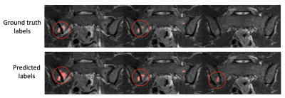 |
83 | Simplified, ‘single-step’ segmentation and quantification of bone marrow oedema using deep learning
Timothy JP Bray1, Alexis JP Jones2, Margaret A Hall-Craggs1, and Hui Zhang3
1Centre for Medical Imaging, University College London, London, United Kingdom, 2Rheumatology, University College London Hospital, London, United Kingdom, 3Centre for Medical Image Computing, University College London, London, United Kingdom Keywords: Bone, Inflammation We present a deep learning workflow which segments and quantifies bone marrow oedema (BMO) in a single deep learning step. Detection and quantification of BMO plays a crucial role in diagnosis and monitoring of patients with inflammatory diseases of the skeleton, including spondyloarthritis (SpA), and various segmentation methods have been developed to facilitate BMO quantification. However, previous attempts have used multiple algorithms in sequence to achieve satisfactory performance. To improve on this, we propose a simplified approach that avoids the need for such sequential algorithms, thus simplifying the workflow, eliminating potential error sources and reducing the need for human input. |
|
2257.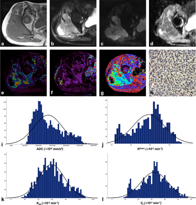 |
84 | Whole-tumor histogram analysis of DWI and DCE-MRI for soft tissue sarcoma: Correlation with HIF-1alpha expression
Xiangwen Li1, Qing Li2, and chen shuang3
1Huashan hospital Fudan university, Shang hai, China, 2MR Collaborations, Siemens Healthcare Ltd., Shang hai, China, 3Radiology, Huashan hospital Fudan university, Shang hai, China Keywords: MSK, Diffusion/other diffusion imaging techniques In this study, we initially revealed the feasibility of conventional MRI features and whole-tumor histogram features of diffusion-weighted imaging (DWI) and dynamic contrast-enhanced MRI (DCE-MRI) parameters in assessing hypoxia-inducible factor 1-alpha (HIF-1α) expression of soft tissue sarcomas (STS). On this basis, we performed a short-term survival analysis of the study population. The findings suggest that MRI morphological features and histogram features of quantitative parameters help predict higher HIF-1α expression and may significantly predict prognosis in patients with STS. |
|
2258.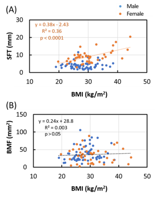 |
85 | Sex differences in subcutaneous fat thickness and bone marrow fat area and their relationship to BMI and age
Talon Johnson1, Jimin Ren1,2, and Anke Henning1,2
1Advanced Imaging Research Center, University of Texas Southwestern Medical Center, Dallas, TX, United States, 2Department of Radiology, University of Texas Southwestern Medical Center, Dallas, TX, United States Keywords: Bone, Fat, Metabolism, diabetes This study evaluated the sex factor in affecting fat distribution in bone marrow and subcutaneous tissue in human lower extremity. Bone marrow fat (BMF) was characterized by the cross-sectional area in the fibula bone and subcutaneous fat (SF) was measured by the thickness of SF (SFT) in the periphery of calf muscle. It is found that SFT and BMF are statistically significantly correlated to BMI and age, respectively, in women (n = 43), but not in men (n = 59). For a subgroup of subjects with thinner SFT (<15 mm, n = 87), SFT is negatively correlated to BMF. |
|
2259. |
86 | Intravoxel incoherent motion and dynamic contrast-enhanced magnetic resonance imaging for neoadjuvant chemotherapy response evaluation in patients with osteosarcoma
Ping Yin1 and Nan Hong1
1Department of Radiology, Peking University People's Hospital, Beijing, China Keywords: MSK, Multimodal, Osteosarcoma In this study, we simultaneously compared the predictive value of ADC, IVIM and semi-quantitative and quantitative DCE-MRI parameters for the efficacy of NACT. We found that D-Bi, D*-Bi, and f-Bi post-NACT and ΔD-Bi had statistical differences between the good response group and the poor response group. ROC curve showed that f-Bi post-NACT had the best performance in all parameters, with AUC of 0.769, sensitivity of 1, and specificity of 0.538. Correlation analysis showed that the efficacy of NACT was negatively correlated with D-Bi, D*-Bi post-NACT and ΔD-Bi, and was significantly positively correlated with f-Bi post-NACT.
|
|
2260.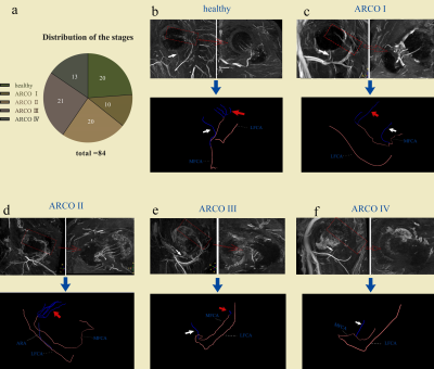 |
87 | Evaluating blood supply changes in the osteonecrosis of the femoral head using gadobutrol-based steady-state MRA
Zhenhong Liao1 and Xiaoyong Zhang2
1Deyang People's Hospital, Sichuan, China, 2Philips Healthcare, Chengdu, China Keywords: Joints, Vessels, osteonecrosis of the femoral head Dynamic evaluation of retinacular arteries is essential for understanding blood supply changes occurring in osteonecrosis of the femoral head (ONFH). The steady-state (SS) magnetic resonance angiography (MRA) may be an effective method as our previous study suggested, however its feasibility is yet to be determined. Therefore, this study intends to use gadobutrol-based SS MRA to evaluate the blood supply of ONFH. |
|
2261.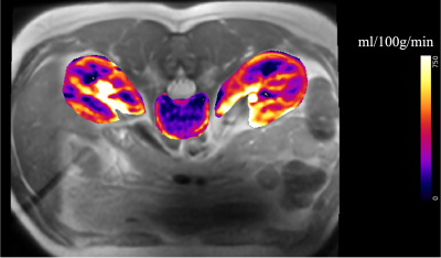 |
88 | Imaging perfusion in the vertebral bone marrow and subchondral bone using FAIR ASL
L. Tugan Muftuler1 and Matthew D. Budde1
1Medical College of Wisconsin, Milwaukee, WI, United States Keywords: MSK, Perfusion, lumbar spine, low back pain Some patients with chronic low back pain (cLBP) do not show any diagnosable conditions in conventional imaging methods. Studies suggested that pain could be associated with the degeneration of vertebral endplate region in those nonspecific cLBP patients. The endplates around degenerating discs experience trauma, which leads to inflammation, angiogenesis, neurogenesis and infection. Currently, there is no established method to diagnose such pathologies. We previously reported associations between increased endplate perfusion and experience of cLBP using DCEMRI. In this study we explored Arterial Spin Labeling to avoid using contrast agents. Results show that FAIR could be used to study endplate perfusion. |
|
2262.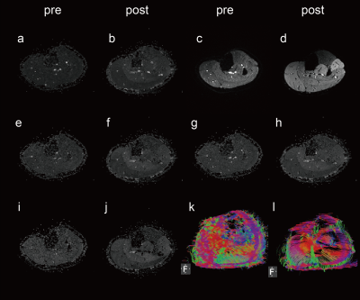 |
89 | Exercise-stimulated muscle hyperemia: a preliminary investigation with diffusion tensor imaging on calf muscles
Xin Mu1, Meng Tian2, Huahui Xu3, Chang Ni1, Yanbin Li4, Xiangwei Kong1, Xiaoli Gu3, and Jeff L Zhang1
1Vascular and Physiologic Imaging Research (VPIR) Lab, School of Biomedical Engineering, ShanghaiTech University, Shanghai, China, 2School of Biomedical Engineering, Shanghai Jiao Tong University, Shanghai, China, 3Department of Radiology, Shanghai Guanghua Hospital of Integrative Medicine, Shanghai, China, 4Central Research Institute, UIH Group, United Imaging Healthcare, Shanghai, China Keywords: Skeletal, Muscle Diffusion tensor imaging (DTI) has been shown to be highly sensitive to subtle pathological alterations caused by multiple diseases. Utilizing this capability of DTI, in this study we characterized post-exercise hyperemic responses of calf muscles. We found that multiple DTI measures, including ADC, λ1-3, and RD, increased significantly in activated muscles, and more than in those in non-activated muscles. Of particular interest was the significant increase in λ3 and RD of the activated muscle, indicating a relatively higher degree of increase in water diffusion along the cross-section direction of muscle fibers. |
|
2263.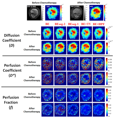 |
90 | Comparison of Penalty-based IVIM analysis Methods for Predicting Response to Neoadjuvant Chemotherapy in Osteosarcoma
Esha Baidya Kayal1, Devasenathipathy Kandasamy2, Kedar Khare1, Raju Sharma2, Sameer Bakhshi2, and Amit Mehndiratta1,2
1Indian Institute of Technology Delhi, New Delhi, India, New Delhi, India, 2All India Institute of Medical Sciences, New Delhi, India, New Delhi, India Keywords: MSK, Cancer, Intravoxel Incoherent motion, Chemotherapy response prediction, Performance comparison Predictive performance of penalty-based IVIM analysis methodologies BE+TV and BE+HPF for response to Neoadjuvant Chemotherapy have been evaluated using clinical dataset with osteosarcoma in comparison with existing IVIM analysis methods. IVIM datasets before and after chemotherapy were analyzed using 5 IVIM analysis methods – Bi-exponential model and two of its segmented variants and penalty-based BE+TV and BE+HPF methods. Results showed, IVIM parameters estimated by BE+TV and BE+HPF methods produced improved prediction of response to chemotherapy in osteosarcoma than the existing IVIM analysis methods at both the time-points, before and after chemotherapy. |
|
2264.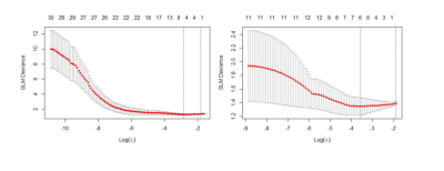 |
91 | Radiomics based on conventional MRI sequences to differentiate inert fibrogenic tumors and invasive fibrogenic tumors of trunk and limbs
Peng Gao1, Weisheng Zhang1, and Hexin Feng2
1Dalian Medical University, Dalian, China, 2China Medical University, Shenyang, China Keywords: Muscle, Tumor 区分不同亚型的纤维化肿瘤仍然具有挑战性,但具有重要的临床意义。本研究的目的是评估基于常规 MRI 序列的影像组学在区分惰性纤维化肿瘤和躯干和四肢浸润性纤维化肿瘤的价值。预测模型基于 T1 图像的逻辑回归在区分惰性纤维瘤和侵袭性纤维瘤方面具有最好的预测效率,对 T1 和 T2 预测模型贡献最大的特征分别是 idn 和最小轴长。病变的大小和深度对判断肿瘤的性质有一定的参考价值。 |
|
2265.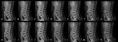 |
92 | Compressed SENSE AI enhanced mDixon-Quant MR Imaging in the Lumber Spine Study
Linzhe Li1, Junhong Duan1, Muqi Liu1, Yunjie Liao1, Pengzhi Hu1, and Chen Thomas Zhao2
1Department of Medical Imaging, the Third Xiangya Hospital, Central South University, Changsha, China, 2Philips Healthcare, Guangzhou, China Keywords: MSK, Fat, CS AI, Bone Marrow Fat, mDIXON-quant Compressed SENSE (CS) has been suggested to speed up MRI acquisition in clinical studies, while reducing artefacts and improving image quality. To date, the optimal acceleration factor (AF) for Compressed SENSE AI (CS AI) versus conventional compressed SENSE (CS) on lumber spine images remains unclear. In this study, the impact of CS AI technique with different acceleration factors compared with conventional CS on the utility of measuring lumber spine fat was investigated. Results of this study showed that CS AI not only shortened MRI acquisition time, but also ensured image quality, as well as clinical diagnostic accuracy and clinical throughput. |
|
2266.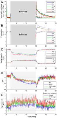 |
93 | Evaluating age-related changes of vascular function and oxidative energy metabolism in the lower leg during a plantar flexion exercise
Alfredo Liubomir Lopez Kolkovsky1, Béatrice Matot1, Harmen Reyngoudt1, Benjamin Marty1, Ericky Caldas de Almeida Araujo1, and Yves Fromes1
1NMR Laboratory , Neuromuscular Investigation Center, Institute of Myology, Paris Cedex 13, France Keywords: Muscle, Aging, Multi-contrast, Metabolism, Non-proton, Spectroscopy The age-related loss of muscle mass, strength and quality is a multifactorial process whose mechanisms are incompletely understood. NMR allows investigating numerous physiological and biochemical variables in vivo dynamically. Here, 51 physically active subjects performed a normalized plantar flexion exercise to evaluate vascular response (blood flow, T2*, water compartmentalization), tissue oxygenation and oxidative energy metabolism (pH, mitochondrial oxidative capacity, work to [ADP] ratio). Results showed that the vascular and metabolic function was preserved in old subjects relative to young and middle-aged participants. We conclude that vascular and oxidative metabolism impairments are not a necessary condition of old age. |
|
2267.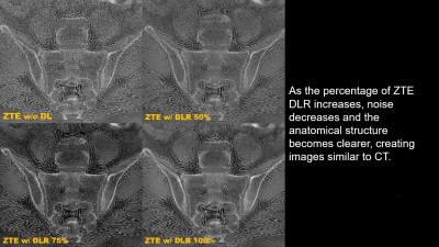 |
94 | Deep learning reconstruction of zero echo time imaging: bone erosion detection in axial spondyloarthritis
Seok Hahn1, Jisook Yi2, Ho-Joon Lee2, Sunggun Lee3, Sekyoung Park4, Joonsung Lee5, Jose de Arcos6, and Maggie Fung7
1Department of Radiology, Haeundae Paik Hospital, Inje University College of Medicine, Busan, Korea, Republic of, 2Department of Radiology, IHaeundae Paik Hospital, Inje University College of Medicine, Busan, Korea, Republic of, 3Division of Rheumatology, Department of Internal Medicine, Haeundae Paik Hospital, Inje University College of Medicine, Busan, Korea, Republic of, 4Department of Radiology, Kosin University Gospel Hospital, Kosin University College of Medicine, Busan, Korea, Republic of, 5GE Healthcare, Seoul, Korea, Republic of, 6GE Healthcare, Little Chalfont, United Kingdom, 7GE Healthcare, New York, NJ, United States Keywords: Joints, MSK, sacroiliac joint, zero echo time imaging, deep learning reconstruction The aim of this study was to compare the diagnostic performance of DLR of ZTE imaging for bone erosion in axial spondyloarthritis, using CT as the reference standard. Twenty-three patients with suspicion of sacroiliitis underwent both CT and MR scans of sacroiliac joints included for analysis. ZTE with or with DLR showed higher correlation coefficients than T1WI for two readers. Inter-reader agreements showed moderate to substantial agreement. ZTE DLR improves diagnostic performance in the detection of SIJ bone erosion in patients with axial spondyloarthritis compared with T1WI and ZTE without DLR. |
|
2268. |
95 | Bone marrow fat composition assessed by fast and simple 1D MRS combined with intermolecular double-quantum coherence on 3.0 T
Jianfeng Bao1, Xiao Wang2, Yuchuan Zhuang3, Liangjie Lin4, Yong Zhang2, and Jingliang Cheng2
1Department of Magnetic Resonance Imaging, The First Affiliated Hospital of Zhengzhou University, zhengzhou, China, 2Department of Magnetic Resonance Imaging, The First Affiliated Hospital of Zhengzhou University, Zhengzhou, China, 3Department of Electronic Science, University of Rochester, ROCHESTER, NY, United States, 4Philips Healthcare, Beijing, China Keywords: Bone, Fat, intermolecular double-quantum coherence,fat composition,magnetic resonance spectroscopy Unsaturated fatty acids in the bone marrow may play a critical role in bone metabolism, however, these peaks cannot be well resolved by conventional MRS techniques due to local intensive B0 inhomogeneities. We introduced intermolecular double-quantum coherence to enhance the spectrum resolution on clinical 3.0 T within one minute and more unsaturated peaks can be identified. |
|
2269. |
96 | DCE-MR Lymphangiography Demonstrates Significantly Different Lymphatic Drainage in Patients with a FOXC2 Gene Mutation Compared to Controls
Michael Mills1, Greta Brezgyte1, Bernard Ho2, Julian Pearce2, Kristiana Gordon1,2, Peter S Mortimer1,2, Pia Ostergaard1, and Franklyn A Howe1
1St George's University of London, London, United Kingdom, 2St George's University Hospitals NHS Foundation Trust, London, United Kingdom Keywords: Vessels, Genetic Diseases, lymphatic MRI can investigate lymphatic dysfunction and lymphoedema (a chronic swelling of interstitial fluid), however little quantitative research has been published. Dynamic Contrast-Enhanced Magnetic Resonance Lymphangiography (DCE-MRL) allows identification of lymph vessels in both healthy and lymphoedematous limbs, and was used to study the oedematous limbs of patients where the lymphoedema was associated with a mutation of the FOXC2 gene. Signal measured in leg lymphatics of these patients (n=6) peaked significantly earlier than in non-oedematous limbs (n=6) and demonstrated higher peak signals, despite an impaired lymphatic system and therefore an expectation of more sluggish drainage. |
|
2270.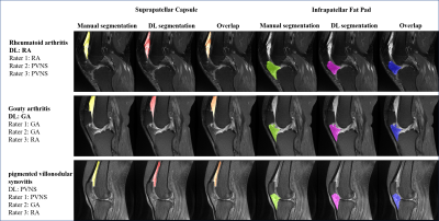 |
97 | Deep Learning Based on Knee MRI for Fully Automated Segmentation and Precision Diagnosis of Rheumatoid Arthritis, Gout and PVNS
Qizheng Wang1, Meiyi Yao2, Yandong Liu3, Xinhang Song2, Xiaoying Xing1, Yongye Chen1, Ke Liu1, Weili Zhao1, Xiaoguang Cheng3, Shuqiang Jiang2, and Ning Lang1
1Peking University Third Hospital, Beijing, China, 2Institute of Computing Technology, Chinese Academy of Sciences, Beijing, China, 3Department of Radiology, Beijing Jishuitan Hospital, Beijing, China Keywords: Joints, Segmentation Segmentation of synovial-related structures in MRI images can help assess synovitis-effusion, infrapatellar fat pad (IPFP) changes, and response to treatment, which is important for the clinical diagnosis of knee disease. However, segmenting images manually, which depends on the skill and experience of the physician; furthermore, it is time-consuming for radiologists. In this study, a deep learning pipeline for the 3D segmentation of the suprapatellar capsule (SC) and IPFP and knee synovitis classification were developed using proton density (PD)-weighted images of sagittal fat-suppressed knees, the most commonly used sequence in clinical practice, to support clinical decision-making. |
|
2271.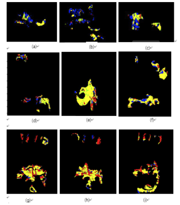 |
98 | Accurate and quantitative assessment of rheumatoid joint synovitis based on DCE-MRI pixel-level analysis
Jinling Mao1, Zhongqi Zhu1, Yinghao Li1, Hongzhi Wang1, and Jie Shi2
1East China Normal University, Shanghai, China, 2Shanghai Guanghua Hospital of Integrated Traditional Chinese and Western Medicine, Shanghai, China Keywords: Vessels, Inflammation, synovitis;Pannus;Wrist joint In this paper, 18 cases of wrist magnetic resonance enhancement were collected, a pixel-by-pixel time-intensity curves (TIC) analysis method was proposed, four types of enhancement curves were customized, and compared with the synovitis score, the results showed that the area of the rapidly rising and descending pixel area was extremely correlated with the synovitis score (r=0.812). |
|
2272.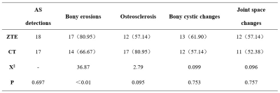 |
99 | The value of zero echo time MR imaging for demonstrating bony change of sacroiliac joint in ankylosing spondylitis: a comparative study with CT
Ziwei Zhang1, Lingling Song1, Weixin He1, Qi Zeng1, Lisha Nie2, Xiaocheng Wei2, Jiawei Wang1, He Sui1, Zhaoshu Huang1, Xia Zhu1, Chen Liang1, and Yu Li1
1The Affiliated Hospital of Guizhou Medical University, GuiYang, China, 2GE Healthcare, MR Research China, BeiJing, China Keywords: Skeletal, Skeletal The current study aims to compare the detection ability of zero echo time(ZTE) MRI and CT in presenting bony change of sacroiliac joint in ankylosing spondylitis. It was concluded that the ankylosing spondylitis detection rate was comparable between ZTE MRI and CT, where the detection rate of bony erosions by ZTE MRI was higher than that by CT. Our observations indicated that in the future, the ZTE MRI may potentially replace the ionization-related CT method by providing a more comprehensive evaluation of sacroiliitis in ankylosing spondylitis patients. |
|
| 2273. | 100 | Prediction of Histopathological Subtypes of Dermatofibrosarcoma protuberans Based on MRI Radiomics Machine Learning Model
Siyu Liu1, Yishi Wang2, and Songtao Ai1
1Shanghai Ninth People's Hospital, Shanghai, China, 2Philips Healthcare, Beijing, China, Beijing, China Keywords: Muscle, Cancer Dermatofibrosarcoma protuberans (DFSP) is a rare low to intermediate grade soft tissue sarcoma of skin, but the fibrosarcomatous DFSP (FS-DFSP) is a clearly malignant pathological subtype. The identification of malignant pathological subtypes by radiomics plays an important role preoperatively. The non-invasive machine learning method based on T1WI and FS-T2WI imaging is potential prognostic tool by distinguishing different levels of DFSP pathological subtypes before operation in this study to provide a new idea for the diagnosis and treatment of DFSP. |
|
 Back to Meeting Home
 Back to the Program-at-a-Glance
The International Society for Magnetic Resonance in Medicine is accredited by the Accreditation Council for Continuing Medical Education to provide continuing medical education for physicians.
 Back to Meeting Home
 Back to the Program-at-a-Glance
 View Presentation Video