Digital Poster
Knee Imaging in Osteoarthritis
ISMRM & ISMRT Annual Meeting & Exhibition • 03-08 June 2023 • Toronto, ON, Canada

| Computer # | |||
|---|---|---|---|
2451.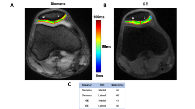 |
101 | Intra- and Inter-vendor Reproducibility of T1ρ Measurements in the Healthy Human Knee Joint
Ryan Robert Armbruster1, Anjali Talluru2, Arijitt Barthakur3, Warren Bilker4, Ravinder Reddy3, and Susanta Sarkar5
1Bioengineering, University of Pennsylvania, Philadelphia, PA, United States, 2University of Pittsburg, Pittsburg, PA, United States, 3Radiology, University of Pennsylvania, Philadelphia, PA, United States, 4Biostatistics and Epidemiology, University of Pennsylvania, Philadelphia, PA, United States, 5Cadenzamed LLC, Berwyn, PA, United States Keywords: Cartilage, Osteoarthritis Osteoarthritis (OA) is a debilitating disease that results in cartilage loss and pain, but no drug is currently available for OA, largely due to the absence of a reliable index for measuring disease modifying effects of drug candidates in clinical trials. T1ρ MRI promises to be a reliable method because it provides a measure of cartilage degradation. However, it is critical to establish its reproducibility across MRI vendors before its universal use in clinical studies. Here we report the establishment of a clinically relevant reproducibility error in T1ρ in 25 healthy volunteer knees within and across 3T MRI scanners. |
|
2452.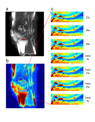 |
102 | Ultrafast T2 mapping of knee articular cartilage under compression using a radial turbo-spin-echo sequence at 3T - initial results
Veronika Janacova1, Vladimir Juras 1, Pavol Szomolanyi1,2, Diana Bencikova1, and Siegfried Trattnig1,3,4,5
1High Field MR Centre, Department of Biomedical Imaging and Image-guided Therapy, Medical University of Vienna, Vienna, Austria, 2Institute of Measurement Science, Slovak Academy of Sciences, Bratislava, Slovakia, 3CD Laboratory for MR Imaging Biomarkers (BIOMAK), Vienna, Austria, 4Austrian Cluster for Tissue Regeneration, Ludwig Boltzmann Institute for Experimental and Clinical Traumatology, Vienna, Austria, 5Institute for Clinical Molecular MRI in the Musculoskeletal System, Karl Landsteiner Society, Vienna, Austria Keywords: Cartilage, Quantitative Imaging, Ultra-fast Quantitative T2 maps have been used for more than a decade in musculoskeletal MR research as a tool for cartilage assessment. In this work, we compared T2 mapping of knee cartilage with ultrafast radial turbo-spin-echo sequence to conventional multi-echo spin echo sequence. We used radial turbo-spin-echo T2 maps to track dynamic changes in knee cartilage during compression. Correlation between the two methods was found in multiple cartilage regions. Dynamic change in T2 values was observable on T2 maps during first two minutes of compression. We demonstrated that radial-TSE sequence allows for fast determination of T2 values of the knee cartilage. |
|
2453.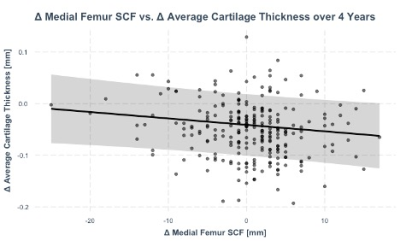 |
103 | Associations between Weight Change, Knee Subcutaneous Fat and Cartilage Thickness: 4-Year Data from the OAI
Gabby B Joseph1, Melia Takakusagi1, Gino Arcilla1, John Lynch2, Valentina Pedoia1, Sharmila Majumdar1, Nancy E. Lane3, Michael C Nevitt2, Charles E McCulloch2, and Thomas M. Link1
1Department of Radiology and Biomedical Imaging, UCSF, San Francisco, CA, United States, 2Department of Epidemiology and Biostatistics, UCSF, San Francisco, CA, United States, 3Department of Rheumatology, UC Davis, Davis, CA, United States Keywords: Osteoarthritis, Cartilage This study assessed the relationship between 4-year changes in: (1) body weight and outcomes of joint-adjacent subcutaneous fat (SCF) and cartilage thickness, and (2) joint-adjacent SCF and knee cartilage thickness. A total of 399 individuals from the Osteoarthritis Initiative with >10% weight gain and >-10% weight loss over 4 years were compared with controls with less than 3% weight change. Weight gain and loss were associated with increases, and decreases, respectively in joint-adjacent SCF. Increases in SCF adjacent to the medial femur were associated with decreases in average cartilage thickness and in several medial and lateral locations in the knee. |
|
 |
2454.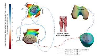 |
104 | Preserved patellar cartilage at the expense of anterior femoral cartilage damage in Hip Osteoarthritis: An Inter-Limb Inter-Joint qMRI analysis
Rupsa Bhattacharjee1, Johanna Luitjens1, Misung Han1, Rafeek Thahakoya1, Koren E. Roach1, Richard B. Souza1,2, Valentina Pedoia1, and Sharmila Majumdar1
1Department of Radiology and Biomedical Imaging, University of California, San Francisco (UCSF), San Francisco, CA, United States, 2Department of Physical Therapy and Rehabilitation Science, University of California, San Francisco (UCSF), San Francisco, CA, United States Keywords: Cartilage, Osteoarthritis, Bilateral, Hip, Knee It is well documented that osteoarthritis often affects multiple joints in patients with the disease. However, direct associations between limbs and across the joints of the lower extremity remains understudied. With bilateral hip and knee MRI of 18 patients, this study utilizes automated segmentation and VBR-atlas-based methods to explore the associations between twelve femoral and acetabular sub-compartments with trochlear and patellar cartilage T1rho, T2 values. Significant negative correlations between the patellar T2 and anterior hip femoral T1rho and T2 values, suggest a probable ongoing gait dysfunction. The data-distribution indicates possibility of a subgroup of patients having different progression and compensatory trajectories, to be explored further with gait pattern analysis. |
2455.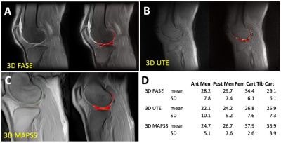 |
105 | T1rho Sequences with FASE, UTE, and MAPSS Acquisitions for Knee Evaluation
Vadim Malis1, Yoshimori Kassai2, Mitsue Miyazaki1, and Won Bae1,3
1Radiology, University of California, San Diego, La Jolla, CA, United States, 2Canon Medical Systems Corp, Otawara, Japan, 3VA San Diego Healthcare System, San Diego, CA, United States Keywords: Cartilage, Tissue Characterization, T1rho Three T1rho sequences based using 3D FASE, 3D UTE, and 3D MAPSS acquisitions were developed and compared by imaging agarose phantoms and normal human knees. The sequences utilize 4 to 6 spin lock times, and can be acquired in about 5 min or less. On phantoms, all three sequences resulted in consistent T1rho values that decreased with increasing agarose concentration, similar to past reports. T1rho values of cartilage and menisci were also in line with previously reported values. This work will be useful for evaluating knee osteoarthritis and other musculoskeletal diseases. |
|
2456.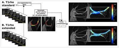 |
106 | Assessing the effects of spin lock time duration on T1ρ relaxation times for imaging of the articular cartilage in the knee
ALLEN A CHAMPAGNE1, TAYLOR M ZULEGER2,3,4,5, DANIEL R SMITH2,4,5, ALEXIS B SLUTSKY-GANESH2,4,5,6, SHAYLA M WARREN2,4,5, LEXIE M SENGKHAMMEE2,4,5, SAGAR MANDAVA7, HONGJIANG WEI8, DAVIDE D BARDANA9, GREG D MYER2,4,5,10, and JED A DIEKFUSS2,4,5
1School of Medicine, Queen's University, Kingston, ON, Canada, 2Emory Sports Performance And Research Center, Flowery Branch, GA, United States, 3Neuroscience Graduate Program, University of Cincinnati, Cincinnati, OH, United States, 4Emory Sports Medicine Center, Atlanta, GA, United States, 5Department of Orthopaedics, Emory University School of Medicine, Atlanta, GA, United States, 6Department of Kinesiology, University of North Carolina at Greensboro, Greensboro, NC, United States, 7GE Healthcare, Atlanta, GA, United States, 8School of Biomedical Engineering, Shanghai Jiao Tong University, Shanghai, China, 9Department of Orthopaedics, Queen's University, Kingston, ON, Canada, 10The Micheli Center for Sports Injury Prevention, Waltham, MA, United States Keywords: Cartilage, Quantitative Imaging, T1rho, knee There is limited standardization of T1ρ acquisition parameters with respect to spin lock times (TSLs). Shorter TSL durations are susceptible to underestimating knee cartilage T1ρ relaxation times, warranting optimization for maximal measurement stability. Additionally, vertical estimation of T1ρ relaxation times throughout cartilage layers is limited. Here, we utilized 40 healthy knees to demonstrate enhanced T1ρ relaxation time measurement stability with maximum TSLs of 90 to 120ms compared to shorter TSL durations. Depth-specific differences in T1ρ were further compartmentalized into deep and superficial knee cartilage layers, highlighting the potential for T1ρ to indirectly and non-invasively evaluate proteoglycan content across cartilaginous layers. |
|
2457.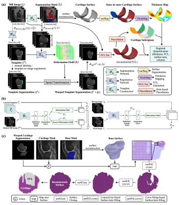 |
107 | Deep-Learning-Based Knee Articular Cartilage Morphometrics
Yongcheng Yao1 and Weitian Chen1
1Department of Imaging and Interventional Radiology, The Chinese University of Hong Kong, Shatin, Hong Kong Keywords: Cartilage, Cartilage, morphometrics We proposed a deep-learning-based system for automatic knee articular cartilage morphometrics. It produces regional metrics including full-thickness cartilage loss (FCL), mean thickness, surface area, and volume. The proposed system comprises deep learning models and algorithms that work collaboratively. We have trained convolutional neural networks for tissue segmentation, template construction, and image registration. We designed modules and pipelines for cartilage thickness mapping, cartilage lesion quantification, and cartilage parcellation. Results shows superior accuracy of the thickness mapping method and robustness of the cartilage parcellation method. The proposed FCL estimation method filled the gap in automatic cartilage lesions quantification. |
|
2458.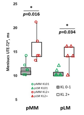 |
108 | Meniscus UTE-T2* Measured Prior to Anterior Cruciate Ligament Reconstruction Predicts Radiographic Osteoarthritis 11 Years Later
Ashley A. Williams1, Nicholas P Drain2, Yongxian Qian3, Kathryn J. Stevens4, and Constance R. Chu1
1Orthopaedic Surgery, Stanford University, Stanford, CA, United States, 2Orthopaedic Surgery, University of Pittsburgh, Pittsburgh, PA, United States, 3Department of Radiology, New York University Grossman School of Medicine, New York, NY, United States, 4Department of Radiology, Stanford University, Stanford, CA, United States Keywords: Osteoarthritis, Relaxometry, UTE-T2*, meniscus, radiography This study examines whether compositional degeneration of the meniscus is a potential predictor of future radiographic knee OA. Mensical UTE-T2* maps acquired prior to anterior cruciate ligament reconstruction (ACLR) were compared to knee radiographs collected 11 years later in 11 ACLR patients. On average, pre-operative posterior medial and lateral meniscal UTE-T2* values were 39% and 33% higher in knees with KL grade ≥ 2 compared to knees with KL grades 0-1 at 11-year follow-up (p=0.016, 0.034). Thus, elevated meniscal UTE-T2* in the context of an ACLR knee may be a warning of increased risk for radiographic osteoarthritis many years later. |
|
2459.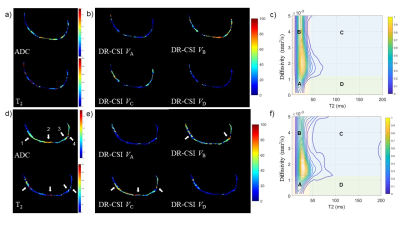 |
109 | Enabling Early Detection of Knee Osteoarthritis Using Diffusion-Relaxation Correlation Spectrum Imaging
Peng Luo1, Wentao Hu2, Yongming Dai2, and Guanwu Li1
1Department of Radiology, Yueyang Hospital of Integrated Traditional Chinese and Western Medicine, Shanghai University of Traditional Chinese Medicine, Shanghai, China, 2Central Research Institue, United Imaging Healthcare, Shanghai, China Keywords: Cartilage, Diffusion/other diffusion imaging techniques Early OA is subclinical for anatomic change of cartilage, making it difficult for conventional MRI detection. This study is aimed to apply diffusion-relaxation correlation spectrum imaging (DR-CSI) to knee early-stage OA detection. DR-CSI compartment volume fractions VA, VB and VC had correlation with the modified Whole-Organ MR Imaging Scores (WORMS). VC had better ability than VA, VB, VD, T2 and ADC to discriminate early OA patients from healthy controls. The results illustrated that DR-CSI compartment volume fractions may be sensitive indicators for detecting early-stage degeneration in knee articular cartilage. |
|
2460.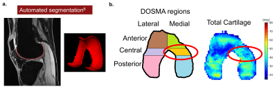 |
110 | Preoperative Femoral Cartilage T2 Relaxation Time Predicts Change in PROs in Patients with Degenerative Meniscal Tears
Jessica Lauren Asay1, Anthony A Gatti1, Arjun D Desai1, Emily J McWalter2, Shannon N Edd3, Thomas P Andriacchi3, Nicholas J Giori4,5, and Garry E Gold1
1Radiology, Stanford University, Stanford, CA, United States, 2Mechanical Engineering and Division of Biomedical Engineering, University of Saskatchewan, Saskatoon, SK, Canada, 3Mechanical Engineering, Stanford University, Stanford, CA, United States, 4VA Palo Alto Health Care System, Palo Alto, CA, United States, 5Orthopedic Surgery, Stanford University, Stanford, CA, United States Keywords: Cartilage, Joints, Meniscus, Knee, Patient Reported Outcomes Meniscectomies are common but not always successful in treating degenerative meniscus tears. Preoperative patient reported outcomes (PROs) and quantitative MRI could help surgeons decide candidacy for meniscectomy. This study demonstrated that pre-operative T2 relaxation times in the medial central femoral cartilage are correlated to pre-operative PROs in patients with degenerative meniscal tears and are predictive of 1 and 2-year changes in PROs post-operation. This study suggests patients who have longer T2 relaxation times pre-operation might not benefit as much from a meniscectomy. |
|
2461.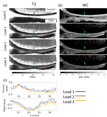 |
111 | Water content and T2 changes in ex vivo bovine knee cartilage under compressive strain
Andrew C Yung1, Emily Sullivan2,3, Jessica C Küpper2,4, Kirsten Bale1, Piotr Kozlowski1, and David Wilson2,4
1UBC MRI Research Centre, Radiology, University of British Columbia, Vancouver, BC, Canada, 2Centre for Hip Health and Mobility, Vancouver, BC, Canada, 3School of Biomedical Engineering, University of British Columbia, Vancouver, BC, Canada, 4Orthopaedics, University of British Columbia, Vancouver, BC, Canada Keywords: Cartilage, Osteoarthritis, Strain We present a novel approach to cartilage multi-echo data where T2 reflects focal changes in strain along the cartilage depth, and the signal amplitude reflects a measure of water content which presents a simpler pattern of change that reflects strain. Ex vivo bovine knee samples were scanned at 9.4T while under load, with high resolution along the cartilage depth to allow direct measurement of strain. Changes in water content were found to be more spatially uniform than the complex depth dependence shown by T2, and showed a high degree of correlation to whole-cartilage averages of bulk strain. |
|
2462.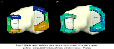 |
112 | Evaluation of whole compartment and regional meniscal T2* values within elite athletes
Erin C Argentieri1, Sara E Sacher1, Ek Tsoon Tan1, Garry Gold2, Hollis G Potter1, Sharmila Majumdar3, and Matthew F Koff1
1Radiology and Imaging, Hospital for Special Surgery, New York, NY, United States, 2Stanford University, Stanford, CA, United States, 3University of California San Francisco, San Francisco, CA, United States Keywords: Cartilage, Quantitative Imaging Results of the current study demonstrate compartment- and region-specific differences in medial and lateral meniscal T2* values. Meniscal T2* values may provide a non-invasive means to assess response of the tissue specific to the loading environment and identify the early onset meniscal degeneration. A better understanding of the early changes in meniscal T2* values may help define the progression of meniscal degeneration, prior to the onset of gross morphologic defects. |
|
2463.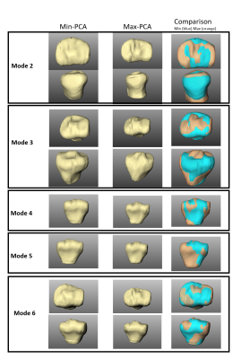 |
113 | Evaluation of the relationship between meniscal T2* metrics and tibial bone shape
Erin C Argentieri1, Kenneth Gao1, Valentina Pedoia1, Garry Gold2, Matthew F Koff3, Hollis G Potter3, and Sharmila Majumdar1
1Radiology, University of California San Francisco, San Francisco, CA, United States, 2Radiology, Stanford University, Stanford, CA, United States, 3Radiology and Imaging, Hospital for Special Surgery, New York, NY, United States Keywords: Cartilage, MSK Features of tibial bone morphology such as tibial slope and tibial spine volume have a significant impact on overall joint mechanics and, results of the current study indicate that these feature of tibial bone shape impact both medial and lateral meniscal T2* metrics. |
|
2464.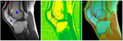 |
114 | Quantitative assessment of the adipose tissue around the knee joint in patients with KOA using IDEAL-IQ and T2 mapping sequences with 3.0 T MRI
Lin Qiu1, Long Qian2, and Xiang-Ran Cai1
1Medical Imaging Center, First Affiliated Hospital, Jinan university, Guangzhou, China, 2MR Research, GE Healthcare, Guangzhou, China Keywords: Cartilage, Osteoarthritis IDEAL-IQ and T2 mapping sequences are capable of objectively assessing fat and water content in the adipose tissue around the knee joint. The severity of KOA is related to the fat and water content of knee adipose tissue. |
|
2465.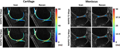 |
115 | Scan-Rescan Repeatability of 10-Minutes Long MAPSS Sequence for Simultaneous MR T2 and T1ρ Mapping of Cartilage and Meniscus at 7 Tesla
Stefan Zbyn1,2,3, Richard Lartey1,2, Jeehun Kim1,2, Carl S. Winalski1,2,3, and Xiaojuan Li1,2,3
1Program of Advanced Musculoskeletal Imaging (PAMI), Cleveland Clinic, Cleveland, OH, United States, 2Department of Biomedical Engineering, Lerner Research Institute, Cleveland Clinic, Cleveland, OH, United States, 3Department of Diagnostic Radiology, Imaging Institute, Cleveland Clinic, Cleveland, OH, United States Keywords: Cartilage, Relaxometry, Meniscus The aim of this study was to evaluate the in vivo scan-rescan repeatability of T2 and T1ρ quantification in knee cartilage and meniscus of patients with knee pain at 7 Tesla. Four subjects were scanned-rescanned on the same day using the 10-minutes-long MAPSS sequence for simultaneous T2 and T1ρ mapping. Coefficients of variation and Bland-Altman plots showed excellent scan-rescan repeatability of in vivo T2 and T1ρ quantification in cartilage and meniscus at 7 Tesla. Our results indicate that fast T2 and T1ρ mapping with four echo and four spin-lock times remains sensitive to cartilage degeneration. |
|
2466.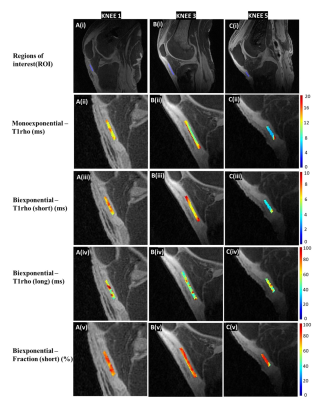 |
116 | Bi-exponential 3D UTE-T1rho relaxation mapping of ex-vivo human knee patellar tendon at 3T
Bhavsimran Malhi1, Dina Moazamian1, Michael Carl2, Jiyo Athertya1, Saeed Jerban1, Hyungseok Jang1, Yajun Ma1, Eric Chang1,3, and Jiang Du1,4,5
1Department of Radiology, University of California, San Diego, USA, San Diego, CA, United States, 2GE Healthcare, San Diego, USA, San Diego, CA, United States, 3Veterans Affairs San Diego Helath Care system, San Diego, USA, San Diego, CA, United States, 4Veterans Affairs San Diego Health Care System, San Diego, CA, USA, San Diego, CA, United States, 5Department of Bioengineering, University of California, San Diego, USA, San Diego, CA, United States Keywords: Tendon/Ligament, Tendon/Ligament, UTE, MRI UTE sequences have been previously used to image short T2 tissues. T1ρ has shown promising results in assessing macromolecules and has been proposed as a potential biomarker for tissue degeneration. In this study, we aimed to demonstrate bi-exponential relaxation of the patellar tendon at 3T, using the 3D ultrashort echo time T1ρ (UTE-T1ρ) sequence. Five ex-vivo knee joints were scanned, and we were able to demonstrate better fitting with a bi-component model and better discern the long and short relaxation components of the tissue, along with their respective fractions |
|
2467.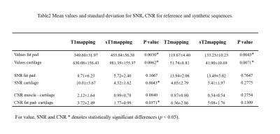 |
117 | QRAPMASTER for Quantitative evaluation of knee cartilage: ready for clinical daily practice?
Lin Mu1, Qianting Wang1, Jiahui Fu2, Dong Dong2, Yueluan Jiang3, Lin Li4, and Huimao Zhang2
1The First Hospital of Jilin University, Changchun, China, 2Radiology, The First Hospital of Jilin University, Changchun, China, 3MR Scientific Marketing, Diagnostic Imaging, Siemens Healthineers Ltd, Beijing, China, 4Radiology, The First Hospital of Jilin University, changchun, China Keywords: Cartilage, MSK Quantitative MRI assigns absolute quantification of T1,T2 and PD based on tissue characteristics, such as the T2 metric values a surrogate marker for cartilage collagen integrity. The QRAPMASTER technology enables inline generation of quantitative T1maps and T2 maps and scans of high image quality, representing a promising synthetic MRI option that appears clinically feasible and may eventually facilitate the time neutral acquisition of quantitative T1 maps, T2 maps in the knee MRI. |
|
2468.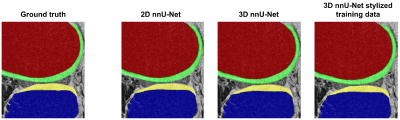 |
118 | Generalizability of nnU-Net for automatic segmentation of knee MRI
Heather Hanegraaf1, Rianne A. van der Heijden1,2, Edwin H.G. Oei1, Marienke van Middelkoop3, Stefan Klein1, and Jukka Hirvasniemi1
1Department of Radiology & Nuclear Medicine, Erasmus MC University Medical Center, Rotterdam, Netherlands, 2Department of Radiology, University of Wisconsin-Madison, Madison, WI, United States, 3Department of General Practice, Erasmus MC University Medical Center, Rotterdam, Netherlands Keywords: Osteoarthritis, Segmentation To investigate the generalizability of deep learning segmentation models, three different nnU-Nets (2D, 3D, and ensemble) were trained on the OAI dataset and tested on a different dataset. In addition to the nnU-Nets trained on the original OAI data, the style of the test set was transferred to the training set using a CycleGAN method and the nnU-Nets were trained again. Depending on the tissue, the 3D nnU-Net or the ensemble trained on the original or stylized training data had the highest segmentation accuracy in the test set. The results indicate that nnU-Net may generalize well to independent datasets. |
|
2469.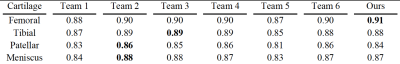 |
119 | Automated Deep Learning Segmentation of Human Knee Cartilage from 3T MRI with Boundary Information
Zhisen Hu1, Peter J Lally1, and Neal K Bangerter1
1Department of Bioengineering, Imperial College London, London, United Kingdom Keywords: Osteoarthritis, Segmentation Knee Osteoarthritis (OA) is serious and prevalent today. Image segmentation of high-resolution MRI scans measuring cartilage volume and thickness is useful to track knee OA progression in the early stages and avoid joint replacement. In this work, we developed a cheap and efficient automated technique based on U-Net for knee cartilage segmentation, paying more attention to boundary information. Our model outperforms many existing models for segmentation of the femoral cartilage and performs as well as other techniques for other cartilage compartments. The boundary loss appears to improve cartilage segmentation for the edge slices with smaller cartilage volume. |
|
2470.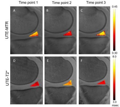 |
120 | Ultrashort Echo Time Magnetization Transfer Imaging of Meniscus After a Marathon
Yijie Fang1, Dantian Zhu1, Wenhao Wu1, Shaolin Li1, Long Qian2, and Yajun Ma3
1Department of Radiology,Fifth Affiliated Hospital, Sun Yat-Sen University, Zhuhai, China, 2MR Research, GE Healthcare, Beijing, China, 3University of California, San Diego, Department of Radiology, San Diego, CA, United States Keywords: MSK, Joints In this study, a 3D UTE-MT preparation will be used to detecting changes in the meniscus of amateur marathon runners before and after a marathon. In this prospective cohort study, 23 amateur marathon runners were enrolled. There were three MRI scans (pre-race, 2 days post-race, and 4 weeks post-race) using the UTE-MT and UTE-T2* sequences. UTE-MTR values decreased 2 days post-race and increased after 4 weeks of rest. Conversely, the UTE-T2* values increased 2 days post-race and increased after 4 weeks of rest. UTE-MTR is a promising biomarker for the detection of changes in the meniscus after a marathon. |
|
 Back to Meeting Home
 Back to the Program-at-a-Glance
The International Society for Magnetic Resonance in Medicine is accredited by the Accreditation Council for Continuing Medical Education to provide continuing medical education for physicians.
 Back to Meeting Home
 Back to the Program-at-a-Glance
 View Presentation Video