Digital Poster
Imaging of the Articular Cartilage
ISMRM & ISMRT Annual Meeting & Exhibition • 03-08 June 2023 • Toronto, ON, Canada

Digital Poster
Imaging of the Articular Cartilage
| Computer # | |||
|---|---|---|---|
4362.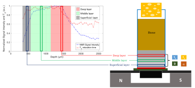 |
101 |
A single-sided NMR approach to study structural differences of
the articular cartilage tissue
Carlo Golini1,
Claudia Testa1,
Anastasiia Nagmutdinova2,
Villiam Bortolotti2,
and Leonardo Brizi1
1Department of Physics and Astronomy "Augusto Righi", University of Bologna, 40127 Bologna, Italy, 2Department of Civil, Chemical, Environmental, and Materials Engineering, University of Bologna, 40134 Bologna, Italy Keywords: Cartilage, Relaxometry, Single-sided NMR In the last decade, few preliminary low-field relaxometry studies were conducted on the articular cartilage using the single-sided NMR-MOUSE device that can be useful for osteoarthritis characterization. This study aims to develop a procedure capable of quantitatively evaluating the structure of the three cartilage layers. Forty osteochondral cylindrical bovine knee specimens were analyzed obtaining four NMR parameters T1, T2, D, and α (extracted from a Double-Quantum-like sequence for solid/liquid estimation) for each cartilage layer. Significant discrimination of the three layers was found by all the parameters, making the NMR dataset sensitive to structural differences and changes due to cartilage diseases. |
|
4363.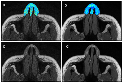 |
102 |
An application of parallel imaging and deep-learning
reconstructed T2 mapping in nasal cartilage
Weiyin Vivian Liu1,
Yufan Gao2,
and Yunfei Zha2
1GE Healthcare, Beijing, China, 2Department of Radiology, Renmin Hospital of Wuhan University, Wuhan, China Keywords: Cartilage, Cartilage Rhinoplasty is a reversible surgery; MRI became a non-invasive post-surgery tracking approach to assess the influence of implant-compressed on nasal cartilage such as mechanical stress, cellular organization and tissue degeneration, etc. Widely-used T2 mapping in musculoskeletal imaging such as knee, shoulder and pelvics takes long acquisition time and thus deters clinical utility. With greater number of multi-channel coils and parallel imaging techniques, scan time shortens but noise enhances. With the additional deep-learning reconstruction, T2 mapping with parallel imaging was stable with small RMS CV% = 0.077% to parallel imaging only image data sets. |
|
4364.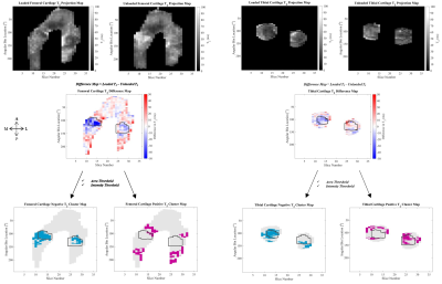 |
103 |
T2 Relaxation Time Decreases in Regions of Loaded Contact in
Superficial and Deep Layers of Articular Cartilage
Natasha M. Bzowey1,
Marianne S. Black2,
Lumeng Cui3,
Chelsey S. Thorson1,
Madeline M. Martel1,
and Emily J. McWalter1
1Mechanical Engineering, University of Saskatchewan, Saskatoon, SK, Canada, 2Mechanical Engineering, University of Victoria, Victoria, BC, Canada, 3Siemens Healthcare Ltd., Burnaby, BC, Canada Keywords: Cartilage, Joints The aim of this work was to identify focal changes in T2 relaxation times inside and outside regions of tibiofemoral contact in loaded cadaver knee articular cartilage whole, superficial, and deep layers. We used a cluster analysis approach to identify regions of change in T2 relaxation time. We found that 1) decreases in T2 relaxation time with load appeared predominantly inside of the femoral and tibial cartilage contact areas, and 2) greater decreases in T2 within the tibial and femoral cartilage contact area were observed in the superficial layer of cartilage. |
|
4365.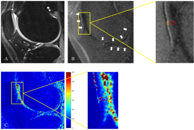 |
104 |
Comparing the effect of body weight, BMI and physical activity
intensity on calcified and uncalcified cartilage using
quantitative UTE MRI
Hanqi Wang1,
Qing Li2,
Stefan Sommer3,4,5,
and Yong Lu1
1Ruijin Hospital, School of Medicine, Shanghai Jiao Tong University, Shanghai, China, 2MR Collaborations, Siemens Healthineers Ltd., Shanghai, China, Shanghai, China, 3Siemens Healthineers International AG, Zurich, Switzerland, Zurich, Switzerland, 4Swiss Center for Musculoskeletal Imaging (SCMI), Balgrist Campus, Zurich, Switzerland, Zurich, Switzerland, 5Advanced Clinical Imaging Technology (ACIT), Siemens Healthineers International AG, Lausanne, Switzerland, Lausanne, Switzerland Keywords: Cartilage, Cartilage UTE technique was utilized to quantitatively assess calcified cartilage (CC) and uncalcified cartilage (UC). T2* values of UC in patella, medial tibia and lateral femur positively moderately correlated with body weight, while T2* values of CC showed no significant correlation. T2* values of UC showed no significant correlation with BMI, while T2* values of CC in medial femur and lateral tibia showed negatively strong and positively moderate correlation, respectively. T2* values of CC and UC showed no significant correlation with physical activity level. The mechanical loading effect performs different between CC and UC, indicating their different bio-mechanical properties. |
|
| 4366. | 105 |
Deep-learning based fast spin echo magnetic resonance imaging
improved clarity of nasal cartilage
yufan Gao1,
yunfei zha1,
and Weiyin Vivian Liu2
1renmin hospital of wuhan university, wuhan, China, 2GE Healthcare, MR Research China, beijing, China Keywords: Cartilage, Cartilage In this study, a comparative study of the original images (T1WI, T2WI) and ARDL images based on deep learning reconstruction (T1WI-DL, T2WI-DL) was conducted for magnetic resonance imaging of nasal cartilage. We aim to evaluate the feasibility and performance of a novel deep learning based MRI reconstruction method to show the structures of nasal cartilage, especially lower lateral cartilage. The results showed that applying ARDL to nasal cartilage MRI showed significant improvement in both SNR and CNR. we conclusion that the DL-based reconstruction algorithm was feasible to be applied to nasal cartilage MRI and improve the image quality. |
|
4367.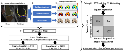 |
106 |
Automated Principal Component Analysis of cartilage thickness,
T2 times and bone shape as interpretable model for
osteoarthritis progression
Felix G Gassert1,
Gabrielle Hoyer1,
Jenny Lee1,
Kenneth Gao1,
Aniket Tolpadi1,
Valentina Pedoia1,
and Sharmila Majumdar1
1Radiology, Center for Intelligent Imaging, UCSF, San Francisco, CA, United States Keywords: Osteoarthritis, Cartilage In this study we used fully automatic segmentations of the bones and the cartilage of the knee to perform a principal component analysis of cartilage thickness, T2-relaxation times and bone shapes in the Osteoarthritis Initiative Dataset. We extracted and visualized the principal components which significantly contributed to a model for prediction of osteoarthritis progression to (i) show that besides mean values of these parameters also their distribution is crucial for osteoarthritis progression and (ii) prove the feasibility of visual interpretation of these components for better understanding factors associated with the disease. |
|
4368.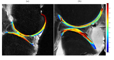 |
107 |
Relaxation Anisotropy Mapping of Articular Cartilage at 3 T
Ville Kantola1,2,
Jouni Karjalainen1,2,
Victor Casula1,2,
Mikko J. Nissi3,
and Miika T. Nieminen1,2,4
1Research Unit of Health Science and Technology, University of Oulu, Oulu, Finland, 2Medical Research Center, University of Oulu, Oulu, Finland, 3University of Eastern Finland, Kuopio, Finland, 4Department of Diagnostic Radiology, University of Oulu, Oulu, Finland Keywords: Cartilage, Relaxometry, Relaxation anisotropy The study of T2 and T1ρ anisotropy in musculoskeletal tissues, arising from the orientation of collagen fibrils in tissue, has so far been limited to experimental MRI systems. Here we present a method for measuring relaxation anisotropy ex vivo on a clinical MRI system. Furthermore, we aimed to quantify and compare the degrees of orientation dependence in bovine articular cartilage through anisotropy mapping. T2 and T1ρ mapping was performed on bovine stifle joint samples at multiple sample orientations. After coregistration anisotropy maps were created using Michelson contrast. The results showed large variations in anisotropy across cartilage thickness and topographical locations. |
|
4369.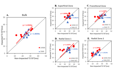 |
108 |
Assessment of articular cartilage in response to an impact
injury using µMRI
Amanveer Singh1,
Hannah Mantebea2,
Farid Badar2,
Syeda Batool2,
Gabrielle Abdelmessih3,
Talia Sebastian3,
Michael Newton4,
and Yang Xia2
1Department of Physics and Center for Biomedical Research, Oakland University, Rochester, MI, United States, 2Department of Physics and Center for Biomedical Research, Oakland University, Rochestor, MI, United States, 3Department of Chemistry, Oakland University, Rochestor, MI, United States, 4Beaumont Hospital, Royal Oak, MI, United States Keywords: Cartilage, Osteoarthritis, Osteoarthritis, Cartilage, Impact injury This project aimed to study the properties of articular cartilage in response to mechanical injury in rabbit knees using μMRI. Femoral cartilage bone plugs were excised 2 and 14 weeks after impact and imaged using μMRI at a resolution of 11.7 μm/pixel. Higher T2 relaxation values in affected specimens illustrated deterioration in the cartilage after impact. Furthermore, the higher T2 values in impacted samples of the 14-week batch compared to the 2-week batch indicate progressive cartilage degradation over time. |
|
4370.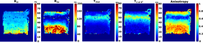 |
109 |
Orientation-independent T2 relaxation parameters and
biomechanics of healthy and degenerated human articular
cartilage
Henri P.P. Leskinen1,2,
Juuso Tuppurainen1,3,
Jiri Jäntti1,3,
Janne T.A. Mäkelä1,3,
and Mikko J. Nissi1
1Department of Applied Physics, University of Eastern Finland, Kuopio, Finland, 2Mikkeli Central Hospital, Mikkeli, Finland, 3Kuopio University Hospital, Kuopio, Finland Keywords: Cartilage, Osteoarthritis Variation in T2 relaxation has been linked to changes in articular cartilage degenerative status. Most prominently to the collagen network integrity. However, T2 is greatly anisotropic in highly organized tissues, e.g. articular cartilage. Previous results show that using re-orientation experiments, orientation-independent T2 components can be defined. This study assesses the potential of these parameters to serve as biomarkers for osteoarthritis via comparing the orientation-independent T2 parameters to biomechanical testing results in human articular cartilage. Changes in the anisotropic component of T2 in the superficial and transitional zones are observed along with the biomechanical degeneration of the articular cartilage. |
|
4371.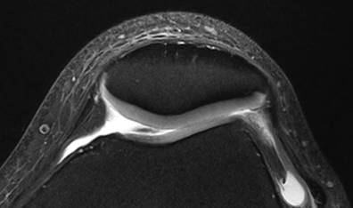 |
110 |
Improved identification of cartilage lesion in chondromalacia
patellae (CMP) with 7.0 T MRI compared with 3.0T MRI: a
preliminary study
Yan Wang1,
Jiayu Huang1,
Jianxun Qu2,
Xiangbing Bian1,
Caohui Duan1,
Chunbao Li3,
Jing Zhang1,
and Xin Lou1
1Radiology, PLA general hospital, Beijing, China, 2MR Collaboration, Siemens Healthineers Ltd, Beijing, China, 3Orthopaedics, PLA general hospital, Beijing, China Keywords: Cartilage, Cartilage To compare the ability in revealing subtle changes of patellar cartilage in suspected CMP between 7.0T and 3.0T MRI. We documented the total number of identified cartilage lesions by two radiologists and compared the number of cartilage lesions identified between 3.0T and 7.0T MRI. The result showed good consistency between readers. The mean number of cartilage lesions identified on 3.0T images was significantly less than 7.0T images. Considering the preliminary and small-sample feature of the study, we conclude conservatively that compared with 3.0T MRI, 7.0T MR images reveal more lesion of the patellar cartilage in CMP patients. |
|
4372.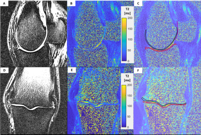 |
111 |
T2 and T1rho mapping of equine metacarpophalangeal joint
cartilage
Carly Lockard1,
Brad Sutton2,
Bruce Damon1,
Xiaojuan Li3,
and Annette McCoy4
1Stephens Family Clinical Research Institute, Carle Clinical Imaging Research Program, Carle Health, Urbana, IL, United States, 2The Grainger College of Engineering, University of Illinois at Urbana-Champaign, Urbana, IL, United States, 3Lerner Research Institute, Cleveland Clinic, Cleveland, OH, United States, 4Department of Veterinary Clinical Medicine (College of Veterinary Medicine), University of Illinois, Urbana, IL, United States Keywords: Cartilage, Quantitative Imaging Horses are an excellent translational model of orthopedic disease with high relevance to human disease. T2 and T1rho mapping can provide information about early cartilage changes in a horse post-traumatic osteoarthritis model. We investigated the feasibility of using MAPSS-based T2 and T1rho mapping in the horse metacarpophalangeal joint cartilage in cadaver specimens and live horses with and without joint pathology. MAPSS-based T2 and T1rho mapping of equine metacarpophalangeal joint cartilage is feasible and provides realistic cartilage relaxometry values. |
|
4373.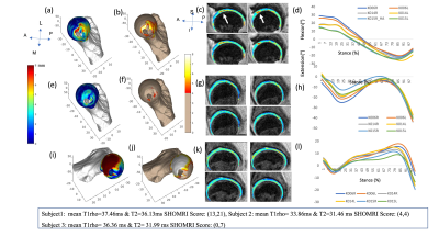 |
112 |
Evaluating the relationship of proximal femoral bone shape
asymmetry with cartilage health and biomechanics in patients
with hip OA
Rafeek Thahakoya1,
Valentina Pedoia1,
Kenneth Gao1,
Rupsa Bhattacharjee1,
Felix Gassert1,
Johanna Luitjens1,
Koren Roach1,
Eric Hammond1,
Richard Souza1,2,
and Sharmila Majumdar1
1Radiology and Biomedical Imaging, University of California San Francisco, San Francisco, CA, United States, 2Department of Physical Therapy and Rehabilitation Science, University of California San Francisco, San Francisco, CA, United States Keywords: Bone, Osteoarthritis This current study evaluated the role of bone shape asymmetry with other biomarkers of hip OA such as T1rho/T2 relaxation time and gait biomechanics data. We observed similar relationship trend in higher bone asymmetry values in the AMS region correlates with lesser number of repetitions performed 30s chair rise test, validating the common understanding of performance. This is one of the first feasibility studies to be explored which conveys the potential of bone asymmetry to be considered as a strong biomarker for understanding hip OA |
|
4374.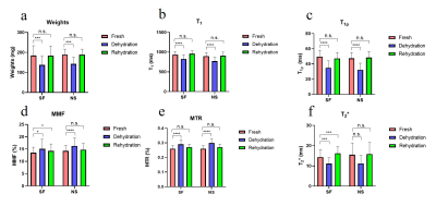 |
113 |
Evaluation of the effect on quantitative ultrashort echo time
(UTE) biomarkers with cartilage dehydration and rehydration
Lidi Wan1,
Adam Searleman2,
Yajun Ma2,
Jonathan H Wong3,
Guangyu Tang1,
Jiang Du2,
and Eric Y Chang3
1Radiology, Tenth People's Hospital of Tongji University, Shanghai, China, 2Radiology, University of California, San Diego, San Diego, CA, United States, 3Radiology, VA San Diego Healthcare System, San Diego, CA, United States Keywords: Cartilage, Quantitative Imaging Ex vivo and cadaveric articular cartilage samples often undergo freeze and thaw cycles and sample preparation in air. These procedures will result in cartilage dehydration and degradation, which may affect the results of UTE-biomarkers. This study was aimed to evaluate the effect of cartilage dehydration and rehydration on UTE-biomarkers and to compare the rehydration capability of synovial fluid and normal saline. Cartilage dehydration resulted in significant changes in all evaluated UTE-biomarkers. Rehydrating with normal saline had non-significant effect on UTE-biomarkers while synovial fluid resulted in significant changes for MMF and T2*. Rehydrating with normal saline is better than synovial fluid. |
|
4375.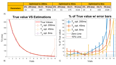 |
114 |
CRLB minimization for optimal Multi Spin-Echo T2-mapping of the
cartilage using dictionary-based estimation
Tiago Timoteo Fernandes1,2,
Sairam Geethanath2,
and Rita G Nunes3
1nstitute for Systems and Robotics - Lisboa and Department of Bioengineering, Instituto Superior Técn, Lisboa, Portugal, 2Accessible Magnetic Resonance Laboratory, Biomedical Imaging and Engineering Institute, Department of Diagnostic, Molecular and Interventional Radiology, Icahn School of Medicine at Mount Sinai, New York, NY, United States, New York, NY, United States, 3Institute for Systems and Robotics - Lisboa and Department of Bioengineering, Instituto Superior Técnico – Universidade de Lisboa, Lisbon, Portugal, Lisboa, Portugal Keywords: Cartilage, Quantitative Imaging .We compared Cramer-Rao Lower Bounds of Multi Spin-Echo sequences’ derived T2 values using a dictionary approach for musculoskeletal relaxometry. The refocusing flip angle, inter-echo spacing and echoe number were varied, constraining the B1+rms to 5μT and the exam time to 9 minutes, keeping TR to a minimum. Designed sequences were tested in NIST/ISMRM phantom and ex vivo, and compared against the vendor’s MSE. A repeatability study was performed during 5 days. An optimized sequence with target T2 value (45ms), representative of the cartilage, scored below 10% for Within-case Coefficient of Variation in 9 out of 14 vials. |
|
4376.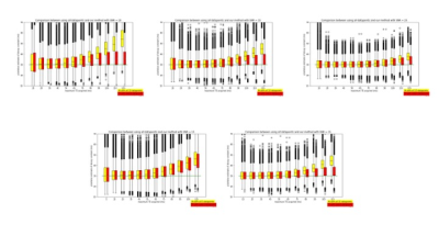 |
115 |
Increased accuracy in relaxometry estimates through statistical
determination of the noise floor
Laurel Hales1,
Ananya Goyal1,
and Feliks Kogan1
1Radiology, Stanford University, Stanford, CA, United States Keywords: Cartilage, Relaxometry, low SNR Including data below the noise floor in estimating relaxometry values leads to overestimation. It is possible to identify and remove data below the noise floor by considering the set of voxel data in an ROI at each echo time as a separate but related dataset, and then comparing the data from each echo time statistically. Removing data below the noise floor, reduces error in the estimates of relaxometry values. |
|
4377.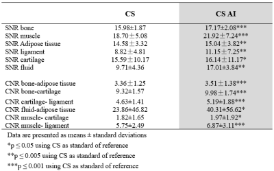 |
116 |
Quantitative MRI T2mapping in the knee joint using deep
learning-based reconstruction for Compressed sensing
jiahui fu1,
chinting wong1,
lin mu1,
dong dong1,
ying qiu1,
yi Zhu2,
Ke Jiang2,
and huimao zhang1
1The First Hospital of Jilin University, Changchun, China, chang chun, China, 2Philips Healthcare, Beijing, China, Beijing, China, China Keywords: Osteoarthritis, Machine Learning/Artificial Intelligence MRI T2 mapping has been recommended as a noninvasive biomarker of knee cartilage lesions. However, due to the long acquisition time, it hasn’t been widely used in the clinical setting. Recently, deep learning-based acceleration of compressed sensing (CS) has shown promising results without losing image quality. The purpose of this study was to explore the feasibility of quantitative knee T2-mapping accelerated by deep learning-based compressed sensing (CS-AI), and compare the image quality and diagnostic performance with conventional CS. The results demonstrates that quantitative knee T2 mapping with reconstruction by CS-AI was feasible, suggesting better diagnostic performance without extra time consuming. |
|
4378.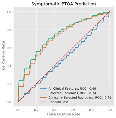 |
117 |
RADIOMICS FEATURES OF INFRAPATELLAR FAT PAD ASSOCIATED WITH
POST-TRAUMATIC OSTEOARTHRITIS AT 10+ YEARS AFTER ACL
RECONSTRUCTION
Sameed Khan1,2,3,
Richard Lartey1,2,
Nancy Obuchowski1,4,
Sibaji Gaj1,2,
Jee Hun Kim1,2,
Mei Li1,2,
Brendan Eck1,3,
Carl S. Winalski1,2,3,
Faysal Altahawi1,3,
Morgan H Jones5,
Laura J Huston6,
Kevin D Harkins7,
Michael V Knopp8,
Christopher C Kaeding9,
Kurt Spindler1,10,
and Xiaojuan Li1,2,3
1Program of Advanced Musculoskeletal Imaging, Lerner Research Institute, Cleveland, OH, United States, 2Department of Biomedical Engineering, Lerner Research Institute, Cleveland, OH, United States, 3Department of Diagnostic Radiology, Imaging Institute, Cleveland, OH, United States, 4Department of Quantitative Health Sciences, Lerner Research Institute, Cleveland, OH, United States, 5Department of Orthopedic Surgery, Brigham and Women's Hospital, Boston, MA, United States, 6Department of Orthopedics and Rehabilitation, Vanderbilt University, Nashville, TN, United States, 7Department of Biomedical Engineering, Vanderbilt University, Nashville, TN, United States, 8Wright Center of Innovation in Biomedical Imaging, Ohio State University, Columbus, OH, United States, 9Department of Orthopedic Surgery, The Ohio State University, Columbus, OH, United States, 10Department of Orthopedic Surgery, Cleveland Clinic, Cleveland, OH, United States Keywords: Osteoarthritis, Fat ACL injury is a major risk factor for post-traumatic osteoarthritis (PTOA), however mechanisms are not fully understood. In this work, we use radiomics from quantitative MRI imaging to associate subvisual changes in the infrapatellar fat pad (IPFP) to both symptomatic and radiographic PTOA. MRI was collected from 113 patients at least 10 years post-ACLR. 1690 radiomics and 11 clinical features were extracted and selected using a gradient-boosted decision tree classifier model. A subset of texture-associated features of IPFP and no clinical features were associated with symptomatic PTOA with an AUROC of 0.74 and radiographic PTOA with an AUROC of 0.84. |
|
4379.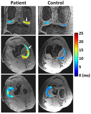 |
118 |
Quantitative 3D Assessment of Meniscus in Patients with
Posterior Root Tears and Healthy Controls Using T2* Mapping at
7T
Abdul Wahed Kajabi1,2,
Stefan Zbyn1,2,3,
Jesse Smith1,2,
Morgan Homan4,
Hasan Abbasguliyev5,
Ariel N. Rodriguez4,
Gregory J. Metzger1,
Robert F. F. LaPrade4,
and Jutta M. Ellermann1,2
1Center for Magnetic Resonance Research, University of Minnesota, Minneapolis, MN, United States, 2Department of Radiology, University of Minnesota, Minneapolis, MN, United States, 3Department of Biomedical Engineering, Lerner Research Institute, Ohio, OH, United States, 4Twin Cities Orthopedics, Edina, MN, United States, 5Department of Diagnostic and Interventional Radiology, Ataturk University Research Hospital, Erzurum, Turkey Keywords: MSK, Quantitative Imaging, Meniscus Evaluating 3D T2* mapping of the meniscus at ultra-high field 7T provides necessary high resolution to better characterize tissue changes in the meniscus. This study was designed to measure the 3D structure of the meniscus with T2* mapping in a clinically acceptable scan time at 7T, and to evaluate meniscus degeneration in twelve patients with arthroscopically verified tears of the posterior horn of the medial meniscus and nine age, sex and body mass index matched healthy controls. T2* mapping at 7T provided comprehensive 3D evaluation of meniscus and detected degenerative alterations caused by the root tears in the patients. |
|
4380.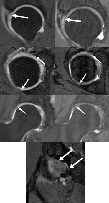 |
119 |
Isotropic 3D MRI determined labral tear extent correlates with
hyaline cartilage loss in hip dysplasia patients
Shuda Xia1,
Avneesh Chhabra2,
Gaurav Sharan2,
Uma Thakur2,
Holden Archer2,
Yin Xi2,
and Joel Wells2
1University of Texas Southwestern Medical Center, Plano, TX, United States, 2University of Texas Southwestern Medical Center, Dallas, TX, United States Keywords: Joints, Joints This study is a novel investigation into the clinical correlations between parameters measured through 3D MRI and outcomes for patients with hip dysplasia (HD). Although there were no statistically significant correlations seen between labral tear length and PROMs (p>0.05), a few non-significant correlation trends were observed. Furthermore, labral tear lengths directly positively correlated with worsening cartilage damage, the presence of subchondral bone cysts, para-labral cysts, and the number of labral tears seen. |
|
4381.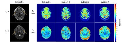 |
120 |
Relaxometric mapping of normal tissue using magnetic resonance
fingerprinting (MRF)
Enlin Qian1,
Amaresha Shridhar Konar2,
Tiago Fernandes3,
Ramesh Paudyal2,
Amita Shukla-Dave2,4,
Lawrence Schwartz5,
and Sairam Geethanath3
1Columbia Magnetic Resonance Research Center, Columbia University, New York, NY, United States, 2Department of Medical Physics, Memorial Sloan Kettering Cancer Center, New York, NY, United States, 3Accessible MR Laboratory, Biomedical Engineering and Imaging Institute, Dept. of Diagnostic, Molecular and Interventional Radiology, Icahn School of Medicine at Mt. Sinai, New York, NY, United States, 4Department of Radiology, Memorial Sloan Kettering Cancer Center, New York, NY, United States, 5Department of Radiology, Columbia University Irving Medical Center and New York Presbyterian Hospital, New York, NY, United States Keywords: Muscle, Muscle We explored the feasibility of T1 and T2 mapping of normal tissue (masseter muscle) in the head and neck (HN) region using magnetic resonance fingerprinting (MRF) and evaluated its repeatability. Our study included fingerprinting data from four healthy volunteers and four repeats each, along with acquiring B0 maps. The T1 and T2 estimation of masseter muscle by MRF is similar to previous literature values. We report wCV<4.3% for T1 measurements and wCV<7% for T2 measurements for all subjects. No significant B0 variations were observed in the masseter muscle regions. |
|
 Back to Meeting Home
 Back to the Program-at-a-Glance
The International Society for Magnetic Resonance in Medicine is accredited by the Accreditation Council for Continuing Medical Education to provide continuing medical education for physicians.
 Back to Meeting Home
 Back to the Program-at-a-Glance
 View Presentation Video