Digital Poster
Imaging of Bone & Marrow
ISMRM & ISMRT Annual Meeting & Exhibition • 03-08 June 2023 • Toronto, ON, Canada

| Computer # | |||
|---|---|---|---|
4524.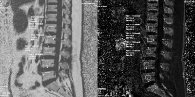 |
101 |
Correlation of R2* with fat fraction and bone mineral density
and its role in quantitative assessment of osteoporosis
zhenghua Liu1
1Radiology, Honghui Hospital Affiliated Xi’an Jiaotong University, xi an, China Keywords: Bone, Quantitative Imaging, Osteoporosis; Bone density In this study, we investigated the relationship between vertebral R2* and FF based on IDEAL-IQ sequences and BMD based on QCT, and then performed diagnostic experiments. The results revealed a definite but weak linear relationship between R2* with FF and BMD, which has limited value as a diagnostic indicator for OP and osteopenia, but has some potential as a complement to FF and BMD for fine quantification of bone marrow conversion and bone mineral loss. |
|
4525.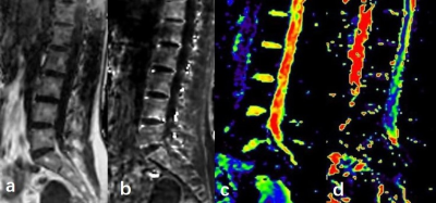 |
102 |
Assessment of vertebral and intervertebral disc microenvironment
changes in postmenopausal women using IVIM and fat-water MRI
Shuo Zhang1,
Qingwei Song1,
Hongbo Feng1,
Qianrui Guo2,
and Yang Yang3
1The First Affiliated Hospital of Dalian Medical University, Dalian, China, 2Dalian Medical University, Dalian, China, 3Beijing United Imaging Research Institute of Intelligent Imaging, Beijing, China Keywords: Bone, Data Acquisition Intravoxel incoherent motion (IVIM) and fat-water magnetic resonance imaging (FWMRI) are rarely used for vertebral and intervertebral disc. To search for predictive value of them in assessing the lumbar spine microenvironment changes, we performed a study with postmenopausal women combined the parameters with bone mineral density (BMD) and major fracture risk assessment (FRAX) score. We report that fat fraction (FF) has significant correlation with FRAX, FF was moderately negatively correlated with BMD; ADCslow of intervertebral discs showed moderately negatively correlation with FRAX score and FF. IVIM and FWMRI have potential to become new biomarkers in predicting lumbar spine microenvironment changes. |
|
4526.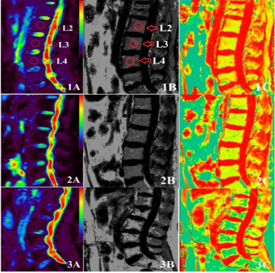 |
103 |
Application of Multiecho Dixon Technique and IVIM-DWI in
Patients with Primary Osteoporosis : A Preliminary Study with
3.0 T MRI
fan qiu ju1,
tan hui1,
yu nan 1,
and wang shao yu2
1Affiliated Hospital of Shaanxi University of Chinese Medicine,, xianyang, China, 2MR senior Scientific Marketing Specialist,Siemens Healthineers, Shang hai, China Keywords: Bone, Quantitative Imaging, Multiecho Dixon Technique , IVIM-DWI Bone mineral density (BMD) is a common standard for evaluating osteoporosis, but it can only reflect the change of bone mass, not the microscopic nodules of tissueChange of structure.Multi-echo Dixon and IVIM-DWI were used to evaluate the changes of spinal bone marrow microstructure in osteoporosis, osteopenia and normal subjects.The results showed that there were significant differences in FF, D and D* between osteoporosis, osteopenia and normal subjects.These results indicate that IVIM-DWI can quantitatively reflect the changes of lumbar microcirculation and fat content, and can be used as a biomarker for the progression of osteoporosis. |
|
4527. |
104 |
A clinical application of ZTE-MRI in diagnosis of knee using
joint space and angles
Hui Wang1,
Weiyin Vivian Liu2,
Wen Chen3,
Jingyu Jiang4,
Ling Sang3,
Peng Zhang3,
and Hu Chen3
1Hubei University of Medicine, Department of Radiology, Taihe Hospital,, Hubei, China, 2GE Healthcare, Beijing, China, 3Shiyan Taihe Hospital, Hubei, China, 4Biomedical Engneering College, Hubei University of Medicine, Hubei, China Keywords: Bone, Bone X-ray showed no significant abnormalities at early stage of OA, but obvious manifestation appeared in the middle and late stages. Our study demonstrated that joint space and joint angle can be measured on ZTE-MRI in evaluation of knee mechanical function and further support anatomical evaluation using Kellgren-Lawrence grading system that is the most widely used method for osteosurgeons in diagnosis of knee osteoarthritis. We also found both JS and CD were correlated with KLZTE score and both showed significant difference between mild/moderate and advanced groups. Therefore, ZTE-MRI has great potential in displaying structure abnormality. |
|
4528.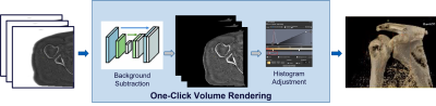 |
105 |
Intelligent volume rendering of ZTE MR Bone images
Vineet Mehta1,
Deepthi Sundaran1,
Jignesh Dholakia1,
Maggie Fung2,
and Michael Carl3
1GE Healthcare, Bengaluru, India, 2GE Healthcare, New York City, NY, United States, 3GE Healthcare, San Diego, CA, United States Keywords: Bone, Visualization, Volume Rendering This study presents a new Deep-Learning (DL) based processing pipeline to enable one-click volume rendering of MR Bone Zero TE (ZTE) images. It comprises of DL based background subtraction followed by histogram adjustment. A comparison with existing algorithms demonstrates that the proposed method prevails in accuracy of background subtraction (DICE = 0.97) and eliminates the tedious, error-prone manual steps of segmentation and histogram adjustment required to prepare the data for usable volume rendering. It can also be used universally across anatomies as opposed to other methods requiring anatomy-specific tuning. |
|
4529.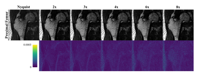 |
106 |
Quantification of the effects on proximal femur trabecular bone
biometrics by parallel imaging and compressed sensing
reconstruction
Brian-Tinh Duc Vu1,2,
Brandon C. Jones1,2,
Rajiv S. Deshpande1,2,
Hyunyeol Lee2,3,
Trevor J. Chan1,2,
Nada Kamona1,2,
Sabrina Ripsman2,
Dilini Ranaweera2,
and Chamith S. Rajapakse2,4
1Department of Bioengineering, University of Pennsylvania, Philadelphia, PA, United States, 2Department of Radiology, University of Pennsylvania, Philadelphia, PA, United States, 3School of Electronics Engineering, Kyungpook National University, Daegu, Korea, Republic of, 4Department of Orthopaedic Surgery, University of Pennsylvania, Philadelphia, PA, United States Keywords: Bone, Bone, Osteoporosis The high resolution needed for quantification of trabecular bone biometrics in the proximal femur results in a prohibitively long scan time for clinical screening. Parallel imaging and compressed sensing (PICS) can reduce scan times by undersampling k-space while producing images comparable in quality to that of the Nyquist-sampled scan. Here, we show PICS recovers information on the trabecular microarchitecture at a quality comparable to that of Nyquist-sampled 3D balanced steady-state free precession. We quantify several parameters of trabecular bone for various acceleration factors to investigate the upper limit of PICS acceleration for femoral imaging. |
|
4530.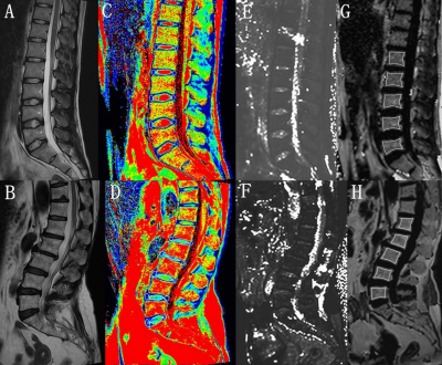 |
107 |
Q-Dixon and T2 Mapping for assessment of intervertebral disc
degeneration in lower back pain
Futing Feng1,
Wei Wang1,
Meining Chen2,
Lusi Liu1,
Mixue Sun1,
and Min Luo1
1Department of Radiology, Zigong Fourth People's Hospital, Zigong, Sichuan, China, 2Department of MR Scientific Marketing,, Siemens Healthineers, Shanghai, China Keywords: Bone, Degenerative The diagnostic performance of MR T2w-based Pfirrmann classification decreases in early and severe stages in patients with lower back pain(LBP). The MR mapping technique, including q-Dixon and T2 mapping, allows for visualization and quantification of the discs and vertebrae in patients with LBP. The changes of bone marrow fat (BMF), T2 and T2* values can be seen in different pain levels and Pfirrmann classification, with great potential in quantifying biochemical degeneration of the lumbar spine. |
|
4531.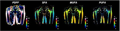 |
108 |
Toward an Imaging Biomarker for Proximal Femur Fracture Risk
Using 3T Chemical Shift-Encoded MRI Radiomic Of Bone Marrow
Dimitri Martel1,
Benjamin Leporq2,
Anmol Monga1,
Stephen Honig3,
and Gregory Chang1
1Radiology, NYU Langone, New York, NY, United States, 2Université de Lyon; CREATIS CNRS UMR 5220, Inserm U1206, INSA-Lyon, Villeurbanne, France, 3Osteoporosis Center, Hospital for Joint Diseases, NYU Langone, New York, NY, United States Keywords: Bone, Radiomics Recent studies using Chemical Shift Encoded-MRI have used textural analysis to assess vertebral marrow heterogeneities in postmenopausal women and in paraspinal muscle to predict its strength. Radiomics provide another means to quantify bone marrow on MR images, specifically by analyzing texture, shape, and intensity distribution in the region of interest. Our aim was to investigate whether radiomic features in fatty acid composition maps of the proximal femur bone marrow improve the prediction of osteoporotic fracture. |
|
4532.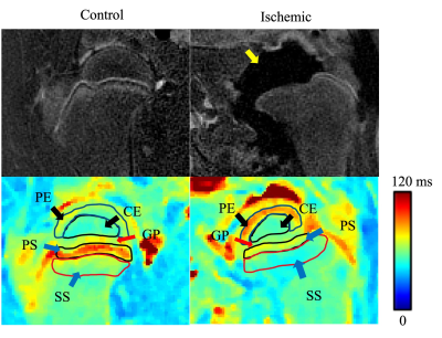 |
109 |
3D T2 and T1ρ Mapping of Ischemic Injury to the Femoral Head: An
In Vivo Piglet Model Study
Erick O. Buko1,2,
Douglas Albrecht1,
Alexandra R. Armstrong1,
Ferenc Tóth1,
and Casey P. Johnson1,2
1Veterinary Clinical Sciences, University of Minnesota, Saint Paul, MN, United States, 2Center for Magnetic Resonance Research, University of Minnesota, Minneapolis, MN, United States Keywords: Bone, Ischemia Legg-Calvé-Perthes disease (LCPD) is a pediatric hip disorder caused by femoral head ischemia and osteonecrosis. In this work, we investigated whether 3D T2 and T1ρ relaxation time mapping are sensitive to ischemic injury to the bone and marrow of the femoral epiphysis and metaphysis following surgical induction of femoral head ischemia in a piglet model of LCPD. We found that T2 and T1ρ increased in the ischemic femoral epiphysis and decreased in the perfused metaphyseal spongiosa. Our findings support the potential clinical use of T2 and T1ρ mapping to assess the severity of femoral head injury in patients with LCPD. |
|
4533.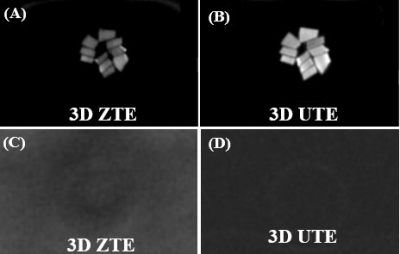 |
110 |
Can Zero Echo Time (ZTE) Magnetic Resonance Imaging Sequences
Detect Signal from Collagen Backbone Protons?
Dina Moazamian1,
Xiaojun Chen1,
Arya Suprana1,
Andrew Andrew Xia1,
Saeed Jerban1,
Bhavsimran Malhi1,
Michael Carl2,
Eric Y Chang1,3,
Yajun Ma1,
Hyungseok Jang1,
and Jiang Du1,3,4
1Radiology, University of California San Diego, San Diego, CA, United States, 2Radiology, GE Healthcare, San Diego, CA, United States, 3VA San Diego Healthcare System, San Diego, CA, United States, 4Bioengineering, University of California San Diego, San Diego, CA, United States Keywords: Bone, Bone Ultrashort echo time (UTE) and zero echo time (ZTE) sequences have been extensively investigated for imaging of short T2 species. The ZTE sequence has a shorter effective TE than UTE and may be superior in imaging ultrashort T2 species. This study investigated whether ZTE could directly image collagen backbone protons in bovine cortical bone and human patellar tendon samples after D2O exchange and freeze-drying. Our experimental results demonstrate that collagen backbone protons are "invisible" with ZTE, which may not be able to directly image species with T2s of ~10 µs. |
|
4534.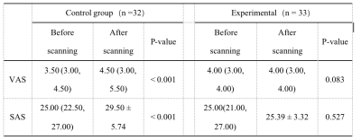 |
111 |
Evaluation of the application of integrated high-quality
intervention mode in large-aperture MRI examination of patients
with knee joint injury
Jun Zhang1,
Lina Zhuang1,
Nan Wang2,
Lihua Chen2,
Qinhe Zhang2,
Shuheng Zhang2,
Yuyin Man2,
Qingwei Song2,
and Ailian Liu2
1Discussion and Conclusions, Dalian, China, 2The First Affiliated Hospital of Dalian Medical University, Dalian, China Keywords: Bone, Bone The integrated high-quality intervention mode is adopted for patients with knee joint injuries, combined with super large aperture magnetic resonance equipment, to effectively relieve the negative emotions of patients, relieve pain, improve scanning efficiency and safety, and enhance scanning efficiency and safety. |
|
4535.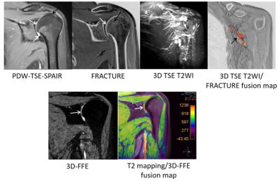 |
112 |
MRI One-stop-shop for Diagnosis of Shoulder Disease
Jienan Wang1,
ShuQing Chen2,
LiFei Ma2,
Rui Zhang2,
and XiaoMeng Wu2
1Department of Radiology, Shanghai Jiao Tong University Affiliated Sixth People‘s Hosptial, Shanghai, China, 2Philips, Shanghai, China Keywords: Bone, MSK, MRI One-stop-shop, T2 mapping, FRACTURE (FFE Resembling A CT Using Restricted Echo - spacing) In this study, shoulder imaging was achieved by MRI one-stop imaging, which contained different sequences. After the post-processing, a variety of fusion images could be formed to comprehensively evaluate the soft tissue, nerve, cartilage and bone of the shoulder, and then gave us a comprehensive view of the shoulder. |
|
4536.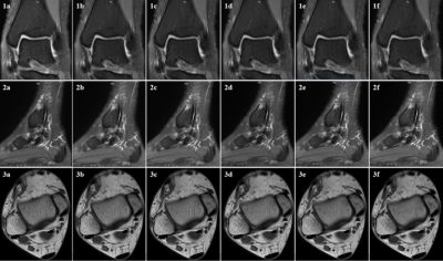 |
113 |
Application of Artificial Intelligence (AI)-assisted compressed
sensing technology in ankle joint
Nan Wang1,
Qingwei Song1,
Ailian Liu1,
Guobin Li2,
Shuheng Zhang2,
Yunfei Zhang3,
and Yongming Dai3
1the First Affiliated Hospital of Dalian Medical University, Dalian, China, 2Shanghai United Imaging Healthcare Co., Ltd, Shanghai, China, 3MR Collaboration, Central Research Institute, United Imaging Healthcare, Shanghai, China Keywords: Bone, Joints ACS combines CS, half Fourier HF and parallel imaging (PI), and introduces deep learning neural network as AI module into the reconstruction process. Millions of fully sampled data are used to train AI models, so as to suppress various reconstruction artifacts introduced by traditional acceleration methods under high acceleration factors without affecting anatomy and pathological structures. ACS is used for noise suppression, artifact reduction and information recovery. ACS can effectively correct any major errors of single acceleration methods, thus providing a higher acceleration level for MRI imaging. |
|
4537.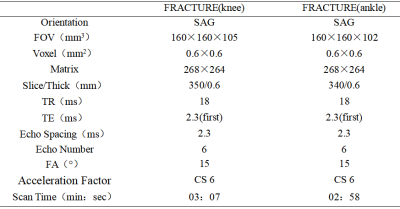 |
114 |
Application value of compressed sensing-based MR FRACTURE
sequence in bone and joint
Nan Wang1,
Qingwei Song1,
Ailian Liu1,
Jiazheng Wang2,
and Liangjie Lin2
1the First Affiliated Hospital of Dalian Medical University, Dalian, China, 2Clinical and Technical Support, Philips Healthcare, Beijing, China Keywords: Bone, Bone FRACTURE sequence does not need to control TE time within 1ms, but uses multi-echo three-dimensional fast field echo, FFE) sequence to perform magnetic resonance imaging with fixed echo interval. After scanning, bone images with high contrast and high signal-to-noise ratio can be obtained through echo accumulation, subtraction and gray scale inversion. |
|
4538.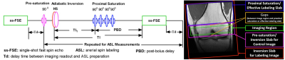 |
115 |
Evaluation of Potential Benefits of 7T for Knee Epiphyseal Bone
Marrow ASL Imaging
Xiufeng Li1,
Jutta Ellermann1,
and Gregory J. Metzger1
1University of Minnesota, Minneapolis, MN, United States Keywords: Bone, Perfusion, ASL, Knee, 7T, Ultrhigh Field Knee epiphyseal bone marrow ASL imaging (called knee ASL imaging in the following) is challenging due to very low perfusion in epiphyseal yellow bone marrow mainly consisting of fat cells with a sparse network of capillaries. In general, it is known that higher magnetic fields (i.e. ≥7T) can specifically benefit ASL imaging. However, the specific potential benefits in the knee have not been systematically evaluated to date. We have performed theoretical simulations to explore the potential gain for knee ASL imaging at 7T compared to 3T and present our preliminary experimental results comparing both field strengths. |
|
4539.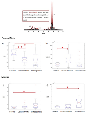 |
116 |
Investigation of the fat-bone-muscle connection in osteoporosis
and osteoarthritis by 1H MRS
Alessandra Maiuro1,2,
Daniele Mattioli2,
Guglielmo Manenti3,
Elena Gasbarra4,
Umberto Tarantino4,
and Silvia Capuani1,2
1Physics, CNR Institute for Complex Systems (ISC), Rome, Italy, 2Physics, Sapienza University of Rome, Rome, Italy, 3Diagnostic and Interventional Radiology, Molecular Imaging and Radiotherapy, Policlinico Tor Vergata Foundation, Tor Vergata University of Rome, Rome, Italy, 4Orthopedics and Traumatology, Policlinico Tor Vergata Foundation, Tor Vergata University of Rome, Rome, Italy Keywords: Bone, Spectroscopy, Osteoporosis, Fatty acids, Osteoarthritis, Muscle The metabolic pathways involved in the development of osteoporosis and osteoarthritis, was investigated by SVS1H MRS. Metabolites and fatty acids of the bone marrow and the adjacent muscle of healthy, osteoporotic and osteoarthritic woman were quantified using LCModel. The increase of E23 resonance in muscles is a potential marker of sarcopenia correlated to osteoporosis. The decrease of the L53 resonance in bone marrow highlights the presence of osteoporosis (together with total lipids increase) or osteoarthritis (together with bone marrow water increase). |
|
4540.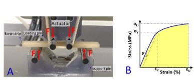 |
117 |
Ultrashort Echo Time, MRI porosity index, and suppression ratio
correlate with the cortical bone microstructural and mechanical
properties
Saeed Jerban 1,2,3,
Yajun Ma1,2,
Salem Alenezi 4,
Dina Moazamian1,
Jiyo Athertya1,
Hyungseok Jang1,2,
Erik Dorthe5,
Darryl Dlima5,
Gina Woods6,
Christine B Chung1,2,
Eric Y Chang1,2,
and Jiang Du1,2
1Department of Radiology, University of California, San Diego, La Jolla, CA, USA, San Diego, CA, United States, 2Radiology Service, Veterans Affairs San Diego Healthcare System, San Diego, La Jolla, CA, USA, San Diego, CA, United States, 3Department of Orthopedic Surgery, University of California, San Diego, La Jolla, CA, USA, San Diego, CA, United States, 4Research and Laboratories Sector, Saudi Food and Drug Authority, Riyadh, KSA, Riyadh, Saudi Arabia, 5Shiley Center for Orthopedic Research and Education at Scripps Clinic, La Jolla, CA, USA, San Diego, CA, United States, 6Department of Medicine, University of California, San Diego, La Jolla, CA, USA, San Diego, CA, United States Keywords: Bone, Bone The cortical bone porous microstructure can be evaluated using ultrashort echo time (UTE) MRI. UTE-MRI-based evaluation of bone has been underutilized partly due to the high cost and time demands of MRI in general. The porosity index (PI) and the suppression ratio (SR) are two rapid UTE-based bone evaluation techniques (~ 5 mins scan time each), which can potentially reduce the time demand and cost in future clinical studies. We have investigated the relationship of PI and SR measures with human cortical bone microstructural and mechanical properties. PI and SR showed significant correlations with microstructural and mechanical properties. |
|
4541.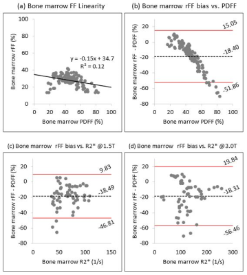 |
118 |
Linearity and Bias of Bone Marrow Relative Fat-fraction Compared
to Proton Density Fat-Fraction
Matthew Mader, MD1,
Diego Hernando, PhD2,
Rianne A. van der Heijden, MD PhD1,
Scott B. Reeder, MD PhD3,
and Ali Pirasteh, MD2
1Radiology, University of Wisconsin-Madison, Madison, WI, United States, 2Radiology and Medical Physics, University of Wisconsin-Madison, Madison, WI, United States, 3Radiology, Medical Physics, Biomedical Engineering, Medicine, Emergency Medicine, University of Wisconsin-Madison, Madison, WI, United States Keywords: Bone, Fat, Fat fraction Bone marrow relative fat-fraction (rFF) measured using dual-echo gradient echo (GRE) in- and opposed phase imaging, has been proposed as an early predictor of treatment response in multiple myeloma. In this work we demonstrate that in patients without known bone marrow pathology (referred for liver fat/iron quantification), bone marrow rFF is unreliable and suffers from large bias when compared with proton density fat-fraction (PDFF), acquired using quantitative chemical-shift encoded (CSE) MRI. Hence, PDFF should be considered for future approaches using bone marrow fat-fraction as a predictor of treatment response in multiple myeloma. |
|
 Back to Meeting Home
 Back to the Program-at-a-Glance
The International Society for Magnetic Resonance in Medicine is accredited by the Accreditation Council for Continuing Medical Education to provide continuing medical education for physicians.
 Back to Meeting Home
 Back to the Program-at-a-Glance
 View Presentation Video