Digital Poster
Imaging of Spine & Neuromuscular Complex
ISMRM & ISMRT Annual Meeting & Exhibition • 03-08 June 2023 • Toronto, ON, Canada

Digital Poster
Imaging of Spine & Neuromuscular Complex
| Computer # | |||
|---|---|---|---|
 |
2588.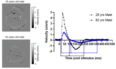 |
61 |
Phase contrast motor unit MRI (PC-MUMRI) to investigate changes
to muscle twitch dynamics in a healthy ageing cohort
Matthew G. Birkbeck 1,2,3,
Linda Heskamp1,
Ian S. Schofield1,
Julie Hall4,
Roger G. Whittaker1,
and Andrew M. Blamire1
1Translational and Clinical Research Institute, Newcastle University, Newcastle upon Tyne, United Kingdom, 2NIHR Biomedical Research Centre, NIHR Biomedical Research Centre, Newcastle upon Tyne, United Kingdom, 3Northern Medical Physics and Clinical Engineering, Newcastle upon Tyne NHS Foundation Trust, Newcastle upon Tyne, United Kingdom, 4Department of Neuroradiology, Newcastle upon Tyne NHS Foundation Trust, Newcastle upon Tyne, United Kingdom Keywords: Muscle, Aging Functional changes to the muscle’s contractile and relaxation properties occur with age. Current techniques which measure twitch dynamics provide limited information about individual muscles. We used phase contrast motor unit MRI (PC-MUMRI) to non-invasively measure the contraction and relaxation times of lower leg muscles in a healthy ageing cohort, relating this to isometric maximum voluntary contraction (MVC). Contraction and relaxation times increased with ageing and were negatively correlated with MVC. PC-MUMRI offers a sensitive method to measure muscle twitch dynamics and could be combined with other MR techniques to characterise functional changes to muscle with ageing. |
2589.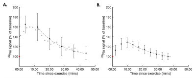 |
62 |
High temporal resolution dynamic 23Na-MRI in skeletal muscle
following exercise
Mary A Neal1,2,
Isobel K Townsend1,2,
Harry Wightman1,2,
and Peter E Thelwall1,2
1Newcastle Magnetic Resonance Centre, Newcastle University, Newcastle upon Tyne, United Kingdom, 2Translational and Clinical Research Institute, Newcastle University, Newcastle upon Tyne, United Kingdom Keywords: Muscle, Non-Proton, 23Na, Sodium Atypical Na+ regulation following exercise can be indicative of skeletal muscle dysfunction. Dynamic 23Na-MRI of signal recovery following exercise therefore holds potential to provide a sensitive, quantitative, and non-invasive tool for physiological monitoring of disease progression or response to interventions. 23Na-MRI sequences with temporal resolutions ranging from 2-8 minutes were acquired in the lower leg before and repeatedly after strenuous isometric dorsiflexion. Our data characterised sodium dynamics during recovery from exercise, and demonstrated an initial increase in 23Na signal intensity from baseline levels after exercise ceased. These findings may provide additional metrics for monitoring skeletal muscle dysfunction. |
|
2590.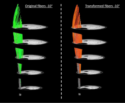 |
63 |
A novel registration strategy to transform skeletal muscle
architecture during passive shortening
Melissa T. Hooijmans1,2,3,
Carly Lockard2,
Crystal Coolbaugh1,
Mark George1,
Xingyu Zhou1,2,4,
and Bruce Damon1,2,4
1Institute of Imaging Sciences, Vanderbilt University, Nashville, TN, United States, 2Carle Health, Stephens Family Clinical Research Institute, Urbana, IL, United States, 3Radiology and Nuclear Medicine, University of Amsterdam, Amsterdam Movement Sciences, Amsterdam UMC, Amsterdam, Netherlands, 4Biomedical Engineering, Vanderbilt University, Nashville, TN, United States Keywords: Muscle, Diffusion Tensor Imaging, Muscle architecture In this study we explored if displacement fields, derived from registration of high-resolution anatomical images in different foot positions, could be used to transform muscle fiber architecture from plantar-flexed to dorsi-flexed foot position (passive shortening). Our data revealed that muscle fiber architecture from one foot position can be transformed in the other foot position and that the original and transformed fibers demonstrate similar architectural characteristics, i.e. fiber lengths, pennation angle, curvature, and physiological cross-sectional area. |
|
2591.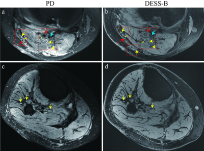 |
64 |
Evaluation of the peripheral nerves of the knee using
Double-Echo Steady-State MRI at 7T
Pinzhen Chen1,
Zhiming Zhen2,
Ting Yin3,
and Wei Chen2
1The first affiliated hospital ofArmy Medical University, Chongqing, China, 2The first affiliated hospital, Army Medical University, Chongqing, China, 3MR Collaborations, Siemens Healthineers Ltd., Chengdu, China Keywords: MSK, Nerves, 7T, DESS, peripheral nerves This study investigated the feasibility of DESS sequence to assess the knee peripheral nerve structures in healthy subjects and patients. The results showed that the nerve imaging quality of DESS sequence was superior to PD sequence in healthy adults. Further, only the isotropic 3D-DESS protocol could clearly visualize the distal peroneal nerve branches and their anatomical course. In patient with peripheral nerves damages, combining the PD and DESS imaging could provide more pathological and anatomical information about the knee peripheral nerve. This suggests that DESS sequence can reliably image the knee peripheral nerves and effectively provide precise knowledge about it. |
|
2592.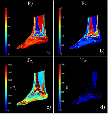 |
65 |
Multi-parametric measures of in-vivo T2* mapping with δUTE and
macromolecule exchange ZAP/CEST of cartilage, tendon, and muscle
tissue in foot.
Vadim Malis1,
Won Bae1,2,
Diana Vucevic1,
Yoshimori Kassai3,
and Mitsue Miyazaki1
1Radiology, UC San Diego, San Diego, CA, United States, 2VA San Diego Healthcare System, San Diego, CA, United States, 3Canon Medical, Ōtawara-shi, Japan Keywords: Tendon/Ligament, CEST & MT, UTE, Ultra-short TE A δUTE sequence allows for collecting multiple echoes with short echo time intervals, without the restriction of in-phase TEs. This study utilizes data collected with δUTE for bi-exponential T2* mapping of cartilage, muscle and tendon tissues in a human foot. The same anatomies are also evaluated using Z-spectrum Analysis Protons (ZAP) and Chemical exchange saturation transfer (CEST). While ZAP imaging provides overall exchange proton quantification of free and restricted exchange protons in a wide range of frequencies, CEST focuses on specific hydroxy (-OH, at 1.0 ppm), amine (-NH2, at 2 ppm), and amide (-NH, at 3.5 ppm) protons. |
|
2593.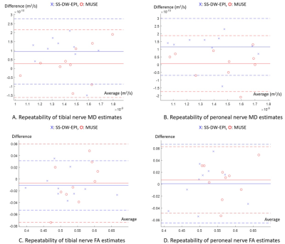 |
66 |
Feasibility of high-resolution multi-shot DW-EPI for DTI of
peripheral nerves in the knee
Daehyun Yoon1,
Brian Hargreaves1,
and Amelie Lutz1
1Radiology, Stanford University, Stanford, CA, United States Keywords: Neurography, Diffusion Tensor Imaging, Multi-shot DW-EPI, repeatability, feasibility DTI of peripheral nerves for detecting nerve damage has been very challenging due to limited resolution and imaging artifacts of the conventional single-shot diffusion-weighted echoplanar imaging (SS-DW-EPI). Here, we have evaluated the feasibility of improved DTI with multi-shot DW-EPI (MUSE) by comparing mean diffusivity (MD) and fractional anisotropy (FA) estimates of tibial and peroneal nerves with those from SS-DW-EPI in ten healthy subjects. MUSE showed lower within-subject coefficient of variation in diffusion estimates, indicating improved repeatability, while correlation analysis showed noticeable differences between FA estimates. Our results show the feasibility of MUSE for in vivo evaluation of peripheral nerves. |
|
2594.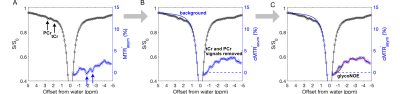 |
67 |
MRI monitors the abnormal muscle glycogen and creatine kinase
activity in Pompe disease mice
Shaowei Bo1,2,
Chongxue Bie1,3,
Nirbhay N. Yadav4,5,
Peter C.M. van Zijl4,5,
Jiadi Xu4,5,
Chao Zou1,
Hairong Zheng1,
and Yang Zhou1
1Shenzhen Institute of Advanced Technology, Chinese Academy of Sciences, Shenzhen, China, 2Medical Imaging Center, The First Affiliated Hospital of Jinan University, Guangzhou, China, 3Department of Information Science and Technology, Northwest University, Xian, China, 4F.M Kirby Research Center, Kennedy Krieger Institute, Baltimore, MD, United States, 5Department of Radiology, Johns Hopkins University School of Medicine, Baltimore, MD, United States Keywords: Muscle, CEST & MT Pompe disease (PD) is a glycogen storage disease, characterized by progressive lysosomal glycogen accumulation and changes in energy metabolite levels. There is a lack of methods to noninvasively assess PD progression and treatment response. We demonstrate that saturation transfer MRI can detect changes in glycogen, total creatine (tCr), and phosphocreatine (PCr) in a mouse model of PD and show more than doubling of muscle glycogen levels and a gradual decrease of tCr and PCr levels with age. The simultaneous mapping of these energy metabolites with MRI has potential to assess PD and other diseases involving mitochondrial or energy metabolic disorders. |
|
2595.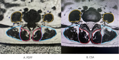 |
68 |
Feasibility study of paraspinal muscle fat infiltration assessed
by lumbar disc degeneration grade
Jiujun Lan1,
Wei Wang1,
Xiaocheng Wei2,
and Junrong Chen1
1Sichuan Province Orthopedic Hospital, Chengdu, China, 2GE Healthcare China, Beijing, China Keywords: Muscle, Fat, Quantitative imaging, Lumbar disc herniation Previous study found that low back pain (LBP) and Modic changes were closely associated with fatty infiltration and cross-sectional area (CSA) of the paraspinal muscles. However, conflicting evidences were reported. This study investigated the association between intervertebral discs degeneration and lumbar paraspinal muscles CSA and fat infiltration, and explored the relationship between Modic change and intervertebral discs degeneration. Our results found that lumbar disc’s Pfirrmann grading was weakly correlated with CSA of multifidus, fat fraction of multifidus and erector spinae, and the incidence rate of Modic changes increased in higher Pfirrmann grading. |
|
2596.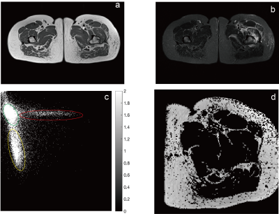 |
69 |
Decipher the simultaneous progress of fat infiltration and
muscle atrophy with fat-clustering analysis of MRI
Xin Mu1,
Huahui Xu2,
Jialin Wang3,
Chang Ni1,
Meng Tian4,
Xiaoli Gu2,
and Jeff L Zhang1
1Vascular and Physiologic Imaging Research (VPIR) Lab, School of Biomedical Engineering, ShanghaiTech University, Shanghai, China, 2Department of Radiology, Shanghai Guanghua Hospital of Integrative Medicine, Shanghai, China, 3Central Research Institute, UIH Group, Central Research Institute, UIH Group, Shanghai, China, 4School of Biomedical Engineering, Shanghai Jiao Tong University, Shanghai, China Keywords: Osteoarthritis, Aging Fat content and clustering are linked to muscle function in various disorders and are of therapeutic importance because they reflect the physical activity. Thus, we studied the fat content and clustering in three groups: hip-joint replacement surgery (HJRS) patients, ankylosing spondylitis (AS) patients who received medicine, and healthy controls. Although the intramuscular fat of unaffected side remained same in HJRS patients, Moran's index declined. Meanwhile, the Moran index of HJRS patients' affected side decreased significantly when fat content decreased. Besides, the drug treatment for AS patients resulted in a decrease in both fat content and Moran's index. |
|
2597.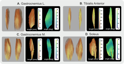 |
70 |
Feasibility of muscle template creation using tensor based
registration
Martijn Froeling1
1Radiology, UMC Utrecht, Utrecht, Netherlands Keywords: Muscle, Diffusion Tensor Imaging In this study, the aim is to show the feasibility of generating a per-muscle architecture template using a combination of muscle shape and muscle tensor registration. Additionally, the use of a generalized muscle template to identify between-subject differences will be explored. The tensor-based registration does allow for better alignment of internal muscle structures which is not possible using registration based on distance maps alone. |
|
2598.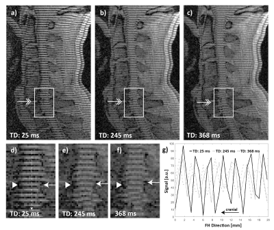 |
71 |
Magnetic-Resonance Tag Imaging of Cervical Spinal-Cord Motion in
Patients with Segmental Stenosis
Constantin von Deuster1,2,
Shila Pazahr3,
Nikolai Pfender4,
Armin Curt4,
Reto Sutter3,
and Daniel Nanz2
1Advanced Clinical Imaging Technology, Siemens Healthineers International AG, Zurich, Switzerland, 2Swiss Center of Musculoskeletal Imaging, Balgrist Campus AG, Zurich, Switzerland, 3Radiology, Balgrist University Hospital, Zurich, Switzerland, 4Spinal Cord Injury Center, Balgrist University Hospital, Zurich, Switzerland Keywords: MSK, Skeletal, Spine, Tagging The reference standard for spinal-cord motion measurement is phase-contrast (PC) imaging. This approach, however, requires a dedicated phase correction during post processing, which renders clinical application very challenging. The objective of this work was to employ MR-tag imaging to directly visualize cervical spinal-cord motion in patients with spinal-canal stenosis and compare results with PC imaging. Among all patients, a good correlation between tag imaging and matched PC measurements was found (R2 = 0.84). Hence, MR-tag imaging may well complement standard static imaging within clinical routine of patients with spinal-canal stenosis, reflecting dynamic spinal cord distress. |
|
2599.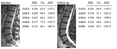 |
72 |
Quantitative assessment of longitudinal changes in
intervertebral discs
L. Tugan Muftuler1,
Brendan Bych1,
Jeffrey A. King1,
and Jordan A. Gliedt1
1Medical College of Wisconsin, Milwaukee, WI, United States Keywords: Skeletal, Degenerative, intervertebral disc Chronic low back pain is usually associated with degeneration of the intervertebral discs, although the disc itself may not be the source of pain. Unfortunately, there are no objective measures of the disc degeneration process. The gold standard relies on visual assessment of MRI scans, which may not capture complex physiological changes in degenerating discs. We used quantitative MRI metrics to study longitudinal changes disc degeneration over a period of 2 years. Although most subjects did not show visible changes in discs, there were significant losses in disc height and some discs also showed a reduction in ADC and T1ρ. |
|
2600.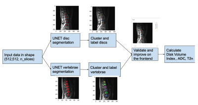 |
73 |
Automated quantification of intervertebral disc volume loss, ADC
and normalized T2 using convolutional neural networks
L. Tugan Muftuler1,
Tomas Rokos2,
Srivishnu Appalaraju2,
Tyler Tran2,
Joshua Goldshteyn2,
Garin Jankowski2,
and John Bukowy2
1Medical College of Wisconsin, Milwaukee, WI, United States, 2Milwaukee School of Engineering University, Milwaukee, WI, United States Keywords: MSK, Quantitative Imaging, spine, lumbar, intervertebral disc degeneration Intervertebral disc degeneration is the leading cause of chronic low back pain (CLBP). However, there are no objective measures of the disc degeneration process. The current gold standard for grading disc degeneration relies on visual assessment of discs using MRI, which may not adequately capture complex physiological changes in degenerating discs. We developed a deep convolutional neural network to automatically segment vertebral structures and calculate quantitative MRI metrics. Results show significant changes in ADC and normalized T2 values with disc volume loss. |
|
2601.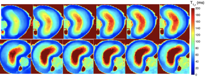 |
74 |
T1rho Dispersion in Proteoglycan-depleted Swine Spine Disc
Mitchell J. Christiansen1,2,
Mark C. Preul3,
Steven Marsh3,
Jay D. Turner4,
Juan Uribe4,
Muhammad Nadeem2,
Elliott J. Mufson2,
Richard D. Dortch2,
John C. Gore5,
and Ping Wang2
1Creighton University School of Medicine, Phoenix, AZ, United States, 2Translational Neuroscience, Barrow Neurological Institute, Phoenix, AZ, United States, 3Neurosurgery, Barrow Neurological Institute, Phoenix, AZ, United States, 4Sonntag Spine Center, Barrow Neurological Institute, Phoenix, AZ, United States, 5Radiology and Radiological Sciences, Vanderbilt Institute of Imaging Science, Nashville, TN, United States Keywords: MSK, Degenerative, Intervertebral disc T1rho imaging has been used to probe relatively slow macromolecular processes, making it a practical tool to gain information about water spin dynamics and interactions with endogenous macromolecules. We measured the dispersion of T1rho with different spin-lock fields in the intervertebral disc of swine spine specimens before and after the treatment to induce proteoglycan loss, a sign of early disc degeneration. Analysis revealed elevated T1rho values in all proteoglycan-depleted discs at each spin-lock field, and that post-treatment T1rho changes increase at a higher spin-lock field. |
|
2602.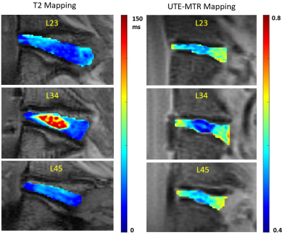 |
75 |
Assessment of Rabbit Intervertebral Disc Degeneration Using
Quantitative MR Imaging
Jiyo S Athertya1,
James Lo1,2,
Alicia Ji1,
Charles Ding1,
Arya Suprana1,2,
Kiersten Red3,
Yujia Ge3,
Madeline Brown 3,
Jiang Du1,2,4,
Eric Y Chang1,4,
Koichi Masuda 3,
and Yajun Ma1
1Radiology, UC San Diego, San Diego, CA, United States, 2Bioengineering, UC San Diego, San Diego, CA, United States, 3Orthopedic Surgery, UC San Diego, San Diego, CA, United States, 4Radiology, Veterans Affairs San Diego Healthcare System, San Diego, CA, United States Keywords: MSK, Joints, Intervertebral disc Intervertebral disc degeneration is a leading cause of disability. In this study, quantitative T2 and ultrashort echo time magnetization transfer ratio (UTE-MTR) measurement techniques are utilized to assess biochemical component changes for better understanding of the mechanism of disc degeneration using a rabbit spinal intervertebral disc degeneration model. Both T2 and UTE-MTR values of nucleus pulposus (NP) are significantly different between normal and degenerated discs, indicating water and/or proteoglycan content changes. |
|
2603.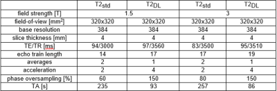 |
76 |
Deep Learning Reconstructed T2-weighted Dixon Imaging of the
Spine: Impact on Acquisition Time and Diagnostic Performance
Thierno D. Diallo1,
Zeynep Berkarda1,
Simon Wiedemann1,
Caroline Wilpert1,
Ralph Strecker2,
Gregor Koerzdorfer3,
Dominik Nickel3,
Fabian Bamberg1,
Matthias Benndorf1,
and Jakob Weiß1
1Radiology, University Medical Center Freiburg, Freiburg, Germany, 2EMEA Scientific Partnerships, Siemens Healthcare GmbH, Erlangen, Germany, 3MR Application Predevelopment, Siemens Healthcare GmbH, Erlangen, Germany Keywords: MSK, Machine Learning/Artificial Intelligence Magnetic resonance imaging of the spine is considered one of the most commonly performed examinations in clinical routine. The raising demand for high quality imaging of the spine creates the need for tailored examination protocols, especially with regard to increasingly limited scanner capacities. Deep Learning based imaging reconstruction has emerged as promising novel technique to accelerate MR imaging while maintaining image quality. This study analyzed a novel deep learning accelerated T2-weighted Dixon sequence of the spine in terms of diagnostic performance. The results suggest that the here presented sequence is feasible with a diagnostic performance comparable to standard imaging. |
|
2604.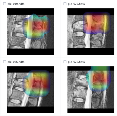 |
77 |
Deep learning system for automated detection of posterior
ligamentous complex injury in patients with thoracolumbar
fracture on magnetic resonance imaging
Sang Won Jo1,
Eun Kyung Khil1,
Seun Ah Lee1,
Jihe Lim1,
Jae Hyeok Lee2,
and Yu Sung Yoon3
1Radiology, Hallym University Dongtan Sacred Heart Hospital, Hwaseong, Korea, Republic of, 2Deepnoid, Seoul, Korea, Republic of, 3Radiology, Soonchunhyang University Bucheon Hospital, Bucheon, Korea, Republic of Keywords: MSK, Machine Learning/Artificial Intelligence, thoracolumbar fracture This study was aimed to develop a deep learning algorithm for automated detection and localization of posterior ligamentous complex (PLC) injury on magnetic resonance imaging and evaluate its diagnostic performance. The sensitivity, the specificity and AUC of inception-ResNet V2 architecture of second step were 88%, 82% and 0.928, for the internal test set and 86%, 74% and 0.916 for the external test set, respectively. A deep learning algorithm detected PLC injury in patients with thoracolumbar fracture with a high diagnostic performance which was validated using external data set. |
|
2605.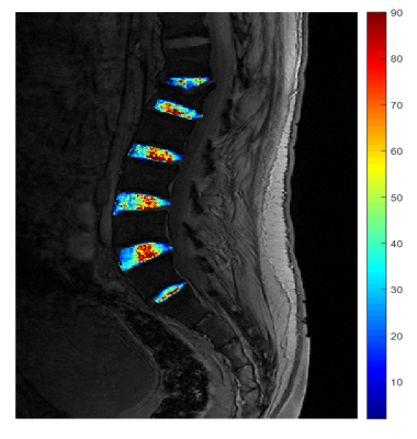 |
78 |
A pilot study of quantitative T2* mapping in intervertebral
lumbar disc patients with lower back pain
Adiraju Karthik1,
Apoorwa Devappa2,
Aakaar Kapoor3,
Dharmesh Singh4,
and Dileep Kumar4
1Department of Radiology, Sprint Diagnostics, Jubilee Hills, Hyderabad, India, 2Department of Radiology, Mahadevappa Rampure Medical College, Kalaburagi, India, 3Department of Radiology, City X-Rays Scan & Clinical Private Limited, New Delhi, India, 4Central Research Institute, Global Scientific Collaborations, United Imaging Healthcare, New Delhi, India Keywords: Skeletal, Spinal Cord, Intervertebral lumbar disc patients , T2* mapping The human lumbar spine is comprised of multiple tissue components that serve to offer structural stability as well as optimal nutrition. Traditional magnetic resonance (MR) imaging techniques have been beneficial in assessing intervertebral disc (IVD). Quantitative T2* mapping has recently been used to diagnose and characterize abnormalities associated with IVD, particularly water content variations. However, this approach remains in research settings, and its clinical application has not been thoroughly investigated. This study quantitatively evaluate the clinical value of T2* mapping of intervertebral lumbar disc patients with lower back pain using 3T magnetic resonance imaging (MRI). |
|
2606.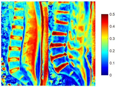 |
79 |
Association of Lumbar Trabecular Bone Ultrashort Echo Time
Magnetization Transfer Ratio (UTE-MTR) with Intervertebral Disc
Degeneration
Jian-Bang Zhang1,
Jin Liu1,
Jia-Xin Feng1,
Jianwei Liao1,
Wei Li1,
Xiao-Jun Chen1,
Long Qian2,
Ya-Jun Ma3,
and Shao-Lin Li1
1Department of Radiology, The Fifth Affiliated Hospital of Sun Yat-Sen University, Zhuhai, China, 2GE Healthcare, Beijing, China, 3Department of Radiology, University of California San Diego, San Diego, CA, United States Keywords: Bone, Skeletal Ultrashort echo time magnetization transfer (UTE-MT) technique can non-invasively quantify the macromolecular component changes. It has great potential to assess intervertebral disc degeneration (IVDD). In this study, we utilized the UTE-MT sequence to investigate the relationship between UTE-MT ratio (UTE-MTR) measurements of lumbar trabecular bone and IVDD. We found that the lumbar trabecular bone UTE-MTR negatively correlated with the IVDD grading. It demonstrates that the UTE-MTR of lumbar trabecular bone may serve as a useful biomarker to assess IVDD. |
|
2607.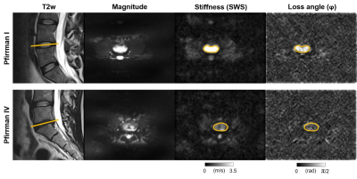 |
80 |
Lumbar spine disc degeneration assessed by in vivo 3T
multifrequency MR elastography
Rolf Reiter1,2,
David Kaufmann3,
Mehrgan Shahryari1,
Janosch Simon Jahn1,
Rebecca Strehle1,
Christian Bayerl1,
Bernd Hamm1,
Jürgen Braun4,
Ingolf Sack1,
and Patrick Asbach1
1Radiology, Charité - Universitätsmedizin Berlin, Berlin, Germany, 2BIH Biomedical Innovation Academy, BIH Charité Digital Clinician Scientist Program, Berlin Institute of Health at Charité – Universitätsmedizin Berlin, Berlin, Germany, 3Radiology, Universitätsklinikum Augsburg, Augsburg, Germany, 4Medical Informatics, Charité - Universitätsmedizin Berlin, Berlin, Germany Keywords: Elastography, MSK Despite the success in detecting intervertebral disc degeneration with high-resolution T2 sequences, conventional MRI is limited by large inter-reader variability as well as lacking stratification of clinical trials and their assessment of treatment responses. Several studies have shown the technical feasibility of MR elastography for the study of the intervertebral disc, but its clinical relevance remains unclear. This study shows an excellent diagnostic performance of high-resolution multifrequency MR elastography in the evaluation of lumbar spine intervertebral disc degeneration. |
|
 Back to Meeting Home
 Back to the Program-at-a-Glance
The International Society for Magnetic Resonance in Medicine is accredited by the Accreditation Council for Continuing Medical Education to provide continuing medical education for physicians.
 Back to Meeting Home
 Back to the Program-at-a-Glance
 View Presentation Video