Digital Poster
Imaging of Non-Cartilaginous MSK Tissues
ISMRM & ISMRT Annual Meeting & Exhibition • 03-08 June 2023 • Toronto, ON, Canada

Digital Poster
Imaging of Non-Cartilaginous MSK Tissues
| Computer # | |||
|---|---|---|---|
2764.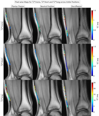 |
61 |
Sensitivity of Bi-Exponential UTE-T2* Mapping to Tendon Laxity
Ananya Goyal1,
Marco Barbieri1,
Valentina Mazzoli1,
and Feliks Kogan1
1Radiology, Stanford University, Stanford, CA, United States Keywords: Tendon/Ligament, Quantitative Imaging, T2*, Bi-exponential, UTE, CONES Tendon laxity can cause pain and increased injury risk. Quantitative MRI using Ultrashort echo time (UTE) sequences can quantify both short and long T2* water components in tendons. This study evaluates the sensitivity of UTE-T2* mapping with a bi-exponential fit model to tendon laxity, in the Achilles tendon under tension and relaxation. T2*long values seemed to increase as tendon load increased, while T2*short values were the opposite. These changes may be caused by water movement due to loading and changes in fiber orientation due to tendon slackening. |
|
2765.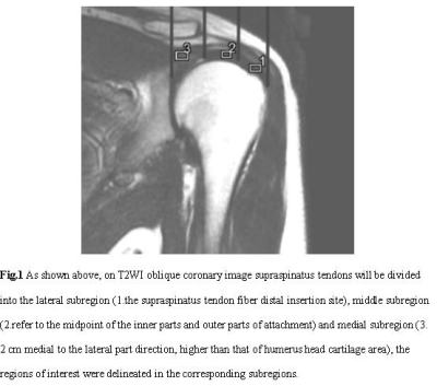 |
62 |
Synthetic MRI in the assessment of the classification of rotator
cuff injury
Ni Yabo1,
Wang zhijun1,
and Tian Zhaorong2
1Ningxia Medical University General Hospital Radiology department, Yinchuan, China, 2Ningxia Medical University General Hospital Radiology department, Yinchuan, China, Yinchuan, China Keywords: Tendon/Ligament, Quantitative Imaging In this study,to investigate the diagnostic value of quantitative parameters of synthetic magnetic resonance image in the grade of rotator cuff injury , we found that the T2 values had high diagnostic efficacy in grading the degree of rotator cuff injury, especially the T2 values of the lateral subregion and the middle subregion had high diagnostic efficacy in differentiating complete tears from partial tears, tendinopathy and normal tendons. |
|
2766.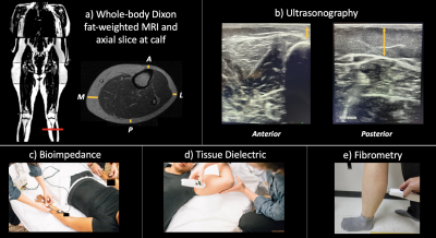 |
63 |
MRI and Ultrasound of lower extremity subcutaneous adipose
tissue: potential translational screening tools for lipedema
Yinan Zheng1,
John A. Beasley2,
Vanessa Crain2,
Shannon L. Taylor3,
Michael D. Pridmore2,
Maria E. Garza2,4,
Sheau-Chiann Chen5,
Aaron W. Aday6,
Joshua A. Beckman6,
Paula M.C. Donahue7,8,
Manus J. Donahue4,9,
and Rachelle L. Crescenzi2,3
1Vanderbilt University School of Medicine, Nashville, TN, United States, 2Radiology and Radiological Sciences, Vanderbilt University Medical Center, Nashville, TN, United States, 3Biomedical Engineering, Vanderbilt University, Nashville, TN, United States, 4Department of Neurology, Vanderbilt University Medical Center, Nashville, TN, United States, 5Department of Biostatistics, Vanderbilt University Medical Center, Nashville, TN, United States, 6Vanderbilt Translational and Clinical Cardiovascular Research Center, Cardiovascular Medicine Division, Vanderbilt University Medical Center, Nashville, TN, United States, 7Department of Physical Medicine and Rehabilitation, Vanderbilt University Medical Center, Nashville, TN, United States, 8Dayani Center for Health and Wellness, Vanderbilt University Medical Center, Nashville, TN, United States, 9Department of Psychiatry, Vanderbilt University Medical Center, Nashville, TN, United States Keywords: MSK, Screening, Translational Studies Lipedema is an underdiagnosed connective tissue disorder characterized by disproportionate subcutaneous adipose tissue (SAT) accumulation in the lower extremities. Lipedema is frequently misdiagnosed as obesity and objective diagnostic tools are lacking for lipedema. Patients with lipedema (n=22) and BMI-matched controls (n=7) underwent lower extremity MRI and ultrasound evaluations of SAT as potential diagnostic modalities. MRI and ultrasound measurement of SAT thickness yielded good concordance between modalities (Pearson’s r=0.95, reproducibility coefficient=3.3 mm), and demonstrated similar moderate effect sizes (Cohen’s d>0.5) for discriminating lipedema from controls. MRI and ultrasound are promising screening modalities for lipedema. |
|
2767.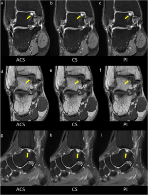 |
64 |
Evaluation of a deep learning-based acceleration technique for
ankle MRI protocol in clinical applications
Qiang Zhao1,
Jiajia Xu1,
Yu Xin Yang2,
Yuqing Zhao1,
Qizheng Wang1,
Yongming Dai3,
and Huishu Yuan1
1Peking University Third Hospital, Beijing, China, 2United Imaging Research Institute of Intelligent Imaging, Beijing, China, 3Central Research Institute, United Imaging Healthcare, Shanghai, China Keywords: Joints, Joints, Machine Learning/Artificial Intelligence A deep learning-based compressed sensing (ACS) technology was recently introduced for an integrative MR acceleration solution. This study assessed the effectiveness of using ACS to evaluate ankle injuries. The ACS acceleration technique allows faster imaging than conventional acceleration methods, providing adequate image quality and diagnosis performance. |
|
2768.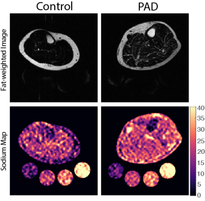 |
65 |
Sodium 23Na-MRI reveals elevated sodium content in
lower-extremity skin and adipose tissue in peripheral arterial
disease
Michael Pridmore1,2,
Emily Shardelow3,
Shannon L. Taylor1,2,4,
Adam Behroozian5,
Manus J. Donahue6,7,
Alex Sullivan3,
Joshua A. Beckman8,
and Rachelle Crescenzi1,2,4
1Institute of Imaging Science, Vanderbilt University Medical Center, Nashville, TN, United States, 2Radiology & Radiological Sciences, Vanderbilt University Medical Center, Nashville, TN, United States, 3Cardiovascular Medicine, Vanderbilt University Medical Center, Nashville, TN, United States, 4Biomedical Engineering, Vanderbilt University, Nashville, TN, United States, 5Cardiovascular Medicine, Scripps Memorial Hospital La Jolla, San Diego, CA, United States, 6Neurology, Vanderbilt University Medical Center, Nashville, TN, United States, 7Psychiatry and Behavioral Sciences, Vanderbilt University Medical Center, Nashville, TN, United States, 8Internal Medicine, University of Texas Southwestern Medical Center, Dallas, TX, United States Keywords: MSK, Atherosclerosis, Multi-Nuclear, Peripheral Disease Peripheral artery disease (PAD) is an atherosclerotic occlusive disease that reduces lower-limb perfusion, causes ambulatory dysfunction, and leg pain. Sodium imaging (23Na-MRI) may reveal areas of increased tissue sodium deposition related to the mechanisms of PAD dysfunction. We found that patients with PAD showed increased tissue sodium content in skin, subcutaneous adipose tissue (SAT), and inter-muscular adipose tissue (IMAT) compared to controls. Participants with PAD had lower SAT volume and fat content, arguing against a positive relationship between fat as the mediator of increased tissue sodium. 23Na-MRI may inform PAD mechanisms and is being investigated in response to PAD therapies. |
|
| 2769. | WITHDRAWN | ||
| 2770. | WITHDRAWN | ||
2771.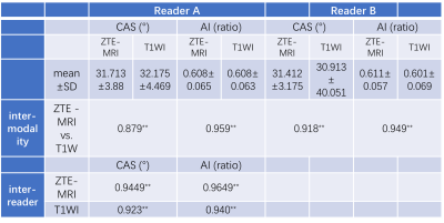 |
66 |
Clinical feasibility of synthetic T1-weighted images in
evaluation of rotator cuff injury using ZTE-MRI as reference
Jingyu Jiang1,
Weiyin Vivian Liu2,
Wen Chen3,
Ling Sang3,
Huizheng Wang3,
Xingyao Yu1,
and Lin Xu3
1Biomedical Engineering College, Hubei University of Medicine, Shiyan Hubei, China, 2GE Healthcare,, Beijing, China, 3Taihe Hospital, Hubei University of Medicine,, Shiyan Hubei, China Keywords: Tendon/Ligament, Aging, synthetic Magnetic resonance imaging is the gold standard for evaluation of rotator cuff injury in non-invasive examination. And 90% occur in supraspinatus tendons. In this study, synthetic MRI (SyMRI) generated both structural and functional images to qualitatively and quantitatively evaluate supraspinatus tendon injury using zero-echo magnetic resonance imaging (ZTE-MRI) as structural reference images. Despite superior quality and inter-reader agreement on critical shoulder angle measurements of ZTE-MRI to SyMRI-generated T1WI, SyMRI showed excellent intra-modality agreement of acromion index and CSA to ZTE-MRI and also provided quantitative information for better identifying injured sites at the early stage of rotator cuff injury. |
|
2772.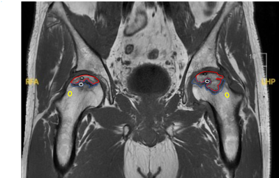 |
67 |
Alteration in microcirculation with osteonecrosis of the femoral
head: Study on Dynamic Contrast-enhanced MRI
Huan Da Wei1,
Yuan Zhao Feng1,
Dou Wei Qiang2,
Wen Qing Qing2,
Liu Shao Wei1,
Lu Chao1,
Zhang Chao1,
Xia Tian Wei1,
and Shen Ji Rong1
1Affiliated Hospital of Nanjing University of Chinese Medicine, Jiangsu Nanjing, China, 2MR Research China, GE Healthcare, Beijing, China Keywords: Joints, DSC & DCE Perfusion Osteonecrosis of femoral head (ONFH) occurs due to a defect in the blood supply to the femoral head. In this study, Contrast-Enhanced Magnetic Resonance Imaging (DCE-MRI) was applied to ONFH patients in different stages to explore the perfusion changes in the progression of the severity of ONFH. It was found that perfusion in the necrotic area was persistently reduced, while in the repair reaction areas, perfusion was progressively enhanced. DCE-MRI can sensitively detect the perfusion changes of ONFH and potentially could predict ONFH development. |
|
2773.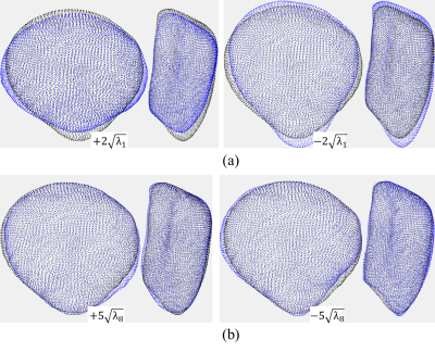 |
68 |
Longitudinal Changes in Patella Bone Shape in Subjects after ACL
Reconstruction
James R Peters1,
Sibaji Gaj1,
Nancy Obuchowski1,
Carl Winalski1,
Naveen Subhas1,
Valentina Pedoia2,
Sharmila Majumdar2,
Hollis Potter3,
Matthew F Koff3,
Kimberly K Amrami4,
Cale Jacobs5,
and Xiaojuan Li1
1Cleveland Clinic, Cleveland, OH, United States, 2University of California, San Francisco, San Francisco, CA, United States, 3Hospital for Special Surgery, New York, NY, United States, 4Mayo Clinic, Rochester, MN, United States, 5University of Kentucky, Lexington, KY, United States Keywords: Osteoarthritis, Joints Acute knee joint injury often leads to the development of post-traumatic osteoarthritis (PTOA); however, the factors which may indicate PTOA progression are not well understood. Bone shape changes have been suggested as potential predictors for OA development. This study investigates longitudinal changes in patella bone shape in subjects who underwent ACL reconstruction. Subject’s patellae were segmented using machine learning methods and a statistical shape model was used to quantify patella bone shape. Data analysis showed longitudinal changes in patella shape between contralateral and ipsilateral sides, between subjects with allografts and autografts, and associations with age, sex, and BMI. |
|
2774.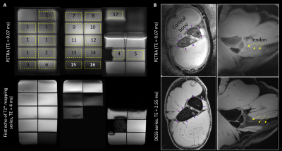 |
69 |
Initial evaluation of PETRA UTE imaging at 7T in a phantom and
musculoskeletal tissue specimen
Carly Lockard1,2,
Bruce Damon1,2,
and Hacene Serrai 1,2
1Stephens Family Clinical Research Institute, Carle Clinical Imaging Research Program, Carle Health, Urbana, IL, United States, 2Carle Illinois Advanced Imaging Center, Urbana, IL, United States Keywords: MSK, High-Field MRI, zero echo time; ultrashort echo time; UTE; ZTE Ultrashort echo-time MRI allow signal acquisition from tissues with ultrashort (<1 ms) T2*. We evaluated repeatability, SNR, acquisition time, and potential for T2* mapping using a modified PETRA sequence at 7T. Signal values were repeatable, and subtraction imaging was both feasible and consistent with those previously reported. T2* mapping had lower repeatability and may require improved modeling of TE-dependent k-space filling strategy effects. Short- and long-TE PETRA imaging was feasible at 7T in a phantom and ex vivo specimen. These initial results will serve as guidance for further optimization/potential applications. |
|
2775.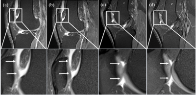 |
70 |
A Comparison of High-Resolution Simultaneous Multi-Slice
Accelerated Turbo Spin-Echo Knee Imaging with Routine Turbo
Spin-Echo Imaging
Yanping Xue1,
Li Wang1,
Hua Gu1,
Jiyang Zhang1,
Chen Zhang2,
Xiuqin Jia1,
and Qi Yang1
1Radiology, Beijing Chao-Yang Hospital, Capital Medical University, Beijing, China, 2MR Scientific Marketing, Siemens Healthineers, Beijing, China Keywords: MSK, Joints MRI has been widely used in the early detection of osteoarthritis (OA), especially in knee, as an important technique. However, the long acquisition time of MRI is a barrier in improving the quality of imaging and the comfort of patients, especially for pediatric and disabled people. Many imaging acceleration factors have been implied in order to improve the condition. This study focuses on the clinical application of simultaneous multi-slice (SMS) imaging, a newly developed acceleration technique using multi-band RF pulses, which can simultaneously excite, acquire, and reconstruct multiple slices and readout with two-dimensional images. |
|
2776.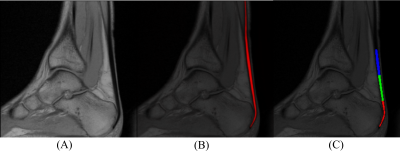 |
71 |
Regional Achilles Tendon Segmentation for Quantitative Magnetic
Resonance Imaging at 1.5 Tesla
Dominik Vilimek1,
Radana Kahankova1,
Radek Martinek1,
Martin Kryl1,
Veronika Janacova2,
Milos Golian3,4,
Jaroslav Uchytil3,
Pavla Hanzlikova5,6,
Pavol Szomolanyi2,7,
Siegfried Trattnig2,8,9,10,
and Vladimir Juras2
1Department of Cybernetics and Biomedical Engineering, VSB Technical University of Ostrava, Ostrava, Czech Republic, 2Department of Biomedical Imaging and Image-guided Therapy, High Field MR Centre, Medical University of Vienna, Vienna, Austria, 3Department of Human Movement Studies, Human Motion Diagnostic Center, University of Ostrava, Ostrava, Czech Republic, 4Department of Radiology, Vitkovice Hospital, Ostrava, Czech Republic, 5Department of Imaging Methods, Faculty of Medicine, University of Ostrava, Ostrava, Czech Republic, 6Department of Radiology, University Hospital Ostrava, Ostrava, Czech Republic, 7Institute of Measurement Science, Slovak Academy of Sciences, Bratislava, Slovakia, 8CD Laboratory fo MR Imaging Biomarkers (BIOMAK), Vienna, Austria, 9Austrian Cluster for Tissue Regeneration, Ludwig Boltzmann Institute for Experimental and Clinical Traumatology, Vienna, Austria, 10Institute for Clinical Molecular MRI in the Musculoskeletal System, Karl Landsteiner Society, Vienna, Austria Keywords: Tendon/Ligament, Machine Learning/Artificial Intelligence Changes in the Achilles tendon composition are associated with an increased risk of tendinopathy which is common in middle-aged overweight patients and is one of the most common sports injuries. However, measuring and quantifying such properties is a challenging task. The purpose of this paper is to introduce an end-to-end pipeline for segmenting Achilles tendon using deep convolutional neural networks and automatic segmentation into three segments. Our model shows promising results outperforming state-of-the-art approaches (Dice 90.6% and Jaccard 84.0%). This is one of the key steps for short and long T2* value analysis from 1.5T data. |
|
2777.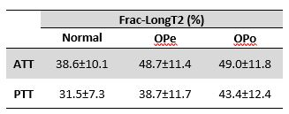 |
72 |
Fast dual-echo ultrashort echo time (UTE) MRI to estimate long
T2 fraction in tendons and detect age-and osteoporosis-related
differences
Saeed Jerban1,2,3,
Amir Masoud Afsahi 1,
Sophia Dwek1,
Jiyo Athertya1,
Bhavsimran Malhi1,
Dina Moazamian1,
Yajun Ma1,2,
Sam Sedaghat1,
Hyungseok Jang1,2,
Gina Woods4,
Christine B Chung1,2,
Jiang Du1,2,
and Eric Y Chang1,2
1Department of Radiology, University of California, San Diego, La Jolla, CA, USA, San Diego, CA, United States, 2Radiology Service, Veterans Affairs San Diego Healthcare System, San Diego, La Jolla, CA, USA, San Diego, CA, United States, 3Department of Orthopedic Surgery, University of California, San Diego, La Jolla, CA, USA, San Diego, CA, United States, 4Department of Medicine, University of California, San Diego, La Jolla, CA, USA, San Diego, CA, United States Keywords: Tendon/Ligament, Tendon/Ligament Tendon and bone comprise a special interacting unit. Bone loss in osteoporosis (OPo) may associate with a reduction in tendon quality that needs to be investigated. We investigated the tendon quality differences between OPo patients, osteopenia (OPe) patients, and healthy volunteers with normal bone (Normal) using the Frac-LongT2 index, a rapid measure performed by dual-echo ultrashort echo time (UTE) MRI. The estimated Frac-LongT2 in the anterior and posterior tibialis tendon (ATT and PTT) were significantly higher in the OPo group compared with the Normal group. This study highlights the potential of this rapid UTE-based technique for in vivo tendon assessment. |
|
2778.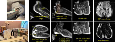 |
73 |
Quantification of soft tissue of the buttocks in a seated
position
Jill M Slade1,
Nkhensani Mogale2,
Daniel Schanz1,
Viktor Ilyasov1,
Justin Scott3,
Albert van Schoor2,
and Tamara R Bush3
1Radiology, Michigan State University, East Lansing, MI, United States, 2University of Pretoria, Pretoria, South Africa, 3Mechanical Engineering, Michigan State Univerisity, East Lansing, MI, United States Keywords: MSK, Muscle MRI is well suited to quantify soft tissue but challenges remain in using MRI to address soft tissue in a seated position and for studying how tissue responds to loading. IDEAL and T1 weighted images were used to quantify apparent fat fraction and soft tissue thicknesses of the buttocks in a side lying position with the tissue unloaded and loaded. Tissue compression during loading predominantly reflected changes in the muscle and tendon. Intramuscular fat was not related to the change in muscle/tendon. These methods may be useful to study soft tissue in populations at risk for pressure ulcer development. |
|
 |
2779.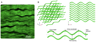 |
74 |
Diffusion Tensor Imaging of tendon and ligament: Influence of
crimping behavior and microstructural variations.
Noel Naughton1,
Amir Ostadi Moghaddam1,
Mariana Kersh1,2,3,
Samni Koyejo4,
Shreyan Majumdar1,
Amy Wagoner Johnson1,2,3,5,
and Bruce Damon1,6
1Beckman Institute for Advanced Science and Technology, University of Illinois at Urbana-Champaign, Urbana, IL, United States, 2Department of Mechanical Science and Engineering, University of Illinois at Urbana-Champaign, Urbana, IL, United States, 3Biomedical and Translational Sciences, Carle Illinois College of Medicine, University of Illinois at Urbana-Champaign, Urbana, IL, United States, 4Computer Science Department, Stanford University, Palo Alto, CA, United States, 5Carle R. Woese Institute for Genomic Biology, University of Illinois at Urbana-Champaign, Urbana, IL, United States, 6Carle Clinical Imaging Research Program, Stephens Family Clinical Research Institute, Carle Health, Urbana, IL, United States Keywords: Tendon/Ligament, Diffusion Tensor Imaging To guide development and optimization of dMRI protocols, here we present a numerical simulation framework for analyzing how changes in collagen microstructure influence the measured diffusion tensor. We experimentally characterize a porcine digital flexor tendon using second harmonic generation (SHG) microscopy to identify characteristic geometric parameters and use these parameter ranges to perform a sensitivity analysis of what microstructural features most influence diffusion tensor imaging (DTI) metrics. We conclude by exploring how geometric changes in collagen crimping due to tissue strain influence DTI metrics. |
2780.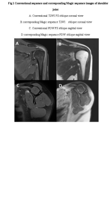 |
75 |
Feasibility study of synthetic MRI in shoulder imaging
Ni Yabo1,
Tian Zhaorong1,
and Wang zhijun1
1Ningxia Medical University General Hospital Radiology department, Yinchuan, China Keywords: Multimodal, Tendon/Ligament In this work,we found that the image quality of shoulder synthetic MRI sequence is not comparable to that of conventional image quality, and the scanning time is long, but once scanning can generate multiple sets of quantitative images, T2 mapping of synthetic sequence can provide a new quantitative method for the diagnosis and differential diagnosis of supraspinatus tendon related diseases. |
|
2781. |
76 |
A preliminary analysis for vertebral marrow fat and disc
herniation or annular tear by simultaneous multi-relaxation-time
Imaging (TXI) method
Limei Han1,2,
Jie Yang1,
Zhiqiang Ming1,
Hao Feng1,
Yantao Huang1,
Hengping Wu1,
Chao Yuan1,
Huan Guo1,
Meining Chen3,
and Jianquan Zhong1
1Department of Radiology, Zigong First People's Hospital, Zigong, China, 2North Sichuan Medical College, Nanchong, China, 3MR Scientific Marketing, SIEMENS Healthineers Ltd., Shanghai, China Keywords: Screening, Degenerative, lumbar disc herniation, annular tear, bone marrow fat Recently T1 mapping, T2* mapping and bone marrow fat fraction (BMFF) has been separately applied to spine. But it hasn't studied the value of those mappings together with intervertebral discs (IVD) degeneration. In this study, we obtained three values (T1, T2* and BMFF value) by simultaneous multi-relaxation-time imaging technique, called TXI. We found that T1 values in abnormal (herniation or annular tear) disc were significantly lower than those in the normal. This study demonstrates that T1 mapping can be helpful in detecting IVD with herniation or annular tear. |
|
2782.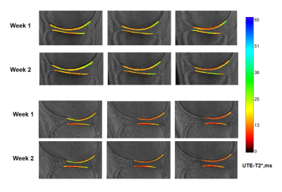 |
77 |
Repeatability of Cones UTE-T2* Mapping of Cartilage
Ashley A. Williams1,
Gordhan B. Mahtani1,
Jade R He1,
Adam L. C. Wadsworth1,
and Constance R. Chu1
1Orthopaedic Surgery, Stanford University, Stanford, CA, United States Keywords: Cartilage, Relaxometry, Repeatability, UTE-T2* This work examines the combined acquisition and segmentation repeatability of Cones UTE-T2* measures in tread mark cartilage regions and in small 2-D cartilage and meniscus ROIs in the knees of uninjured participants. Both intra- and inter-day repeatability assessments showed excellent average CVs of less than 10% in all tread mark ROIs and in all but 2 small 2-D ROIs segmented from only a single slice. ICC estimates were good to excellent (0.79–0.98) in all tread mark ROIs and ranged from moderate to excellent (0.54-0.98) in small 2-D ROIs corresponding to absolute precision errors of less than 1ms in most cases. |
|
2783.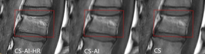 |
78 |
Rapid high-resolution musculoskeletal MR imaging with a super
resolution deep learning constrained Compressed SENSE
reconstruction
Zheng Sun1,
Shan Huang2,
Xiance Zhao2,
Weibo Chen2,
Queenie Chan3,
Yajing Zhang4,
Johannes M. Peeters5,
and Yuehua Li1
1Shanghai Sixth People's Hospital, Shanghai, China, 2Philips Healthcare, Shanghai, China, 3Philips Healthcare, Hong Kong, China, 4Philips Health Technology, Suzhou, China, 5Philips Healthcare, Best, Netherlands Keywords: Skeletal, Bone The integration of compressed-SENSE and artificial intelligence allows to acquire high-resolution images within relatively short scan times The purpose of this study is to compare the image quality of the musculoskeletal images reconstructed with Compressed -SENSE (CS), CS with integrated artificial intelligence (CS-AI) and CS-AI combined with superresolution(CS-AI-HR) . We found that the image quality was significantly improved by using CS-AI-HR compared to CS. This study provides supportive information for the application of CS-AI-HR in routine clinical practice. |
|
 Back to Meeting Home
 Back to the Program-at-a-Glance
The International Society for Magnetic Resonance in Medicine is accredited by the Accreditation Council for Continuing Medical Education to provide continuing medical education for physicians.
 Back to Meeting Home
 Back to the Program-at-a-Glance
 View Presentation Video