Digital Poster
New Frontiers in Brain Imaging I
ISMRM & ISMRT Annual Meeting & Exhibition • 03-08 June 2023 • Toronto, ON, Canada

Digital Poster
New Frontiers in Brain Imaging I
| Computer # | |||
|---|---|---|---|
3291.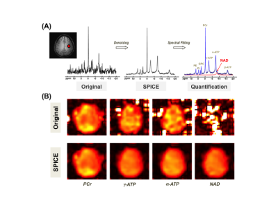 |
61 |
Feasibility of Mapping Intracellular NAD Content in Entire Human
Brain at 7T
Xiao-Hong Zhu1,
Rong Guo2,3,
Hannes M Wiesner1,
Yudu Li3,4,
Yibo Zhao3,5,
Wen Jin3,5,
Zhi-Pei Liang3,5,
and Wei Chen1
1Center for Magnetic Resonance Research, Department of Radiology, University of Minnesota, Minneapolis, MN, United States, 2Siemens Medical Solutions USA, Inc., Urbana, IL, United States, 3Beckman Institute for Advanced Science and Technology, University of Illinois at Urbana-Champaign, Urbana, IL, United States, 4National Center for Supercomputing Applications, University of Illinois at Urbana-Champaign, Urbana, IL, United States, 5Department of Electrical and Computer Engineering, University of Illinois at Urbana-Champaign, Urbana, IL, United States Keywords: Quantitative Imaging, Metabolism, Human Brain NAD Imaging Nicotinamide adenine dinucleotide (NAD) is an important molecule critical for cellular metabolism and signaling. The cerebral NAD concentration is only at submillimolar level, and thus, difficult to measure and imaging in vivo. In this study, we utilize 3D 31P MRSI-based NAD assay in combination with a SPICE-based signal processing method to evaluate the feasibility for non-invasively mapping the NAD contents in healthy human brains at 7T. The results suggest that by taking advantage of ultrahigh field scanner and advanced post-processing, it is possible to obtain whole-brain NAD maps with NAD measurements consistent with the literature reported values. |
|
3292.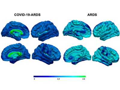 |
62 |
Spectrum of Brain Injury in COVID-19 Acute Respiratory Distress
Syndrome: A Systematic Review of Neuroimaging Findings
Rachel Wagner1,2,
Michael T Jurkiewicz2,3,
Jennifer Chen4,5,
Angela Jerath6,7,
Marat Slessarev2,5,8,
and Udunna Anazodo2,9
1Neuroscience, Western University, London, ON, Canada, 2Lawson Health Research Institute, London, ON, Canada, 3Medical Imaging, London Health Sciences Centre, London, ON, Canada, 4Critical Care Medicine, Western University, London, ON, Canada, 5Western Institute for Neuroscience, Western University, London, ON, Canada, 6Sunnybrook Health Sciences Centre, Toronto, ON, Canada, 7Evaluative Clinical Sciences, Schulich Heart Program, Sunnybrook Research Institute, Toronto, ON, Canada, 8Critical Care Medicine, Schulich School of Medicine & Dentistry, Western University, London, ON, Canada, 9Department of Neurology and Neurosurgery, McGill University, Montreal, QC, Canada Keywords: Infectious disease, COVID-19, Neuroscience While most COVID-19 patients present with mild symptoms, many of those hospitalized with severe infection develop acute respiratory distress syndrome (ARDS)1 and frequently report central nervous system dysfunction2. While previous research has characterized clinical symptomatology, investigation regarding brain changes remains limited. To address this gap, this review summarized brain injuries from 372 COVID-19-ARDS and 370 ARDS patients and pooled findings. COVID-19-ARDS patients presented an unusual pattern of injury preferentially targeting white matter, suggesting impact to brain connectivity. The long-term consequences of white matter injury in COVID-19-ARDS should be taken into account in devising policies for managing patient health. |
|
3293.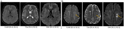 |
63 |
Feasibility of routine brain imaging at 0.55T: initial
experience in a clinical workflow
Anna Lavrova1,
Jacob Richardson1,
Ryo Kurokawa1,
Mariko Kurokawa1,
Pedro Itriago-Leon2,
Vikas Gulani1,
Hero Hussain1,
Katherine Wright1,
Toshio Moritani1,
and Nicole Seiberlich1
1Department of Radiology, University of Michigan, Ann Arbor, MI, United States, 2Siemens Medical Solutions USA Inc., Houston, TX, United States Keywords: Low-Field MRI, Brain The purpose of this study is to assess the quality of brain imaging studies performed on an FDA-approved commercial 0.55T MRI system, and to provide information about the feasibility of using this scanner in a clinical workflow. The image quality of 1356 image series (378 in healthy subjects, 978 in patients) was independently rated by two neuroradiologists. While images from T1w SPACE, DWI/ADC, FLAIR, SWI, and T2w TSE sequences received acceptable image quality scores, contrast-enhanced T1w TSE sequence received lower, although acceptable, average scores. Our results suggest that neurological MRI studies for some indications can be performed at 0.55T. |
|
3294.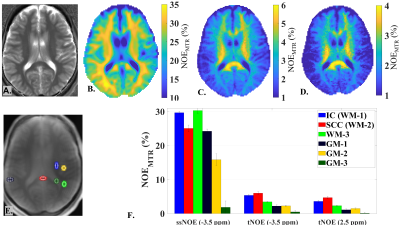 |
64 |
Feasibility and Improved Specificity of Brain Lipid Imaging at 7
Tesla using Transient Nuclear Overhauser Effect (tNOE)
Dushyant Kumar1,
Blake Benyard1,
Narayan Datt Soni1,
Anshuman Swain1,
Neil Wilson1,
and Ravinder Reddy1
1Radiology, University of Pennsylvania, Philadelphia, PA, United States Keywords: Data Acquisition, Brain, Lipid imaging Brain lipid imaging using steady-state nuclear Overhauser effect (ssNOE), though a traditionally popular approach, suffers from multiple confounding non-NOE specific sources, including direct saturation, magnetization transfer (MT), and relevant chemical exchange species. B0, B1+- dependent data are also needed to correct for the effect of (B0, B1+)- inhomogeneities¸ leading to other issues such as patient tolerability due to substantially increased scan time. Here, we demonstrate the feasibility of brain lipid mapping using an easily implementable transient NOE (tNOE) approach for the first time. Advantages include improved specificity, faster scan time and robust quantification with minimal confounding contributions. |
|
3295. |
65 |
Whole-Brain Sub-Millimeter Resolution fMRI using 3D EPI
Accelerated with Temporal Random Walk
Suhyung Park1,
Alexander Beckett2,3,
Suvi Hakkinen3,
Samantha Ma4,
and David Feinberg2,3
1Department of Computer Engineering, Chonnam National University, Gwangju, Korea, Republic of, 2Advanced MRI Technologies, Sebastopol, CA, United States, 3University of California, Berkeley, Berkeley, CA, United States, 4Siemens Medical Solutions USA, Inc, Berkeley, CA, United States Keywords: Data Acquisition, Brain There are significant benefits to segmented 3D EPI fMRI acquisitions, which acquires high spatial and temporal resolution across the whole brain to better understand brain neuronal activity. This can be achieved through accelerated 3D EPI imaging, which provides rapid whole-brain coverage at the cost of SNR efficiency resulting from frame-by-frame reconstruction. Here, we utilize temporal information for whole-brain coverage on 8-fold accelerated 7T fMRI acquisition without altering the subsequent fMRI results. |
|
3296.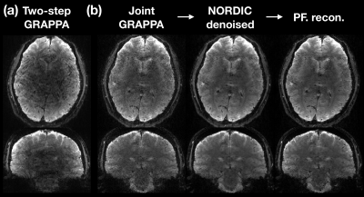 |
66 |
Feasibility of whole-brain functional MRI with 1-mm isotropic
resolution at 3T
Wei-Tang Chang1,2,3,
Stephanie Langella4,
Min Sung Seo5,
Weili Lin2,
and Kelly Giovanello5
1Biomedical Engineering, UNC at Chapel Hill, Chapel Hill, NC, United States, 2Biomedical Research Imaging Center, UNC at Chapel Hill, Chapel Hill, NC, United States, 3Radiology, UNC at Chapel Hill, Chapel Hill, NC, United States, 4Psychiatry, Harvard Medical School, Boston, MA, United States, 5Psychology & Neuroscience, UNC at Chapel Hill, Chapel Hill, NC, United States Keywords: Data Acquisition, Neuro fMRI with ultrahigh resolution has typically been acquired at 7T in order to overcome the inherently low SNR at 3T. However, 7T scanners are inaccessible to the majority of investigators. In this study, we developed new acquisition and reconstruction approaches and employed the denoising method NORDIC to improve the resolution of 3T fMRI to 1 mm isotropic with whole-brain coverage and volumetric scan time of 2 seconds. Our results showed improved tSNR and low spatial blurring. The detection capability of resting-state networks was comparable to that of 7T. BOLD activations in cortical and subcortical regions were observed in task-based fMRI. |
|
3297.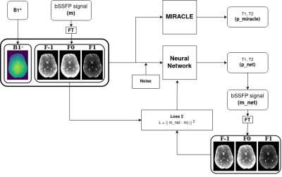 |
67 |
Ultrafast submillimeter model-based NN quantification of
whole-brain T1 and T2 using phase-cycled bSSFP
Florian Birk1,2,
Klaus Scheffler1,2,
and Rahel Heule1,2,3
1Department of High-Field Magnetic Resonance, Max Planck Institute for Biological Cybernetics, Tübingen, Germany, 2Department of Biomedical Magnetic Resonance, University of Tübingen, Tübingen, Germany, 3Center for MR Research, University Children's Hospital, Zurich, Switzerland Keywords: Machine Learning/Artificial Intelligence, Machine Learning/Artificial Intelligence, Model-Based Learning The bSSFP sequence is intrinsically sensitive to T1 and T2, motion robust, and allows highly efficient data acquisition. Slow convergence in qMRI parameter fitting can potentially be mitigated by machine learning, which benefits greatly from the availability of accurate ground truth data. This work presents an unsupervised model-based NN that incorporates the analytical bSSFP signal equation into the training loop, thus avoiding the need for ground truth relaxometry measurements and enabling instantaneous multi-parametric submillimeter whole-brain mapping of T1 and T2. NN performance was compared to MIRACLE quantitatively for in silico noise corrupted data and qualitatively for in vivo data. |
|
3298.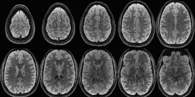 |
68 |
Increasing sampling efficiency using Yarnball sampling in 3D
fast spin echo with long echo trains – TurboYarn
Jeff Snyder1,
Alan H. Wilman1,
and Rob Stobbe1
1Biomedical Engineering, University of Alberta, Edmonton, AB, Canada Keywords: Pulse Sequence Design, Brain An efficient k-space sampling strategy using 3D Yarnball was used in long echo train FSE (TurboYarn). As TurboYarn samples many lines worth of k-space in each readout, fully sampled isotropic 1 mm brain scans were acquired in 40 s. TurboYarn was compared to a time-equivalent, 4x under-sampled 3D FSE sequence in brain of three healthy subjects. Resulting images showed excellent GM/WM contrast for TurboYarn, with CNRs higher than 3D FSE. As each readout samples k-space centre, further work will investigate TurboYarn motion robustness, with additional SNR improvements via trajectory and flip angle train optimizations. |
|
3299.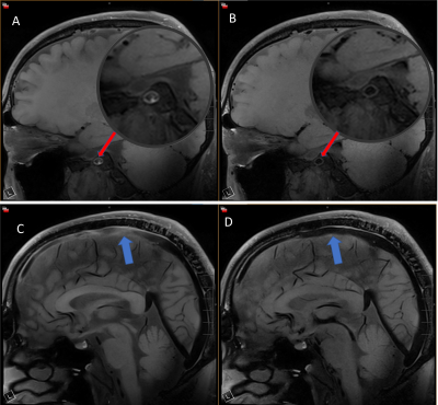 |
69 |
Optimization of T1-Weighted DANTE-SPACE for Intracranial Vessel
Wall Imaging at 7T
Xiangchuang Kong1,
Jun Ma2,
Erin Westerhold1,
Eric H. Middlebrooks1,
Shengzhen Tao1,
Chen Lin1,
and Xiangzhi Zhou1
1Department of Radiology, Mayo Clinic, Jacksonville, FL, United States, 2Siemens Medical Solutions USA, Inc., Jacksonville, FL, United States Keywords: Blood vessels, High-Field MRI, DANTE, SPACE, 7T MR vessel-wall-imaging (VWI) is used clinically for noninvasive characterization of vessel wall pathology. The DANTE preparation module has been combined with conventional 3D-T1W-SPACE to achieve better slow-flow-suppression. 7T could further improve VWI by its increased SNR and spatial-resolution. Here we sought to optimize DANTE preparation module for T1W-SPACE at 7T. Simulations were done to determine the optimal parameters for the DANTE module. Human scans showed that the optimized T1W- DANTE-SPACE at 7T efficiently suppressed slow-blood-flow in both intracranial arteries and veins, and in turn improved wall contrast compared with conventional T1W-SPACE. |
|
3300.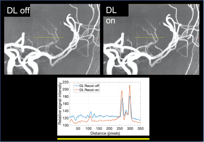 |
70 |
High Resolution Intracranial MR Angiography at 3T and 7T using a
Deep Learning based Image Reconstruction
Naoyuki Takei1,
Baolian Yang2,
Brian Burns3,
Yoichiro Ikushima1,
R Marc Lebel4,
Vince Magnotta5,
Fumiyasu Tsushima6,
Shingo Kakeda6,
Atsushi Nozaki1,
and Tetsuya Wakayama1
1GE Healthcare, Tokyo, Japan, 2GE Healthcare, Waukesha, WI, United States, 3GE Healthcare, Menlo Park, CA, United States, 4GE Healthcare, Calagary, AB, Canada, 5University of Iowa, Iowa City, IA, United States, 6Hirosaki University Graduate School of Medicine, Aomori, Japan Keywords: Blood vessels, Machine Learning/Artificial Intelligence, 7T MRI Ultra-high-field MRA shows the promise to improve visualize the microvasculature and become an important investigation tool for research. However, increasing the consistency in image quality and reliability of small vessel visualization is necessary to demonstrate useful MR applications in clinical practice. We developed a deep learning-based reconstruction method to provide reduced noise and enhanced spatial resolution. The technique was evaluated by applying it to 3DTOF MRA data at 3T and 7T, demonstrating improved small vessel depiction. |
|
3301.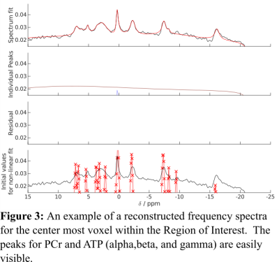 |
71 |
Fast 3D-P31-MRSI Using Custom Rosette Petal Trajectory at 3T
with 4x Accelerated Compressed Sensing
Nicholas Farley1,
Brian Bozymski1,
Ulrike Dydak1,
and Uzay Emir1
1Radiological Health Science, Purdue University, West Lafayette, IN, United States Keywords: Gray Matter, Brain To develop a reasonably fast P-31 3D MRSI sequence utilizing Rosette-Petal trajectories in K-space and compressed sensing reconstruction to acquire 1 mL voxel resolution maps in under 10 minutes with a magnetic field strength of only 3 Tesla |
|
3302.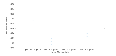 |
72 |
Laminar effective connectivity of the default mode network
assessed using 7T fMRI
Nguyen Phuoc Huynh1 and
Gopikrishna Deshpande1
1Electrical and Computer Engineering, Auburn University, Auburn, AL, United States Keywords: Data Analysis, High-Field MRI, Effective connectivity High-resolution fMRI presents opportunities in the study of fine-scale functional architecture, such as directional interactions between cortical layers. The Default Mode Network (DMN) is a major network in the brain associated with many social behaviors, however, little is known about the feed-forward and feedback information flow between different layers in regions of the DMN. We used high-resolution fMRI combined with effective connectivity to investigate the layer-to-layer interactions of regions in the DMN including medial prefrontal cortex, posterior cingulate cortex, and inferior parietal cortex. The results suggest that the middle layers of PCC are dominant in information flow within the DMN. |
|
3303.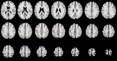 |
73 |
Disruptions in regional homogeneity and functional connectivity
relate to subacute ischemic stroke
Xin Li1,
Yue Qin1,
Yanqiang Qiao1,
Yifan Qian1,
Xiaoshi Li1,
and Lei Wang1
1Xi'an Daxing Hospital, Xi'an, China Keywords: Stroke, fMRI (resting state) The primary aim of the research was to compare the impact of subacute ischemic stroke on brain activity using resting state functional magnetic resonance imaging. In this study, we applied ReHo and FC to investigate the characteristics of brain functional activity in subacute ischemic stroke patients. Our results showed that the subacute ischemic stroke patients showed increased ReHo values in the left postcentral gyrus and significantly decreased FC between the Cerebellum-SUIT_7 and the right caudate, which is beneficial to understand the neurophysiological mechanisms of subacute ischemic stroke. |
|
3304.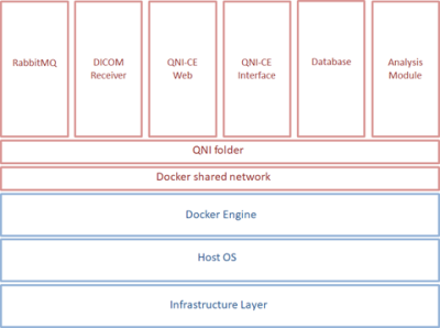 |
74 |
Clinical deployment of novel quantitative neuro-image analysis
techniques using containerisation technology
James Moggridge1,2,
Sjoerd Vos1,3,4,
Ferran Prados3,
Matthew Grech-Sollars1,3,
Daniel Matthews5,
Peter Schmidt5,
Daniel C Alexander3,
John S Duncan6,
Frederik Barkhof1,3,7,
Tarek A Yousry1,2,
and John S Thornton1,2
1Lysholm Department of Neuroradiology, National Hospital for Neurology and Neurosurgery, University College London Hospitals NHS Foundation Trust, London, United Kingdom, 2Department of Brain Repair and Rehabilitation, UCL Institute of Neurology, University College London, London, United Kingdom, 3Centre for Medical Image Computing, Department of Computer Science, University College London, London, United Kingdom, 4Centre for Microscopy, Characterisation, and Analysis, University of Western Australia, Perth, Australia, 5Advanced Research Computing Centre, University College London, London, United Kingdom, 6Department of Clinical and Experimental Epilepsy, UCL QS Institute of Neurology, London, United Kingdom, 7Department of Radiology and Nuclear Medicine, Amsterdam UMC, Amsterdam, Netherlands Keywords: Data Analysis, Software Tools, Containerisation The challenges encountered in development and clinical deployment of quantitative neuroradiological software techniques for in-house use can be overcome to some degree, through the application of a multi-disciplinary approach, and the use of containerisation. However, full life-cycle support including post-market surveillance presents a more significant obstacle. |
|
3305.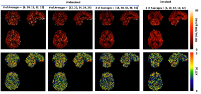 |
75 |
Feasibility of Performing High-Resolution Multi-delay PCASL
Imaging with Intravascular Signal Suppression Within 7 Minutes
Xiaoping Wu1,
Kamil Ugurbil1,
Gregory J. Metzger1,
and Xiufeng Li1
1University of Minnesota, Minneapolis, MN, United States Keywords: Data Processing, Brain, ASL, cerebral perfusion or blood flow To ensure satisfactory perfusion signal-to-noise ratio (SNR) for ASL imaging, intravascular perfusion signals are typically not suppressed (e.g., high-resolution multi-delay SMS/MB-EPI PCASL imaging for Human Connectome Projects) but can induce significant bias for CBF quantification if not addressed sufficiently. Recently proposed image denoising methods may be able to improve ASL imaging quality and SNR, having the potential to making it practical to perform ASL imaging with intravascular signal suppression. Our preliminary study results suggested that with image denoising, high-resolution multi-delay SMS/MB-EPI PCASL imaging with intravascular suppression could be achieved within 7 minutes. |
|
3306.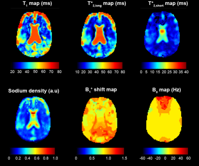 |
76 |
Mapping sodium relaxation parameters in brain using magnetic
resonance fingerprinting at 7T
Lauren F O'Donnell1,
Gonzalo Rodriguez1,
Gregory Lemberskiy1,
Zidan Yu1,2,3,
Olga Dergachyova1,
Martijn A Cloos4,5,
and Guillaume Madelin1
1Department of Radiology, Center for Biomedical Imaging, New York University Grossman School of Medicine, New York, NY, United States, 2Department of Medicine, John A. Burns School of Medicine, University of Hawaii, Honolulu, HI, United States, 3Vilcek Institute for Graduate Biomedical Sciences, NYU Langone Health, New York, NY, United States, 4Centre for Advanced Imaging, The University of Queensland, Brisbane, QLD, Australia, 5ARC Training Centre for Innovation in Biomedical Imaging Technology, The University of Queensland, Brisbane, QLD, Australia Keywords: MR Fingerprinting/Synthetic MR, Brain, Sodium, x - nuclei, relaxometry, 7 Tesla We present a new MRF technique for simultaneous mapping of sodium T1, biexponential T2*, ion density, B+1 shift and B0 in the brain at 7T. This is accomplished using a 23 pulse MRF train combined with a UTE FLORET trajectory readout. Our technique is demonstrated in a 3 compartment phantom and in vivo across four healthy volunteers. Matching to a dictionary with over 800,000 signals produced parameter maps with good agreement to literature values. Flow and partial volume effects may interfere with matching T*2,short in CSF. |
|
3307.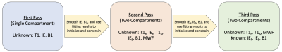 |
77 |
High-Resolution Myelin Water Imaging Using MPnRAGE
Jayse Merle Weaver1,2,
Steven Kecskemeti2,
Jose M Guerrero-Gonzalez1,2,
Andrew L Alexander1,2,3,
and Douglas C Dean III1,2,4
1Medical Physics, University of Wisconsin-Madison, Madison, WI, United States, 2Waisman Center, University of Wisconsin-Madison, Madison, WI, United States, 3Psychiatry, University of Wisconsin-Madison, Madison, WI, United States, 4Pediatrics, University of Wisconsin-Madison, Madison, WI, United States Keywords: Quantitative Imaging, Brain, Myelin A T1-only method to extract a quantitative myelin water fraction, utilizing a 3D radial k-space MPnRAGE sequence that reconstructs hundreds of different inversion time (TI) images in the same scan used for high-resolution T1-weighted morphometry was developed. To address high sensitivities to additive image noise of this complex model, a multi-pass fitting algorithm was prototyped that provides educated initial guesses and constraints on the underlying parameters. |
|
3308.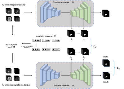 |
78 |
Modality-adaptive Network Learning for Brain Tumor Segmentation
with Incomplete Multi-modal MRI Data
Haoran Li1,
Cheng Li1,
Weijian Huang1,
Yeqi Wang1,2,
Yu Zhang1,
Xue Liu1,
Hairong Zheng1,
and Shanshan Wang1,3,4
1Paul C. Lauterbur Research Center for Biomedical Imaging, Shenzhen Institutes of Advanced Technology, Chinese Academy of Sciences, Shenzhen, China, 2School of Computer and Artificial Intelligence, Zhengzhou University, Zhengzhou, China, 3Peng Cheng Laboratory, Shenzhen, China, 4Guangdong Provincial Key Laboratory of Artificial Intelligence in Medical Image Analysis and Application, Guangdong Provincial People’s Hospital, Guangdong Academy of Medical Sciences, Guangzhou, China Keywords: Segmentation, Brain Automated brain tumor segmentation with multi-modal magnetic resonance imaging (MRI) data is crucial for brain cancer diagnosis. Nevertheless, in clinical applications, it is difficult to guarantee that complete multi-modal MRI data are available due to different imaging protocols and inevitable data corruption. A large test time performance drop could happen. Here, we design a modality-adaptive network learning method to extract common representations from different modalities and make our trained model applicable to different data-missing scenarios. Experiments on an open-source dataset demonstrate that our method can reduce the dependence of deep learning-based segmentation methods on the integrity of input data. |
|
3309.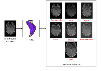 |
79 |
Uncertainty-based Quality Control for Subcortical Structures
Segmentation in T1-weighted Brain MRI
Benjamin Lambert1,2,
Florence Forbes3,
Senan Doyle2,
Alan Tucholka2,
and Michel Dojat1
1Univ. Grenoble Alpes, Inserm, U1216, Grenoble Institut Neurosciences, GIN, Grenoble, France, 2Pixyl, Research and Development Laboratory, Grenoble, France, 3Univ. Grenoble Alpes, Inria, CNRS, Grenoble INP, LJK, Grenoble, France Keywords: Machine Learning/Artificial Intelligence, Artifacts The performance of Deep Learning (DL) models may drastically drop in the presence of characteristics in test images not present in the training set. Then, the automatic detection of these Out-Of-Distribution (OOD) inputs is important to deploy these methods especially for clinical applications. We address this issue in the context of DL-based subcortical structures segmentation on T1w brain MRI. We compare two OOD detection frameworks equipping DL segmentation models, Maximum Softmax Probability and Deterministic Uncertainty Method, and demonstrate the superiority of the latter which allows a robust and versatile identification of artifacts in images. |
|
3310.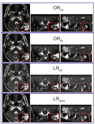 |
80 |
Super resolution permits fast, low resolution bSSFP imaging of
the temporal bone…but do radiologists agree on image quality?
Sarah Reeve1,2,
Alessandro Guida2,3,
Chris Bowen1,2,3,
James Rioux1,2,3,
David Volders3,4,
Jens Heidenreich3,4,
and Steven Beyea1,3,5,6
1Physics and Atmospheric Science, Dalhousie University, Halifax, NS, Canada, 2Biomedical Translational Imaging Centre, QEII Health Sciences Centre, Halifax, NS, Canada, 3Diagnostic Radiology, Dalhousie University, Halifax, NS, Canada, 4Diagnostic Imaging, QEII Health Sciences Centre, Halifax, NS, Canada, 5Biomedical Translational Imaging Centre, IWK Health Centre, Halifax, NS, Canada, 6School of Biomedical Engineering, Dalhousie University, Halifax, NS, Canada Keywords: Machine Learning/Artificial Intelligence, Head & Neck/ENT, Super Resolution Low resolution (LR) balanced steady-state free precession (bSSFP) acquisitions confer decreased acquisition times, reduced patient motion and heating, and increased artifact tolerance due to a decrease in TR. The application of a pre-trained super resolution network to LR bSSFP images of the temporal bone allows for these advantages to be realized, without significantly degrading image quality. In the absence of a matched high resolution image, quality was judged by two radiologists. However, radiologist’s ratings were not in agreement, highlighting the fact that there is no single definition of task-specific image quality, which must be considered when super resolution is performed. |
|
 Back to Meeting Home
 Back to the Program-at-a-Glance
The International Society for Magnetic Resonance in Medicine is accredited by the Accreditation Council for Continuing Medical Education to provide continuing medical education for physicians.
 Back to Meeting Home
 Back to the Program-at-a-Glance
 View Presentation Video