Digital Poster
New Frontiers in Brain Imaging II
ISMRM & ISMRT Annual Meeting & Exhibition • 03-08 June 2023 • Toronto, ON, Canada

Digital Poster
New Frontiers in Brain Imaging II
| Computer # | |||
|---|---|---|---|
3468.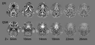 |
61 |
Rapid Neurological QSM measurement based on EPI sequence
XueHua Peng1,
LiFei Ma2,
Rui Zhang2,
and XiaoMeng Wu2
1Radiology Department, WuHan Children’s Hosptial, WuHan, China, 2philips, shanghai, China Keywords: Parkinson's Disease, Quantitative Susceptibility mapping, Rapid QSM measurement, EPI acquisition, Neuro system Quantitative susceptibility mapping(QSM) can be used for quantitatively measurement of neuro system magnetic susceptibility information caused by various diseases, such as Alzheimer's disease (AD), Parkinson's disease (PD) and neuro vascular diseases. In this study, a rapid QSM acquisition based on EPI sequence was proposed to improve clinical feasibility of QSM. The results demonstrated the rapid QSM were very close to those obtained by standard QSM. |
|
3469.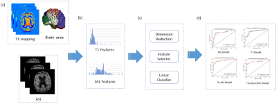 |
62 |
Machine learning models using T1-mapping and arterial spin
labeling images to identify Alzheimer’s disease and mild
cognitive impairment
Shengyong Li1,
Xiaonan Wang2,3,
Yida Wang1,
Yang Song4,
and Guang Yang1
1Shanghai Key Laboratory of Magnetic Resonance, East China Normal University, Shanghai, China, 2Department of Radiology, Beijing Hospital, National Center of Gerontology, Institute of Geriatric Medicine, Beijing, China, 3Graduate School of Peking Union Medical College, Chinese Academy of Medical Sciences, Beijing, China, 4MR Scientific Marketing, Siemens Healthcare, Shanghai, China Keywords: Alzheimer's Disease, Arterial spin labelling To investigate the added value of T1-mapping to arterial spin labeling (ASL) for computer-aided early diagnosis of Alzheimer’s disease (AD). A total of 97 (45 AD/24 mild cognitive impairment (MCI)/38 normal control (NC)) people were enrolled retrospectively. We extracted features from 24 automatically segmented brain regions based on T1-mapping and ASL MR images and constructed three radiomics models to differentiate AD-NC/MIC-NC/AD-MCI, for which the radiomics models achieved a favorable prediction performance with the AUCs of 0.921/0.764/0.727, respectively. |
|
3470. |
63 |
Dedicated head-only high-performance gradients in a 3T scanner
improve image quality compared to a whole-body gradient design
J. Kevin DeMarco1,2,
H. Douglas Morris2,
Robert Y. Shih1,2,
Daniel W. Bess3,
Luca Marinelli4,
Vincent B. Ho1,2,
and Thomas K. F. Foo4
1Walter Reed National Military Medical Center, Bethesda, MD, United States, 2Uniformed Services University of the Health Sciences, Bethesda, MD, United States, 3Fort Belvoir Community Hospital, Fort Belvoir, VA, United States, 4GE Global Research Center, Niskayuna, NY, United States Keywords: Gradients, Brain Ultra-high-performance MAGNUS gradient coils optimized for advanced diffusion imaging were used to obtain routine anatomical brain imaging in a cohort of 31 consecutive subjects enrolled in a mild TBI study. In a blinded review, three neuroradiologists reviewed 3D MPRAGE and 3D T2 FLAIR obtained with MAGNUS and a whole-body 3T scanner (WB3T). Substantial agreement of better overall image quality for MAGNUS vs. WB3T was seen for T2 FLAIR, and mild agreement of better overall image quality for MPRAGE. MAGNUS supports rapid gradient switching and higher gradient strength for additional SNR with 3D T2 FLAIR. |
|
3471.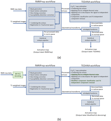 |
64 |
Improving the robustness of independent component analysis for
denoising multi-echo fMRI data
Bahman Tahayori1,
Robert E. Smith1,
David N. Vaughan1,
Chris Tailby1,
Eric Y. Pierre1,
Graeme D. Jackson1,2,
and David F. Abbott1,2
1The Florey Institute of Neuroscience and Mental Health, Heidelberg, Australia, 2Florey Department of Neuroscience and Mental Health, University of Melbourne, Melbourne, Australia Keywords: Data Analysis, fMRI (task based) Multi-Echo fMRI data acquisition has multiple advantages over single-echo acquisition. Principal amongst them is that multiple echoes can distinguish neural activity from artefacts. TE Dependent ANAlysis (TEDANA) is an existing software tool designed to denoise multi-echo fMRI datasets. We evaluated the performance of TEDANA to denoise fMRI data of 120 subjects. Our results demonstrated that TEDANA improved the activation detection at a group level. However, for a subset of subjects TEDANA degraded their individual result substantially. We identified potential causes and proposed a modified framework for multi-echo data analysis that provides reasonable results at an individual subject level. |
|
3472.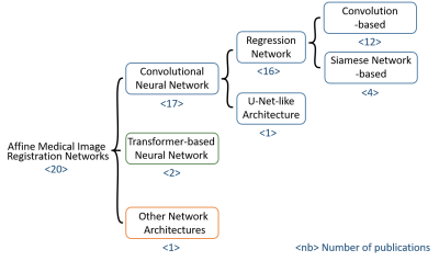 |
65 |
Deep Learning-Based Affine Medical Image Registration - A Review
and Comparative Study on Generalizability
Anika Strittmatter1,2,
Lothar R. Schad1,2,
and Frank G. Zöllner1,2
1Computer Assisted Clinical Medicine, Medical Faculty Mannheim, Heidelberg University, Mannheim, Germany, 2Mannheim Institute for Intelligent Systems in Medicine, Medical Faculty Mannheim, Heidelberg University, Mannheim, Germany Keywords: Machine Learning/Artificial Intelligence, Data Processing, Image Registration In this research we investigated the performance of published neural networks for an affine registration of multimodal medical images and examined the networks' generalizability to new datasets. The neural networks were trained and evaluated using a synthetic multimodal dataset of three-dimensional CT and MRI volumes of the liver. We compared the Normalised Mutual Information, Dice coefficient and the Hausdorff distance across the neural networks described in the papers, using our CNN as a benchmark and the conventional affine registration method as a baseline. Seven networks improved the pre-registration Dice coefficient and are therefore able to generalise to new datasets. |
|
3473.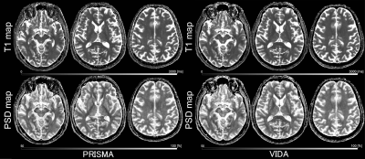 |
66 |
Inter-scanner consistency of T1 map and proton spin density map
derived from strategically acquired gradient echo (STAGE)
imaging
Yasutaka Fushimi1,
Satoshi Nakajima1,
Sachi Okuchi1,
Akihiko Sakata1,
Takuya Hinoda1,
Sayo Otani1,
Azusa Sakurama1,
Krishna Pandu Wicaksono1,
Hiroshi Tagawa1,
Yang Wang1,
Satoshi Ikeda1,
Shuichi Ito1,
Miyuki Takiya1,
and Yuji Nakamoto1
1Department of Diagnostic Imaging and Nuclear Medicine, Kyoto University Graduate School of Medicine, Kyoto, Japan Keywords: Quantitative Imaging, Brain Recently, strategically acquired gradient echo (STAGE) imaging has been developed as a potential standardized brain imaging protocol. STAGE is designed to use two 3D fully flow compensated multi-echo SWI sequences with 2 different flip angles (FAs). We evaluated the inter-scanner consistency of T1 values and proton spin density (PSD) values derived from STAGE imaging for elderly healthy volunteers. We conducted VOI analysis both in Native space and “Mutual space” using AAL3 VOIs. Parcellation was automatically performed with few manipulations, and good ICC and minimal biases were demonstrated between the two scanners. |
|
3474.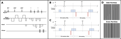 |
67 |
Rapid Frequency Offset Mapping with the Half-Fourier Acquisition
Single-Shot Turbo Spin-Echo with the Selective Parity Approach
Aidin Arbabi1,
Irina De Alba Alvarez1,
Vitaliy Khlebnikov1,
Jose P. Marques1,
and David. G. Norris1
1Radboud University, Nijmegen, Netherlands Keywords: Quantitative Imaging, Brain Frequency Offset maps are widely used to correct image distortion or estimate signal loss in gradient echo signal due to field inhomogeneities. Routinely, multiecho gradient echo sequences are used to collect this information. In this work, we introduce an innovative method which enables frequency offset mapping via one single-shot acquisition. The proposed method offers a significant reduction in the acquisition time without loss of accuracy. |
|
3475.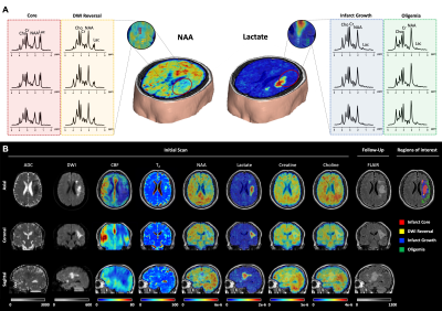 |
68 |
Improved Assessment of Brain Tissue Viability in Acute Stroke
Using Fast High-Resolution 3D-MRSI and Quantitative T2 Imaging
Ziyu Meng1,
Tianyao Wang2,
Bin Bo1,
Yibo Zhao3,4,
Yudu Li3,5,
Rong Guo3,6,
Wen Jin3,4,
Xin Yu7,
Zhi-Pei Liang3,4,
and Yao Li1
1School of Biomedical Engineering, Shanghai Jiao Tong University, Shanghai, China, 2Radiology Department, The Fifth People's Hospital of Shanghai, Fudan University, Shanghai, China, 3Beckman Institute for Advanced Science and Technology, University of Illinois at Urbana-Champaign, Urbana, IL, United States, 4Department of Electrical and Computer Engineering, University of Illinois at Urbana-Champaign, Urbana, IL, United States, 5National Center for Supercomputing Applications, University of Illinois at Urbana-Champaign, Urbana, IL, United States, 6Siemens Medical Solutions USA, Inc, Urbana, IL, United States, 7Department of Biomedical Engineering, Case Western Reserve University, Cleveland, OH, United States Keywords: Stroke, Stroke Accurate assessment of tissue viability is of great importance for the design of therapeutic interventions in acute stroke. 1H-MRSI and quantitative T2 imaging can provide neurochemical biomarkers sensitive to the pathological changes in stroke but their applications have been limited by long scan times. This study introduces a novel approach that combines fast high-resolution 3D-MRSI with quantitative T2 imaging to assess the viability of ischemic tissues in acute stroke. This approach achieved improved discrimination between ischemic brain tissues that ultimately infarcted and those recovered using joint neurometabolite concentrations and quantitative T2 values, in comparison with classical DWI and PWI methods. |
|
3476.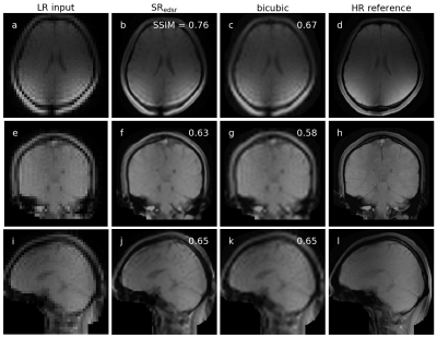 |
69 |
Super-resolution imaging on an MRI-linac to improve real-time
MRI used in MRI guided radiation therapy.
James Grover1,2,
Paul Liu1,2,
Bin Dong2,
Shanshan Shan1,2,
Brendan Whelan1,2,
Paul Keall1,2,
and David Waddington1,2
1Image X Institute, University of Sydney, Sydney, Australia, 2Ingham Institute for Applied Medical Research, Sydney, Australia Keywords: Machine Learning/Artificial Intelligence, Machine Learning/Artificial Intelligence, MRI guided radiation therapy Real-time MRI is limited in its spatiotemporal resolution due to imaging time being proportional to the spatial resolution. Super-resolution imaging was integrated into an MRI-linac to improve the spatiotemporal resolution of images used in real-time adaptive MRI guided radiation therapy. Real-time up-sampling techniques included conventional bicubic interpolation and deep learning-based super-resolution. Up-sampling increased the spatial resolution as characterised by healthy volunteer brain and thorax MRIs with negligible impact on the temporal resolution as measured in a motion phantom tracking experiment. |
|
3477.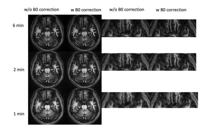 |
70 |
Towards accurate and repeatable 1mm isotropic whole-brain MRF
quantification using a 1-minute scan with optimized processing
pipeline
Quan Chen1,2,
Xiaozhi Cao1,
Congyu Liao1,
Siddharth Srinivasan Iyer3,
Sophie Schauman1,
Nan Wang1,
and Kawin Setsompop1
1Department of Radiology, Stanford University, Stanford, CA, United States, 2standford university, palo alto, CA, United States, 3Department of Electrical Engineering and Computer Science, Massachusetts Institute of Technology, Stanford, CA, United States Keywords: Quantitative Imaging, Brain A rotation-fitting based PV reduction pipeline with B0, B1+ and frequency-based flip angle (FA) scaling corrections are proposed to improve the accuracy and repeatability of magnetic resonance fingerprinting (MRF) in the existence of motion. 5 repeated scans from both stable baseline and big motion positions were acquired and analyzed to compare the repeatability of MRF under different rotation-fitting schemes and B0, B1+ corrections. Rotation before the fitting can substantially reduce the PV effects. The B0/ B1+ and frequency-based FA scaling corrections improve the estimation accuracy. The 2min and 1min 1mm3 whole brain MRF quantitative results also indicate a high repeatability. |
|
3478.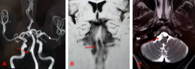 |
71 |
BPAS-MRI combined with MRA to identify intracranial
vertebrobasilar lesions
Xiaohan Guo1,
Pingping Wang1,
Gengming Mu1,
Tianju Jia1,
Kaiguo Zhu1,
Kai Ai2,
and Baoying Chen1
1XI'AN INTERNATIONAL MEDICAL CENTER HOSPITAL, Xi'an, China, 2Philips Healthcare, Xi'an, China Keywords: Blood vessels, Blood vessels Basi-parallel anatomic scanning magnetic resonance imaging (BPAS-MRI) is an MRI technique that can reveal the outer contour of the vertebrobasilar artery, even in the presence of occlusion. A total of 53 patients with 106 segments of blood vessels were scanned by BPAS+MRA, and the results were verified by comparing with VW-MRI. It was found that BPAS+MRA could significantly improve the detection rate of vertebrobasilar lesions, which was of great value for the diagnosis and differential diagnosis of arterial dysplasia, arteriosclerosis and arterial occlusion, and could improve the diagnostic confidence of radiologists. |
|
3479. |
72 |
Altered Glx and APT values in hippocampus of patients with aMCI:
a novel combined imaging diagnostic marker
Xin Chen1,2,
Tao Gong1,
Tong Chen3,
Changyuan Xu1,
Yuchao Li1,
Qingxu Song4,
Liangjie lin5,
Georg Oeltzschner6,7,
Richard A. E. Edden6,
Guangbin Wang1,
and Zhangyong Xia2
1Shandong Provincial Hospital Affiliated to Shandong First Medical University, Jinan, China, 2Liaocheng People’s Hospital, Liaocheng, China, 3Shandong Provincial Hospital, Cheeloo College of Medicine, Shandong University, Jinan, China, 4Qilu Hospital of Shandong University, Jinan, China, 5Philips Healthcare, Beijing, China, 6The Russell H. Morgan Department of Radiology and Radiological Science, Johns Hopkins University School of Medicine, Baltimore, MD, United States, 7F.M. Kirby Research Center for Functional Brain Imaging, Kennedy Krieger Institute, Baltimore, MD, United States Keywords: Alzheimer's Disease, Brain, Amnestic mild cognitive impairment; hippocampus; MEGA-PRESS; amide proton transfer-weighted imaging; imaging diagnostic marker Amnestic mild cognitive impairment (aMCI) is considered as a prodromal stage of Alzheimer's disease (AD). Growing evidence supports the hypothesis that unbalanced excitatory/inhibitory (glutamate and gamma-aminobutyric acid) neurotransmitters and consequent abnormal protein deposition in hippocampus contribute to the pathological process of MCI and AD. We explored the changes of hippocampal Glx/GABA+ levels and APTw in aMCI patients using MEGA-PRESS and APTw imaging. Patients with aMCI exhibited decreased Glx levels (and Glx/GABA+ ratios) and increased APTw values in hippocampus. The combination of Glx and APTw values improved the diagnostic performance for aMCI, suggesting it was a potential imaging diagnostic marker. |
|
 |
3480.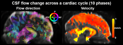 |
73 |
4D CSF flowmetry to map brain-wide slow CSF flow dynamics and
patterns in subarachnoid space
Zijing Dong1,2,
Fuyixue Wang1,2,
Amelia K. Strom1,3,
Korbinian Eckstein4,
Beata Bachrata4,
Simon D. Robinson4,
Bruce R. Rosen1,2,3,
Lawrence L. Wald1,2,3,
Laura D. Lewis5,
and Jonathan R. Polimeni1,2,3
1Athinoula A. Martinos Center for Biomedical Imaging, MGH, Charlestown, MA, United States, 2Department of Radiology, Harvard Medical School, Charlestown, MA, United States, 3Harvard-MIT Health Sciences and Technology, MIT, Cambridge, MA, United States, 4High Field MR Centre, Medical University of Vienna, Vienna, Austria, 5Department of Biomedical Engineering, Boston University, Cambridge, MA, United States Keywords: Neurofluids, Brain Cerebrospinal fluid (CSF) flow is a key component of the brain’s waste clearance system. However, little is known about the brain-wide CSF flow in subarachnoid space (SAS), due to the low sensitivity of MRI-based flow imaging methods for measuring slow flow. Here, we proposed a phase-contrast 4D CSF flowmetry method using a pulsed-gradient-spin-echo EPI sequence, providing high sensitivity and efficiency to measure brain-wide slow CSF flow dynamics in SAS. Whole-brain CSF flow dynamics including velocity and direction changes were measured and our preliminary data suggest that both cardiac pulsation and respiration can drive CSF flow in both ventricles and SAS. |
3481.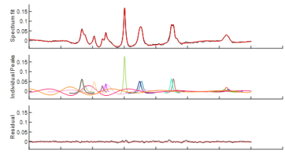 |
74 |
Reproducibility of quantitative 1H-decoupled, NOE-enhanced
31P-MRS in the human brain at 3 T.
Magnus Svensen1,
Christian Dölle2,
Njål Brekke3,
Heidi Espeland4,
Nora Tvedten4,
John Georg Seland4,
Frank Riemer5,
and Charalampos Tzoulis6
1Neuro-SysMed & Mohn Medical Imaging- and Visualization Center (MMIV), Bergen, Norway, 2Neuro-SysMed & University of Bergen, Bergen, Norway, 3Haukeland University Hospital, Bergen, Norway, 4University of Bergen, Bergen, Norway, 5Mohn Medical Imaging- and Visualization Center (MMIV), Bergen, Norway, 6Neuro-SysMed & University of Bergen & Haukeland University Hospital, Bergen, Norway Keywords: Data Analysis, Spectroscopy, 31P 1H-decoupled NOE-enhanced 31P-MRS has promising applications in the diagnosis and stratification of neurodegenerative diseases such as Alzheimer's disease, Parkinson's disease and amytrophic lateral sclerosis (ALS) due to its ability to longitudinally and noninvasively measure changes to cell metabolism and energy conversion, which is heavily involved in these diseases. To aid clinical translation, this study focuses on assessing the reproducibility of 1H-decoupled NOE-enhanced 31P-MRS by measuring the resonances of metabolites involved in neural energy metabolism. |
|
3482.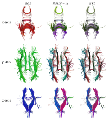 |
75 |
A new directional colormap for DTI fiber tractography and its
application to bundle recognition
Mauro Zucchelli1,
Christos Papageorgakis1,
and Stefano Casagranda1
1Department of R&D Advanced Applications, Olea Medical, La Ciotat, France Keywords: Brain Connectivity, Tractography & Fibre Modelling, Colormap, Diffusion We propose a new tractography colormap based on the HSL model to complement the classical RGB colormap used in the field. Our colormap is based on a new feature, called "streamlines normal", an oriented vector that is computed from the tractography. The streamlines normal in combination with the HSL colormap improves tractography data visualization and can be an asset for better and faster bundle segmentation both manually and automatically. |
|
3483.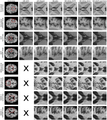 |
76 |
Data Augmentation with Simulated Images for Generalizable Brain
MRI Super-Resolution
Sina Amirrajab1,
Rien Boonstoppel1,
Aymen Ayaz1,
and Marcel Breeuwer1,2
1Biomedical Engineering, Eindhoven University of Technology, Eindhoven, Netherlands, 2MR R&D - Clinical Science, Philips Healthcare, Eindhoven, Netherlands Keywords: Machine Learning/Artificial Intelligence, Brain, Super resolution In many medical applications, high resolution images are required to facilitate early and accurate diagnosis. In this paper, we investigate the potential of data augmentation using simulated brain magnetic resonance (MR) images for training a deep-learning (DL) super resolution model that can generalize to brain data from different publicly available sources. Our qualitative visual evaluation results suggest that data augmentation with simulated images can improve the robustness and generalization of the model and decrease the artifacts of the super-resolved images. |
|
3484.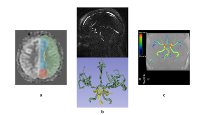 |
77 |
Toward a detailed subject-specific cerebrovascular model using
Arterial Spin Labeling (ASL) Magnetic Resonance Imaging (MRI)
Alireza Sharifzadeh-Kermani1,
Finbar Argus1,
Jiantao Shen1,
Sarah-Jane Guild2,
David Dubowitz2,
Miriam Scadeng2,3,
Paul Condron3,
Eryn Kwon1,2,3,
Samantha Holdsworth2,3,
Gonzalo Daniel Maso Talou1,
and Soroush Safaei1
1Auckland Bioengineering Institute, The University of Auckland, Auckland, New Zealand, 2Faculty of Medical and Health Sciences & Centre for Brain Research, The University of Auckland, Auckland, New Zealand, 3Mātai Medical Research Institute, Tairāwhiti-Gisborne, New Zealand Keywords: Blood vessels, Modelling, Arterial Spin Labeling, ASL, cerebral perfusion, CBF, 4D flow, blood flow Computed flows via haemodynamic models highly depend on peripheral resistances that affect the blood perfusion. To this end, finding a way to estimate these resistances is crucial for having a subject-specific model. Perfusion (CBF) maps produced by Arterial Spin Labeling (ASL)-MRI provide valuable information via which the model can be personalized. This new approach may be promising for providing blood flow measurements of the brain from ASL-CBF data in the absence of a full brain 4D flow sequence. |
|
3485.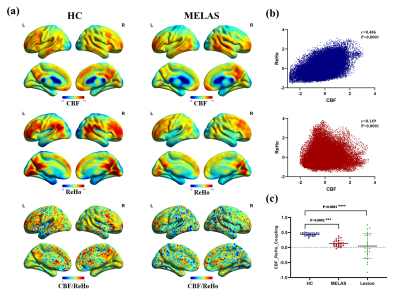 |
78 |
Altered neurovascular coupling in patients with MELAS evaluated
by combining cerebral blood flow and regional homogeneity
Rong Wang1,
Yuxin Li1,
Jie Lin1,
Yong Zhang2,
and Jiankun Dai3
1Huashan Hospital, Shanghai, China, 2GE Healthcare, Shanghai, China, Shanghai, China, 3MR Research, GE Healthcare, Beijing, China, Shanghai, China Keywords: Stroke, fMRI (resting state) This is the first study that used a combined arterial spin labeling (ASL) and resting-state fMRI approach to assess the neurovascular coupling in patients with mitochondrial myopathy, encephalopathy, lactic acidosis and stroke-like episodes (MELAS), which may provide a new mechanistic perspective into understanding numerous brain diseases. |
|
3486. |
79 |
SuperQ: 3D Super-Resolution of Quantitative Susceptibility Maps
Alexandra Grace Roberts1,2,
Yi Wang1,2,
Pascal Spincemaille2,
and Thanh Nguyen2
1Electrical Engineering, Cornell University, Ithaca, NY, United States, 2Radiology, Weill Cornell Medicine, New York, NY, United States Keywords: Machine Learning/Artificial Intelligence, Brain 3D super-resolution of QSM is feasible using the VDSR and U-net architectures using a fraction of the required number of epochs as previous 2D super-resolution networks. Additionally, this method both reduces whole brain MSE and ROI MSE and increases the apparent resolution as compared to the interpolated input |
|
3487. |
80 |
Evaluation of whole-brain oxygen metabolism in Alzheimer's
disease using QSM and quantitative BOLD
Aocai Yang1,2,
Hangwei Zhuang3,4,
Guolin Ma2,
and Yi Wang3,5
1Peking Union Medical College, Beijing, China, 2Department of Radiology, China–Japan Friendship Hospital, Beijing, China, 3Department of Biomedical Engineering, Cornell University, New York, NY, United States, 4Weill Cornell Medical College, Department of Radiology, New York, NY, United States, 5Department of Radiology, Weill Cornell Medical College, New York, NY, United States Keywords: Alzheimer's Disease, Alzheimer's Disease We assessed the whole-brain oxygen metabolism perturbations in AD using QSM and quantitative BOLD. mGRE data and QQ model was employed to calculate OEF and ASL data was used to reconstruct CBF map. The CMRO2 can be estimated from CBF and OEF based on Fick’s principle. Our results demonstrated a characteristic whole-brain hypoperfusion and hypometabolism pattern in AD, predominantly located within default-mode network. Additionally, decreased CBF and CMRO2 in substructures of bilateral hippocampus strongly correlated with global cognition. QQ model-based noninvasively quantitative measurements have a great potential to be complementary biomarkers for evaluating cognitive impairment in AD. |
|
 Back to Meeting Home
 Back to the Program-at-a-Glance
The International Society for Magnetic Resonance in Medicine is accredited by the Accreditation Council for Continuing Medical Education to provide continuing medical education for physicians.
 Back to Meeting Home
 Back to the Program-at-a-Glance
 View Presentation Video