Digital Poster
Signal Modeling
ISMRM & ISMRT Annual Meeting & Exhibition • 03-08 June 2023 • Toronto, ON, Canada

| Computer # | |||
|---|---|---|---|
4596.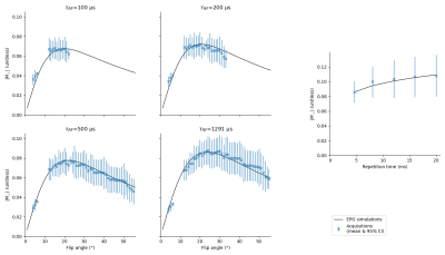 |
1 |
EPG-based optimization of the SNR of the phase-cycled bSSFP in
phase-based electrical conductivity mapping
Julien Lamy1,
Flavy Savigny1,
and Paulo Loureiro de Sousa1
1ICube, Université de Strasbourg-CNRS, Strasbourg, France Keywords: Signal Modeling, Simulations Phase-based electrical conductivity mapping requires high SNR due to the instability of the reconstruction. Electrical conductivity can be measured by the phase-cycled bSSFP, a high-SNR sequence which is also weighted by other biophysical parameters but for which no complete analytical model exist. In this work, we use EPG-based simulations to investigate how the bSSFP signal varies with the flip angle, the phase step and the pulse duration in a diffusive, two-pools model of the brain white matter at 3 T. We show that our simulations agree with in-vivo acquisitions and establish guidelines to optimize the SNR of the phase-cycled bSSFP. |
|
4597. |
2 |
Estimation of the Spatial Gradient of the MR Image from the
Diffusion Profile
Iman Aganj1,2,
Matthew Vera1,
Thorsten Feiweier3,
Andre J. van der Kouwe1,2,
John E. Kirsch1,2,
and Bruce R. Fischl1,2
1Athinoula A. Martinos Center for Biomedical Imaging, Massachusetts General Hospital, Boston, MA, United States, 2Radiology Department, Harvard Medical School, Boston, MA, United States, 3Siemens Healthcare GmbH, Erlangen, Germany Keywords: Signal Modeling, Diffusion Tensor Imaging In the course of diffusion, water molecules experience varying values for the relaxation-time property of the underlying tissue, a factor that has not been accounted for in diffusion MRI (dMRI) modeling. Accordingly, we derive a relationship between the diffusion profile measured by dMRI and the spatial gradient of the image, and subsequently estimate the latter from the former. We test our hypothesized relationship via dMRI of the human brain (a public in vivo image and an acquired ex vivo stimulated-echo image), showing statistically significant results that may be due to our model and/or the confounding factor of “fiber continuity”. |
|
4598.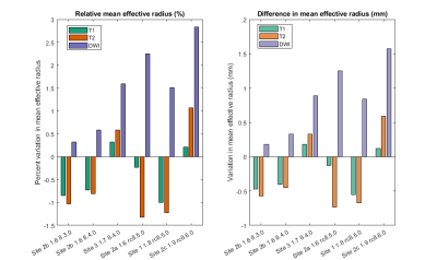 |
3 |
Multi-Continent Analysis of Geometric Distortion in Low-Field
MRI
Stephen E. Ogier1,2,
Annabel Sorby-Adams3,
Kalina Jordanova1,
Andrew M. Dienstfrey4,
Zydrunas Gimbutas4,
Jennifer Guo3,
John Kirsch5,
Steven Schiff6,
Ronald Mulondo7,
Johnes Obungoloch8,
Megan E Poorman9,
John Pitts9,
W. Taylor Kimberly3,
and Kathryn E. Keenan1
1Magnetic Imaging Group, National Institute of Standards and Technology, Boulder, CO, United States, 2Department of Physics, University of Colorado Boulder, Boulder, CO, United States, 3Department of Neurology and the Center for Genomic Medicine, Massachusetts General Hospital and Harvard Medical School, Boston, MA, United States, 4Mathematical Analysis and Modeling Group, National Institute of Standards and Technology, Boulder, CO, United States, 5A. A. Martinos Center for Biomedical Imaging, Massachusetts General Hospital, Charlestown, MA, United States, 6Department of Neurosurgery, Yale University School of Medicine, New Haven, CT, United States, 7CURE Children's Hospital of Uganda, Mbale, Uganda, 8Faculty of Applied Sciences and Technology, Mbarara University of Science and Technology, Mbarara, Uganda, 9Hyperfine, Inc., Guilford, CT, United States Keywords: Artifacts, Low-Field MRI The assessment of brain morphology requires geometric accuracy, and the monitoring of pathologies like dementia requires geometric stability over time and between systems. We aim to assess the geometric accuracy and stability of a head-only 64 mT system by imaging the cylindrical phantom provided by the manufacturer, positioned with a 3D-printed holder for stability, and analyzing the geometric fidelity of the images produced by a variety of product sequences. The initial assessment examines the perimeter of the phantom and how it varies along the S-I direction. |
|
4599.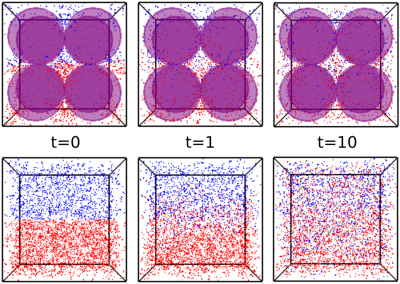 |
4 |
Simultaneous numerical modeling of 13C-MRI enzyme kinetics,
diffusion effects, and RF refocusing
Dylan Archer Dingwell1,2 and
Charles H Cunningham1,2
1Medical Biophysics, University of Toronto, Toronto, ON, Canada, 2Physical Sciences Platform, Sunnybrook Research Institute, Toronto, ON, Canada Keywords: Signal Modeling, Hyperpolarized MR (Non-Gas), 13C Signal Dynamics, 13C MRI, Enzyme Kinetics, Reaction Kinetics, Metabolism In this study, we used a particle-based Brownian dynamics simulator with concurrent MR modeling to characterize diffusion-related effects on 13C signal dynamics. We conducted a spectrophotometric lactate dehydrogenase (LDH) activity assay with and without the presence of glass beads to measure the influence of diffusion constraints and spatial compartmentalization on the reaction kinetics of LDH-mediated conversion of pyruvate to lactate. We also investigated the relationship between diffusion and RF refocusing in a typical fast GRE sequence by comparing simulated signal intensities for RF spoiled and non-spoiled GRE sequences applied under varying diffusion conditions. |
|
4600.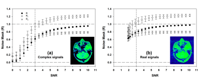 |
5 |
A dual multi-dimensional integration (dMDI) method for reliable
noise masking
Yongquan Ye1
1United Imaging, Houston, TX, United States Keywords: Signal Modeling, Data Processing A dual multi-dimensional integration (dMDI) method was proposed for reliable image noise masking. By adopting a state-of-the-art MDI method, a signal ratio and a reciprocal signal ratio were constructed in the proposed dMDI method. The product of the two signal ratios display a highly reliable dependency of the intrinsic SNR of the signals, which can serve as a voxel-wise noise mask. |
|
4601.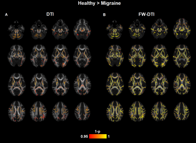 |
6 |
Impact of free-water correction on white matter changes measured
by diffusion tensor imaging in migraine
Irene Guadilla1,2,
Ana Fouto1,
Álvaro Planchuelo-Gómez3,
Antonio Tristán-Vega3,
Amparo Ruiz-Tagle1,
Inês Esteves1,
Gina Caetano1,
Nuno Silva4,
Pedro Vilela5,
Raquel Gil-Gouveia6,7,
Santiago Aja-Fernández3,
Patrícia Figueiredo1,
and Rita Nunes1
1Institute for Systems and Robotics - Lisboa and Department of Bioengineering, Instituto Superior Técnico, Universidade de Lisboa, Lisbon, Portugal, 2Universidad Autónoma de Madrid, Madrid, Spain, 3Laboratorio de Procesado de Imagen, Universidad de Valladolid, Valladolid, Spain, 4Learning Health, Hospital da Luz, Lisbon, Portugal, 5Imaging Department, Hospital da Luz, Lisbon, Portugal, 6Neurology Department, Hospital da Luz, Lisbon, Portugal, 7Center for Interdisciplinary Research in Health, Universidade Católica Portuguesa, Lisbon, Portugal Keywords: Signal Modeling, Diffusion/other diffusion imaging techniques Menstrual migraine affects about 25% of female migraine patients. However, the diagnosis of migraine is particularly difficult because the brain changes associated with migraine are challenging to detect with imaging techniques. Diffusion-weighted MRI (dMRI) permits the detection of alterations in the microenvironment of the brain tissues. We investigate whether removing the contribution of the free water component from the diffusion-signal can provide increased sensitivity to identify white matter changes in migraine using diffusion tensor metrics. |
|
4602.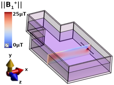 |
7 |
A Simple Analytical Model of B1+ for MRI Measurement of RF
Currents in Wire-like Medical Devices
Chiara Hartmann1,
Mélina Bouldi2,
and Jan M Warnking1
1U1216, Grenoble Institut Neurosciences, Univ. Grenoble Alpes, Inserm, Grenoble, France, 2ZMT Zurich MedTech AG, Zürich, Switzerland Keywords: Signal Modeling, Modelling Safe access to MRI for patients with active implants may be possible by measuring RF currents in implants in individual patients with low-SAR sequences. We present a simple theoretical model of the B1+-field close to a locally straight wire at any angle to B0. MRI signals fitted by our model closely match magnitude and complex signals from electromagnetic simulations with a wire parallel to B0 or tilted at 45° and magnitude experimental signals with parallel wire. RF currents reconstructed from simulated data match ground-truth values to within 6% and current profiles reconstructed from experimental signals follow simulated currents in shape. |
|
4603.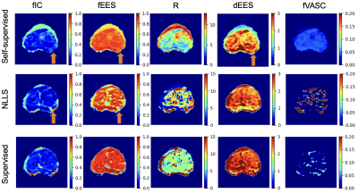 |
8 |
Self-Supervised Model Fitting of VERDICT MRI in the Prostate
Snigdha Sen1,
Saurabh Singh2,
Hayley Pye3,
Caroline Moore4,
Hayley Whitaker3,
Shonit Punwani2,
David Atkinson2,
Eleftheria Panagiotaki1,
and Paddy J Slator1
1Centre for Medical Image Computing, University College London, London, United Kingdom, 2Centre for Medical Imaging, University College London, London, United Kingdom, 3Molecular Diagnostics and Therapeutics Group, University College London, London, United Kingdom, 4Department of Urology, University College London Hospitals NHS Foundation Trust, London, United Kingdom Keywords: Signal Modeling, Microstructure Microstructure models are traditionally fitted via computationally expensive non-linear least squares. Recent model fitting techniques use supervised deep learning algorithms trained on synthetic datasets, however the training data distribution affects parameter estimates. Self-supervised learning can address this by extracting training labels directly from the input data. We introduce a self-supervised machine learning algorithm for fitting the VERDICT MRI model for prostate to diffusion-weighted MRI. On simulated data, our approach improves parameter estimation compared to non-linear least squares and supervised machine learning. We also reveal plausible tumour contrast on in-vivo prostate data. |
|
4604.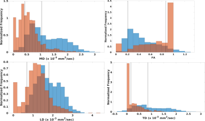 |
9 |
Partial Volume Averaging of Diffusion Tensor Imaging in the
Fornix
Ken Sakaie1,
Ajay Nemani2,
Mark Lowe2,
and Katherine Koenig2
1The Cleveland Clinic, Cleveland, OH, United States, 2Imaging Institute, The Cleveland Clinic, Cleveland, OH, United States Keywords: Signal Modeling, Multiple Sclerosis Diffusion Tensor Imaging of the fornix promises to serve as an imaging biomarker for cognitive decline in multiple sclerosis and Alzheimer’s disease. As the fornix is surrounded by cerebrospinal fluid in the lateral ventricles, partial volume averaging can bias measures of tissue microstructure. Here, we use a high spatial resolution, multishell acquisition to test the methods for combating partial volume averaging in the fornix. |
|
4605.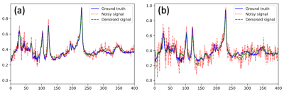 |
10 |
Denoising Magnetic Resonance Spectroscopy Signals with Stack
Auto-encoder Networks
Jing Wang1,
Bing Ji1,
Yang Lei1,
Tian Liu2,
Hui Mao1,
and Xiaofeng Yang1
1Emory University, Atlanta, GA, United States, 2Icahn School of Medicine at Mount Sinai, New York, NY, United States Keywords: Signal Modeling, Spectroscopy This abstract reports a novel and efficient self-supervised deep learning autoencoder network for denoising MRS data and improving signal-to-noise ratio (SNR) of spectra, which may enable rapid MRS data acquisition and improving its clinical workflow and applications. |
|
4606.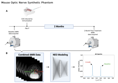 |
11 |
Distinguish Injured from Intact Axons in Coherent Fiber Bundles
Using Network Estimation Operator (NEO)
Jacob Blum1,
Tsen-Hsuan Lin1,
Donsub Rim2,
and Sheng-Kwei Song 1
1Radiology, Washington University School of Medicine in Saint Louis, Saint Louis, MO, United States, 2Department of Mathematics and Statistics, Washington University in Saint Louis, Saint Louis, MO, United States Keywords: Signal Modeling, Diffusion Tensor Imaging Deep Neural Network based Network Estimation Operator (NEO) was developed to differentiate and quantify parallel fibers of different axial diffusivities. Data from both Monte Carlo simulations and an In-Silico imaging phantom were analyzed by both DBSI and NEO. Results reveal that NEO distinguished parallel fibers of different axial diffusivity while DBSI only identified a single fiber. NEO can detect and quantify injured vs. non-injured axons in a coherent fiber bundle. Thus, NEO can quantify the extent of axonal injury and loss more accurately than DBSI and other advanced diffusion MRI models. |
|
4607.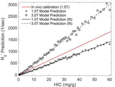 |
12 |
Calibration of R2' relaxation rate and hepatic iron
concentration by Monte Carlo simulations
Xiaoben Li1,
Mengyuan Ma1,
Jinyang Wang1,
Junying Cheng2,
Wenjun Yao3,
and Changqing Wang1
1School of Biomedical Engineering, Anhui Medical University, Hefei, China, 2Department of MRI, The First Affiliated Hospital of Zhengzhou University, Zhengzhou, China, 3Department of Radiology, The Second Affiliated Hospital of Anhui Medical University, Hefei, China Keywords: Signal Modeling, Liver The R2' relaxation rate is expected to become a more specific biomarker in hepatic iron overload because it is not affected by pathological factors. However, calibrating the relationship between R2' relaxation rate and hepatic iron concentration (HIC) at different field strengths is time-consuming. In this work, we develop Monte Carlo simulations to investigate relationship between R2' relaxation rate and HIC in hepatic iron overload at both 1.5T and 3.0T. Results show strong linear relationships between R2' predictions and HIC. The Monte Carlo simulations may greatly shorten the clinical calibration time of MRI relaxation rates and HIC. |
|
4608.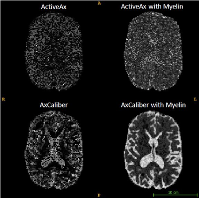 |
13 |
Toward an accurate MR measure of the axon radii by accounting
for myelin-specific attenuation of the diffusion MRI signal in
the brain
Stefania Oliviero1,
Cosimo Del Gratta1,
Andrada Costantina Traeba2,
Tommaso Boccato3,
Caterina Mainero2,
and Nicola Toschi2,3
1Neuroscience, Imaging and Clinical Sciences, and Institute for Advanced Biomedical Technologies, University of Chieti-Pescara G. D'Annunzio, Chieti, Italy, 2A.A. Martinos Center for Biomedical Imaging and Harvard Medical School, Boston, MA, United States, 3Biomedicine and Prevention, University of Rome Tor Vergata, Roma, Italy Keywords: Signal Modeling, White Matter, myelin, diffusion, in silico, MR An accurate, noninvasive measure of axonal radii could play a crucial role in the understanding of healthy and diseased neural processes. Several diffusion-weighted MRI (dMRI) methods report on the axon radii distribution or mean axon radii. However, these measurements systematically overestimate histologically derived values. In this preliminary study, we address this limitation by formulating a model which explicitly includes the myelin contribution to the cerebral dMR signal. In detail, we introduce a myelinic compartment in both the AxCaliber and ActiveAx models and demonstrate a significant improvement of axonal radius estimation using synthetic data simulation. |
|
4609.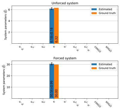 |
14 |
Data-Driven Discovery of mechanical models directly from k-space
data: initial results with an actively forced linear elastic
system.
David G.J. Heesterbeek1,
Max H.C. van Riel1,
Cornelis A.T. van den Berg1,
and Alessandro Sbrizzi1
1Department of Radiotherapy, Computational Imaging Group for MR Therapy and Diagnostics, University Medical Center Utrecht, Utrecht, Netherlands Keywords: Signal Representations, Elastography, Motion estimation, Dynamic system We propose a framework for data-driven discovery of the governing biomechanical equations for soft tissue directly from k-space data. This framework is based on principles from Spectro-dynamics, a novel method to infer dynamical information directly from severely undersampled spectral data with high spatiotemporal resolution, and sparse regression. As a proof-of-concept, the underlying equations of motion are identified for an actively forced linear elastic system using experimentally obtained k-space data. The systems studied in this abstract are successfully identified with errors of less than 2.5% on the system parameters, and their respective displacement fields are reconstructed accurately. |
|
4610.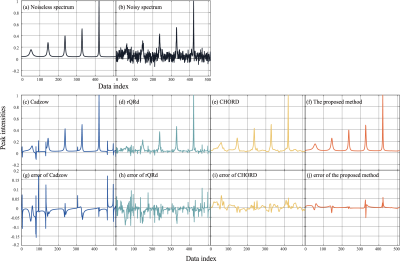 |
15 |
Research on Denoising of Magnetic Resonance Spectrum Based on
Exponential Decomposition Constraint
Jinyu Wu1,
Tianyu Qiu1,
Zhangren Tu1,
Di Guo2,
and Xiaobo Qu1
1Department of Electronic Science, National Institute for Data Science in Health and Medicine, Xiamen University, Xiamen, China, 2School of Computer and Information Engineering, Xiamen University of Technology, Xiamen, China Keywords: Data Processing, PET/MR Nuclear Magnetic Resonance (NMR) has been a frequently-used analytical tool in many areas of modern biology, chemistry and medicine for decades. However, it is usually limited by a low Signal-to-Noise ratio (SNR). In practical applications, Signal Averaging (SA) with repeated samplings is required to improve the signal-to-noise ratio, which greatly increases the scanning time. In this paper, based on the characteristic that NMR time-domain signals can be decomposed exponentially, a model for denoising NMR spectroscopy based on exponential decomposition constraints is proposed. It can effectively improve the denoising ability and therefore save the scanning time. |
|
4611.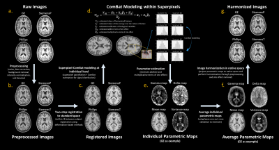 |
16 |
Estimate the Linear Effect of Inter-Scanner Variability: Insight
from Paired Cross-Scanner T1-weighted Images
Chang-Le Chen1,
Mahbaneh Eshaghzadeh Torbati2,
Weiquan Luo3,
Davneet Minhas3,
Charles Laymon3,
Seong Jae Hwang4,
Ciprian Crainiceanu5,
Pauline Maillard6,
Evan Fletcher6,
Charles DeCarli6,
Howard Aizenstein1,7,
and Dana Tudorascu7,8
1Department of Bioengineering, University of Pittsburgh, Pittsburgh, PA, United States, 2Intelligent System Program, School of Computing and Information, University of Pittsburgh, Pittsburgh, PA, United States, 3Department of Radiology, University of Pittsburgh, Pittsburgh, PA, United States, 4Department of Artificial Intelligence, Yonsei University, Seoul, Korea, Republic of, 5Department of Biostatistics, Johns Hopkins University, Baltimore, MD, United States, 6Department of Neurology, University of California Davis, Davis, CA, United States, 7Department of Psychiatry, University of Pittsburgh, Pittsburgh, PA, United States, 8Department of Biostatistics, University of Pittsburgh, Pittsburgh, PA, United States Keywords: Data Processing, Data Processing, Harmonization To broaden our knowledge of inter-scanner variability, we established a pipeline to linearly estimate the site effect by incorporating ComBat modeling and superpixel parcellation. We found that the variation prominently manifested in tissue contrast, noise level, and field inhomogeneity with respect to cross-site data. We further used the estimated parameters to harmonize images. The image quality and structural similarity of cross-scanner data can be improved after the harmonization procedure, and the variation in volumetric measures can also be reduced. This study provides further insight for the research focusing on the development of image harmonization methods. |
|
4612.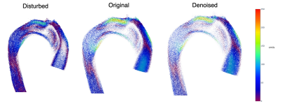 |
17 |
PINN with Divergence-Free Vector Potential for Velocity Fields
Denoising in 4D flow MRI
Javier Bisbal1,2,3,
Joaquin Mura4,
Julio Sotelo1,3,5,
Hernán Mella3,6,
Cristóbal Arrieta1,2,3,
Pablo Irarrazaval1,2,3,7,
and Sergio Uribe1,3,8
1Biomedical Imaging Center, Pontificia Universidad Catolica de Chile, Santiago, Chile, 2Department of Electrical Engineering, Pontificia Universidad Catolica de Chile, Santiago, Chile, 3Millennium Institute for Intelligent Healthcare Engineering, iHEALTH, Santiago, Chile, 4Department of Mechanical Engineering, Universidad Técnica Federico Santa Maria, Santiago, Chile, 5School of Biomedical Engineering, Universidad de Valparaíso, Valparaíso, Chile, 6School of Electrical Engineering, Pontificia Universidad Católica de Valparaiso, Valparaíso, Chile, 7Institute for Biological and Medical Engineering, Pontificia Universidad Católica de Chile, Santiago, Chile, 8Department of Radiology, School of Medicine, Pontificia Universidad Católica de Chile, Santiago, Chile Keywords: Data Processing, Machine Learning/Artificial Intelligence 4D flow MRI suffers from different sources of noise and aliasing artifacts. Recent denoising techniques are time-consuming or dependent of parameter estimation. We developed a physics informed neural network with divergence-free vector potential as a non-parametric denoising technique for 4D flow MRI. Results from simulated pulsatile flow and CFD vascular model shows significant noise reduction and aliasing correction. Future work includes comparison with other techniques on different types of data and uncertainty quantification. |
|
4613.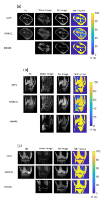 |
18 |
Cluster Based Sparse Variational Minimization for
Multi-Compartment Dictionary Fitting to bSSFP Signal Profiles
Berk Can Acikgoz1,2,
Adèle L.C. Mackowiak1,2,3,
Nils M.J. Plähn1,2,
Yasaman Safarkhanloo4,
Eva S. Peper1,2,
Tom Hilbert3,5,6,
Giulia M.C. Rossi3,
and Jessica A.M. Bastiaansen1,2
1Department of Diagnostic, Interventional and Pediatric Radiology (DIPR), Inselspital, Bern University Hospital, University of Bern, Bern, Switzerland, 2Translation Imaging Center (TIC), Swiss Institute for Translational and Entrepreneurial Medicine, Bern, Switzerland, 3Department of Radiology, Lausanne University Hospital (CHUV) and University of Lausanne (UNIL), Lausanne, Switzerland, 4Advanced Clinical Imaging Technology,Department of Cardiology, Inselspital, Bern University Hospital, Bern, Switzerland, 5Siemens Healthineers International AG, Lausanne, Switzerland, 6LTS5, École Polytechnique Fédérale de Lausanne (EPFL), Lausanne, Switzerland Keywords: Data Processing, Quantitative Imaging, phase-cycled bSSFP, fat fraction A novel cluster-based sparse total-variation minimization (CSTV) dictionary matching algorithm for multi-compartment parameter mapping applied to a phase-cycled balanced steady state free precession (PC-bSSFP) signal profile is presented. A dictionary encoding off-resonance frequency and relaxation time ratio is matched to the measured profiles with the proposed method to recover the underlying fat fraction, proton density, and water-fat-separated images simultaneously. The performance of the proposed method is tested and compared with a Laplacian regularized non-negative least squares dictionary matching method in phantom and in vivo experiments. |
|
4614. |
19 |
Differential quantification of cortical and medullary perfusion
through Gaussian Mixture Model based segmentation on
transplanted renal MRI
Anne Oyarzun-Domeño1,2,
Izaskun Cía 1,
Rebeca Echeverria-Chasco2,3,
María A. Fernández-Seara2,3,
Paloma L. Martin-Moreno2,4,
Nuria Garcia-Fernandez2,4,
Gorka Bastarrika2,3,
Javier Navallas1,2,
and Arantxa Villanueva1,2,5
1Electrical, Electronics and Communications Engineering, Public University of Navarre, Pamplona, Spain, 2Health Research Insitute of Navarra, IdiSNA, Pamplona, Spain, 3Department of Radiology, Clínica Universidad de Navarra, Pamplona, Spain, 4Department of Nephrology, Clínica Universidad de Navarra, Pamplona, Spain, 5Institute of Smart Cities (ISC), Pamplona, Spain Keywords: Data Processing, Perfusion Renal perfusion quantification is of importance in the post-operative surveillance of the allograft in translated patients. Together with cortical perfusion measurement, there is a strong interest in the quantification of medullary perfusion values, which requires an additional segmentation step of renal compartments. We applied Gaussian Mixture Models over renal MRI dataset to automatically extract the labels for each compartment to separately calculate cortical and medullary perfusion values. Proposed method showed performance metrics above 85% against ground truth labels and correlation coefficient above 96% and 58% for cortical and medullary perfusion values comparing with ground truth perfusion values. |
|
4615.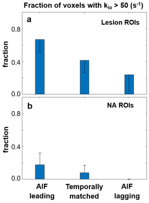 |
20 |
The effect of AIF and tissue time-course temporal alignment on
pharmacokinetic model trans-cytolemmal water exchange
sensitivity
Xin Li1,
Wei Huang1,
William D. Rooney1,
and Charles S Springer1
1Advanced Imaging Research Center, Oregon Health & Science University, Portland, OR, United States Keywords: Data Analysis, DSC & DCE Perfusion Using DCE modeling to quantify prostate transcytolemmal water exchange effect faces the challenges of often insufficient interstitial contrast agent concentration as well as suboptimal temporal resolution. Using ultra-high temporal resolution prostate DCE data, the goal of this work is to investigate the impact of DCE-MRI temporal alignment between the arterial input function (AIF) and the tissue time-courses on water exchange quantification. Results here show that when perfect alignment is not achievable in practice, an earlier AIF bolus arrival bias will generally reduce the capability for water-exchange quantification using DCE-MRI. |
|
 Back to Meeting Home
 Back to the Program-at-a-Glance
The International Society for Magnetic Resonance in Medicine is accredited by the Accreditation Council for Continuing Medical Education to provide continuing medical education for physicians.
 Back to Meeting Home
 Back to the Program-at-a-Glance
 View Presentation Video