Digital Poster
Imaging Neurofluids: New Methods & Applications I
ISMRM & ISMRT Annual Meeting & Exhibition • 03-08 June 2023 • Toronto, ON, Canada

Digital Poster
Imaging Neurofluids: New Methods & Applications I
| Computer # | |||
|---|---|---|---|
2999.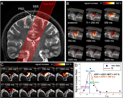 |
121 |
Intrinsic CSF outflow declines with age in healthy humans
detected by spin-labeling MRI
Vadim Malis1,
Won Bae1,2,
Asako Yamamoto3,
Linda McEvoy1,
Marin McDonald1,
and Mitsue Miyazaki1
1Radiology, University of California, San Diego, La Jolla, CA, United States, 2VA San Diego Healthcare System, San Diego, CA, United States, 3Radiology, Teikyo University, Tokyo, Japan Keywords: Neurofluids, Aging, cerebrospinal fluid (CSF), intrinsic CSF outflow, egress pathways Parasagittal dura (PSD) along the superior sagittal sinus (SSS) can be visualized using FLAIR and 3D SSFSE without gadolinium-contrast administration. Visualization of intrinsic CSF outflow from the PSD to the SSS can be achieved in human brains using spin-labeling MRI at clinical 3-T MRI. The spin-labeling MRI shows intrinsically tagged CSF outflow from the upper PSD and lower PSD to SSS. The quantitative intrinsic CSF outflow metrics indicate an age-related decline of the intrinsic CSF outflow at the SSS in human brains. |
|
3000.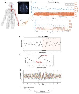 |
122 |
Paravascular Fluid Dynamics Reveal Arterial Stiffness Assessed
using Dynamic Diffusion-Weighted Imaging (dDWI)
Qiuting Wen1,
Adam Wright1,2,
Yunjie Tong2,
Yi Zhao1,
Shannon L. Risacher1,
Andrew J. Saykin1,
Yu-Chien Wu1,
Kalen Riley1,
and Kaustubh Limaye1
1Indiana University, School of Medicine, Indianapolis, IN, United States, 2Weldon School of Biomedical Engineering Department, Purdue University, West Lafayette, IN, United States Keywords: Neurofluids, Aging We recently developped a novel technique, dynamic diffusion-weighted imaging (dDWI), for measuring paravascular cerebrospinal fluid (pCSF) dynamics. In this work, we evaluated the time shifts between the pulsation-driven pCSF waves (measured by dDWI) and finger pulse waves (measured by scanner’s built-in finger pulse oximeter) to calculate brain-finger pulse wave travel time. Our preliminary results of an aging cohort support that the dDWI-derived brain-finger TimeDelay can be a surrogate for arterial stiffness. This method can be used as an add-on analysis to the recently developed dDWI framework to offer information about the participant’s vascular conditions. |
|
3001.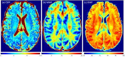 |
123 |
Changes in Brain Water Components in Normal Aging
Liangdong Zhou1,
Yi Li1,
Gloria C Chiang1,
Elizabeth Sweeney2,
Xiuyuan H Wang1,
Susan Gauthier1,
Yi Wang1,
Amy Kuceyeski1,
Mony de Leon1,
and Thanh Nguyen1
1Weill Cornell Medicine, New York, NY, United States, 2University of Pennsylvania, Philadelphia, PA, United States Keywords: Neurofluids, Aging Water homeostasis in brain involves the fluid dynamic through the intracellular fluid, extracellular fluid and CSF, and it’s important to help maintaining the brain function. The change of water compartments with aging may reflect the physiology and pathophysiology of the brain. We mapped the brain CSF water fraction, myelin water fraction and intro-extracellular water fraction using an MR FAST-T2 relaxometry approach. We found the brain CSF water quadratically increases with normal aging. Myelin water increases before the 50s and then decrease in adults. Intro-extracellular water decreases in the adult lifespan. This results are impactful for understanding the glymphatic clearance mechanism. |
|
3002.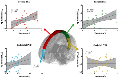 |
124 |
Sequential PET/MR reveals evidence of parasagittal dural
hypertrophy in the setting of elevated Aβ burden in adults with
Alzheimer’s disease
Alexander K Song1,2,
Kilian Hett1,
Jarrod J. Eisma1,
Colin D. Mcknight3,
Jason Elenberger1,
Adam J. Stark1,
Hakmook Kang4,5,
Ciaran M. Considine1,
Manus J. Donahue1,
and Daniel O. Claassen1
1Neurology, Vanderbilt University Medical Center, Nashville, TN, United States, 2Vanderbilt Brain Institute, Vanderbilt University, Nashville, TN, United States, 3Radiology and Radiological Sciences, Vanderbilt University Medical Center, Nashville, TN, United States, 4Biostatistics, Vanderbilt University Medical Center, Nashville, TN, United States, 5Center for Quantitative Sciences, Vanderbilt University Medical Center, Nashville, TN, United States Keywords: Neurofluids, Alzheimer's Disease A pathological hallmark of Alzheimer’s disease (AD) is the elevated aggregation of protein amyloid-β (Aβ) in the cerebrum. Recent studies have suggested a role for the parasagittal dural (PSD) space in cerebrospinal fluid (CSF) egress and associated protein clearance. A fully connected neural network was used to generate PSD segmentation masks from 3D T2-weighted turbo-spin-echo data to assess the relationship between PSD space volume and Aβ burden estimated by 11C-Pittsburgh Compound B in AD participants. PSD space hypertrophy was significantly associated with elevated Aβ levels and was localized to the frontal and parietal subsegments of the PSD. |
|
3003.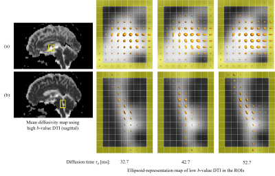 |
125 |
Low b-value DTI of CSF: Resolving Intravoxel Incoherent Motion
into Ordered and Disordered Motions
Yoshitaka Bito1,2,
Hisaaki Ochi1,2,
Ryuji Shirase1,
Wataru Yokohama1,
Kuniaki Harada2,
and Kohsuke Kudo2
1FUJIFILM Healthcare Corporation, Tokyo, Japan, 2Department of Diagnostic Imaging, Hokkaido University Graduate School of Medicine, Sapporo, Japan Keywords: Neurofluids, Diffusion/other diffusion imaging techniques Low b-value DTI (Low-b DTI) has been recently proposed for investigating the CSF motion. Here, an analysis technique using the Low-b DTI was proposed for resolving intravoxel incoherent motion into ordered (linear) and disordered (random) motions of the CSF. A normal-subject study demonstrated that the proposed technique can differentiate characteristics of the complex CSF motion in typical ROIs. The proposed technique can be useful in investigating the dynamics of neurofluids. |
|
3004.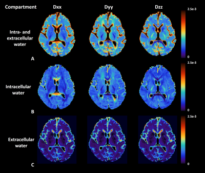 |
126 |
Associations between Extracellular Diffusivity Along
Perivascular Space (eALPS) and Structural Abnormalities in Brain
Sang-Young Kim1,
Eunju Kim1,
Jinwoo Hwang1,
Joo Hyun Kim1,
and Chae Jung Park2
1Health Systems, Philips Healthcare, Seoul, Korea, Republic of, 2Department of Radiology, Yongin Severance Hospital, Yonsei University College of Medicine, Yongin, Korea, Republic of Keywords: Neurofluids, Diffusion Tensor Imaging Non-invasive measure of glymphatic function or flow in vivo has gained a great attention from neuroscience research community. Diffusion tensor imaging (DTI) can provide a mean that measures the diffusivity along perivascular space (ALPS). However, conventional DTI captures diffusion indices from both tissue and free water compartments due to partial volume effects, which may raise a concern about validity of DTI-ALPS index for glymphatic function. In this work, we present a novel method that can extract extracellular diffusivity (i.e., eALPS) using free water eliminated DTI and investigate the relationship between eALPS and structural abnormalities in brain. |
|
3005.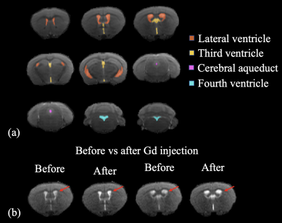 |
127 |
Impaired permeability in the choroid plexus in a tauopathy mouse
model – a pilot study
Yuhan Bian1,2,3,
Ning Wang4,
Di Cao1,2,3,
Yuanqi Sun1,2,3,
Chunming Gu1,2,3,
Yinghao Li1,2,3,
Jiangyang Zhang5,
Peter C.M. Van Zijl1,2,
Xiaobo Mao4,
and Jun Hua1,2
1F.M. Kirby Research Center for Functional Brain Imaging, Kennedy Krieger Institute, Baltimore, MD, United States, 2Neurosection, Division of MRI Research, Department of Radiology, Johns Hopkins, Baltimore, MD, United States, 3Department of Biomedical Engineering, Johns Hopkins, Baltimore, MD, United States, 4Department of Neurology, Johns Hopkins, Baltimore, MD, United States, 5Center for Biomedical Imaging, NYU Grossman School of Medicine, New York, NY, United States Keywords: Neurofluids, Alzheimer's Disease The choroid plexus (CP) is a gateway for the exchange of various metabolites between the microvasculature and brain. Impairment of CP has been reported in various diseases. Recently, dynamic-susceptibility-contrast-in-the-CSF (cDSC) MRI was developed to measure Gd-induced signal changes in the CSF. Here, cDSC MRI was performed with Intraperitoneal Gd-injection in a tauopathy mouse model to examine CP permeability. The maximal Gd-induced signal change was greater in the ventricles in tau mice than WT mice, indicating an increase of Gd leakage from the microvasculature into the ventricles through the CP. |
|
3006.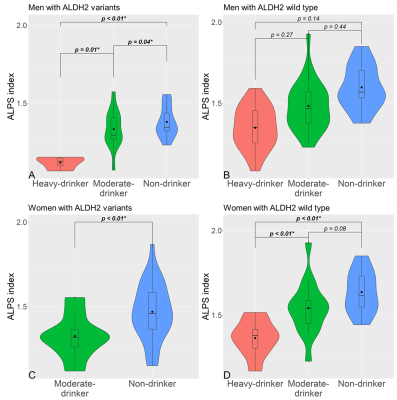 |
128 |
Investigation of the ALDH2 genetic polymorphism effect in
alcohol-related glymphatic dysfunction using DTI-ALPS
Yuichi Morita1,2,
Koji Kamagata1,
Kaito Takabayashi1,
Christina Andica1,3,
Junko Kikuta1,
Shohei Fujita1,2,
Hiroki Tabata4,
Hitoshi Naito5,
Yuki Someya4,6,
Hideyoshi Kaga5,
Toshiaki Akashi1,
Akihiko Wada1,
Yoshifumi Tamura4,5,
Ryuzo Kawamori4,5,
Hirotaka Watada4,5,
Toshiaki Taoka7,
Shinji Naganawa8,
Osamu Abe2,
and Shigeki Aoki1
1Department of Radiology, Graduate School of Medicine, Juntendo university, Tokyo, Japan, 2Department of Radiology, Graduate School of Medicine, The University of Tokyo, Tokyo, Japan, 3Faculty of Health Data Science, Juntendo University, Chiba, Japan, 4Sportology Center, Graduate School of Medicine, Juntendo university, Tokyo, Japan, 5Department of Metabolism & Endocrinology, Graduate School of Medicine, Juntendo university, Tokyo, Japan, 6Graduate School of Health and Sports Science, Juntendo University, Chiba, Japan, 7Department of Innovative Biomedical Visualization, Graduate School of Medicine, Nagoya University, Aichi, Japan, 8Department of Radiology, Graduate School of Medicine, Nagoya University, Aichi, Japan Keywords: Neurofluids, Diffusion Tensor Imaging, Glymphatic system, DTI-ALPS Excessive alcohol intake seriously damages the brain. Previous animal studies reported that the glymphatic system, which is a brain waste clearance system via the cerebral spinal fluid, is affected by chronic high alcohol consumption. Glymphatic dysfunction is related to cognitive impairment. The changes of diffusivity along the perivascular space (ALPS) indices associated with heavy, moderate, and no‑alcohol intake and executive function with or without aldehyde dehydrogenase 2 (ALDH2) rs671 polymorphism was evaluated. The present study revealed the glymphatic function and executive function decline in heavy drinkers. Furthermore, ALDH2 rs671 variants may increase vulnerability to alcohol-induced glymphatic dysfunction. |
|
3007.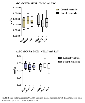 |
129 |
The Application Value of DWI in Differentiating Mega Cisterna
Magna from Cisterna Magna Arachnoid Cyst
Wang Junying1,
Pylypenko Dmytro 2,
and Shi Hao1
1Department of Radiology,, The First Affiliated Hospital of Shandong First Medical University, Jinan, China, 2GE Healthcare,, Beijing, China Keywords: Neurofluids, Diffusion/other diffusion imaging techniques, Arachnoid Cyst;Mega Cisterna Magna The differences between TAC, MCM and CMAC were measured respectively. For TAC and CMAC groups, both ADC and eADC value showed no statistical difference. There was a statistically significant difference in eADC between TAC and MCM, but no significant difference in ADC. |
|
3008.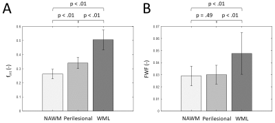 |
130 |
Exploring microstructural changes in patients with vascular
cognitive impairment using multi-b-value diffusion and
T2-relaxometry MRI
Gerhard Drenthen1,
Paulien HM Voorter1,
Maud van Dinther2,
Julie Staals2,
Robert J van Oostenbrugge2,
Walter H Backes1,
and Jacobus FA Jansen1
1Department of Radiology & Nuclear Medicine, School for Mental Health and Neuroscience, Maastricht University Medical Center, Maastricht, Netherlands, 2Department of Neurology, CARIM School for Cardiovascular Diseases, Maastricht University Medical Center, Maastricht, Netherlands Keywords: Neurofluids, Relaxometry, Interstitial fluid Local increases of interstitial fluid (ISF) predates axonal damage in white matter hyperintensities (WMH). However, it is challenging to disentangle these processes on T2w-MRI. By using multi-b-value diffusion and multi-echo T2-relaxometry imaging, we aimed to quantify markers of ISF volume and mobility in patients with vascular cognitive impairment (VCI) and healthy controls (HC). A trend for a higher mobility and volume of ISF was found in VCI patients compared to HC. Moreover, in VCI, elevated mobility of ISF was already present in perilesional tissue compared to normal-appearing white matter, suggesting that it might be an early marker of WMH development. |
|
3009.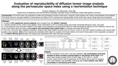 |
131 |
Evaluation of reproducibility of diffusion tensor image analysis
along the perivascular space index using a reorientation
technique
Hiroyuki Tatekawa1,
Shu Matsushita1,
and Yukio Miki1
1Department of Diagnostic and Interventional Radiology, Osaka Metropolitan University, Osaka, Japan Keywords: Neurofluids, Diffusion Tensor Imaging, ALPS index; glymphatic system; Diffusion tensor image analysis along the perivascular space (DTI-ALPS) index is expected to reflect the glymphatic function of the brain, however, head rotation may reduce reliability. This study evaluated whether a reorientation technique, which registers image and vector information of DTI data, improves the reproducibility of the calculation of the ALPS index. A total of 234 cognitively normal subjects were selected from the OASIS-3 dataset. In the evaluation of the intra- and inter-reliability, the reorientation technique enabled to show good to excellent reproducibility (intraclass correlation coefficients > 0.85) in calculating ALPS index even when head rotation existed. |
|
3010.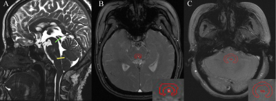 |
132 |
Combining CSF hydrodynamics and 4th ventricle outlet morphology
improves predictive performance of decompression for CM-I
patients
Yawen Xiao1,
Shiqi Chen1,
Zhaotao Zhang1,
Jiankun Dai2,
Yifei Gui1,
and Xinlan Xiao1
1Department of Radiology, The Second Affiliated Hospital of Nanchang University, Nanchang, China, 2GE Healthcare, MR Research China, Beijing, China Keywords: Neurofluids, Velocity & Flow Foramen magnum decompression (FMD) is the most used surgical treatment for Chiari malformation type I patients. However, 25~33% of them have persistent symptoms after surgery. This study aimed at investigating the possibility to predict the FMD outcome by using cerebral fluid (CSF) hydrodynamics and 4th ventricle outlet morphology obtained from magnetic resonance imaging. 27 patients were included and 17 of them had improved outcomes after FMD. Our results showed the peak diastolic velocity of CSF in aqueduct and the width of 4th ventricle outlet were associated with the FMD outcome and combining them can improve the prediction performance. |
|
3011.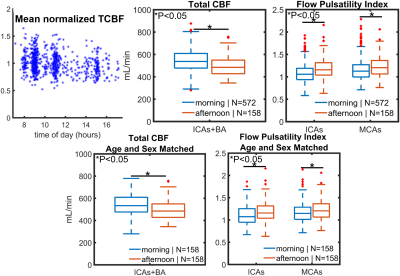 |
133 |
Diurnal Changes in Cerebrovascular Dynamics Measured from
4D-Flow
Leonardo A. Rivera-Rivera1,
Grant S. Roberts1,
Anthony Peret1,
Sterling C. Johnson1,
Oliver Wieben1,
Laura Eisenmenger1,
and Kevin M. Johnson1
1University of Wisconsin, Madison, Madison, WI, United States Keywords: Neurofluids, Velocity & Flow, Aging In this study, we investigated diurnal changes in cerebral hemodynamics from 4D-Flow in a large population of cognitively healthy older adults and in a younger group of healthy volunteers. To separate physiological and technical variability of 4D-Flow measures, volunteers were scanned at 7am, 4pm, and 10pm on the same day three times for each timepoint. Data supports strong cerebral blood flow fluctuations of physiological origin that are much higher than the technical variability. |
|
3012.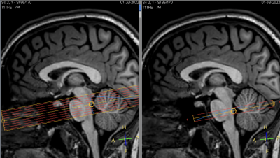 |
134 |
Assessment of CSF pulsatility in the fourth ventricle using EPI
phase contrast and fMRI sequences.
Adrian RUIZ-CHIAPELLO1,
Muriel MESCAM1,
Nathalie VAYSSIERE1,
Isabelle BERRY1,
and Florence REMY-EL BOUSTANI1
1CerCo, UMR 5549, CNRS/Toulouse 3 University, Toulouse, France Keywords: Neurofluids, Velocity & Flow, Cerebrospinal fluid Papers have shown a correlation between CSF flow in the fourth ventricle (V4) and EEG, and also that the low-frequency components (LFC) of EEG were positively correlated with glymphatic system (GS) activity. Analysis of the LFC of CSF flow could be a biomarker of GS degradation. We assessed 6 individuals by using an EPI phase contrast sequence to quantify the time evolution of CSF velocity in V4, and a fMRI sequence with which we exploited the inflow effect phenomenon. EPI-PC highlights the frequencies of physiological phenomena involved in CSF motion, and allows us to quantify LFC, indicative of GS efficiency. |
|
3013.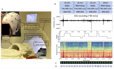 |
135 |
Three-dimensional flow velocity of cerebrospinal fluid (CSF) in
human brain during sleep via simultaneous EEG and dynamic sodium
MRI
Ying-Chia Lin1,
Xingye Chen1,
Simon Henin2,3,
Nahbila-Malikha Kumbella1,
Liz Aguilera1,
Zena Rockowitz3,
Ashley Clayton3,
James Babb1,
Yulin Ge1,
Arjun Masurkar2,3,
Anli Liu2,3,
Yvonne W. Lui1,4,
Fernando E. Boada1,5,
and Yongxian Qian1
1Department of Radiology, NYU Grossman School of Medicine, New York, NY, United States, 2Department of Neurology, NYU Grossman School of Medicine, New York, NY, United States, 3Department of Neurology, NYU Langone Health, New York, NY, United States, 4Department of Radiology, NYU Langone Health, New York, NY, United States, 5Department of Radiology, Stanford University, Stanford, CA, United States Keywords: Neurofluids, Velocity & Flow, Sodium MRI, CSF flow, Sleep Cerebrospinal fluid (CSF) flow plays a key role in clearance of waste proteins from the brain. Current proton (1H) MRI is limited to measurement of CSF flow in specific regions such as the aqueduct, due to the lack of capability distinguishing CSF from water. Sodium (23Na) MRI is uniquely sensitive to CSF, instead of water, and has the potential to measure CSF flow in entire brain. Here, we use a recently-developed dynamic sodium MRI to measure CSF flow velocity in the brain and to understand how it changes with sleep state monitored by MRI-compatible EEG. |
|
3014.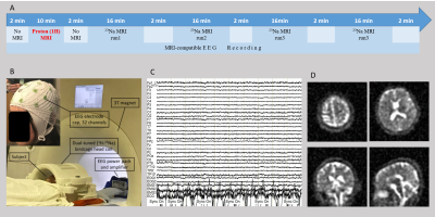 |
136 |
Extracellular Volume Change in the Human Brain During Sleep:
Monitored by MRI-Compatible EEG and Measured by Sodium MRI
Xingye Chen1,
Ying-Chia Lin1,
Simon Henin2,3,
Nahbila-Malikha Kumbella1,
Liz Aguilera1,
Zena Rockowitz3,
Ashley Clayton3,
Shichun Chen4,
Weiying Dai4,
James Babb1,
Yulin Ge1,
Arjun Masurkar2,3,
Yvonne W. Lui1,5,
Fernando E. Boada1,6,
Anli Liu2,3,
and Yongxian Qian1
1Radiology, NYU Grossman School of Medicine, New York, NY, United States, 2Neurology, NYU Grossman School of Medicine, New York, NY, United States, 3Neurology, NYU Langone Health, New York, NY, United States, 4Department of Computer Science, State University of New York at Binghamton, Binghamton, NY, United States, 5Radiology, NYU Langone Health, New York, NY, United States, 6Radiology, Stanford University, Stanford, CA, United States Keywords: Neurofluids, Velocity & Flow, MRI-compatible EEG, Sodium MRI, Sleep, Extracellular Space Sleep is reported in mince studies to increase extracellular space and improve CSF clearance of amyloid beta proteins from the animal brains. However, it is unclear whether sleep also benefits humans. This study used simultaneous MRI-compatible EEG to monitor sleep stage of human study subjects and used sodium MRI to quantify extracellular volume fraction. We seek to find the association between sleep stage and extracellular volume change in the human brain. |
|
3015.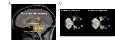 |
137 |
Measurement of changes in cerebrospinal fluid pulsation after
traumatic brain injury using EPI-based functional MRI
Jun-Hee Kim1 and
Sung-Hong Park1
1Korea Advanced Institute of Science and Technology, Daejeon, Korea, Republic of Keywords: Neurofluids, Traumatic brain injury A recently-proposed method of simultaneous CSF pulsation and BOLD activity imaging was applied to the TBI-fMRI dataset. The CSF pulsation was significantly lower in the TBI group compared to that of healthy control group. The CSF pulsation decreased significantly in the first 6 months after TBI and then no significant changes in the later stage, which was consistent with previous studies, where CSF pulsation from TBI patients was lower than that of control subjects and starts to slowly recover thereafter. This study can be expanded to post-TBI fMRI datasets in general to examine functional activity and CSF pulsation simultaneously. |
|
3016. |
138 |
Hierarchical clustering analysis reveals size-dependent
transport pathways of cerebrospinal fluid tracer in mouse brain
Yuran Zhu1,
Guanhua Wang2,
Yuning Gu1,
Huiyun Gao3,4,
Jing Zhang5,
Yunmei Wang3,4,
Xiaofeng Zhu5,
Chris A. Flask1,6,7,
and Xin Yu1,6,8
1Department of Biomedical Engineering, Case Western Reserve University, Cleveland, OH, United States, 2Department of Biomedical Engineering, University of Michigan, Ann Arbor, MI, United States, 3Cardiovascular Research Institute, Case Western Reserve University, Cleveland, OH, United States, 4Department of Medicine, Case Western Reserve University, Cleveland, OH, United States, 5Department of Biostatistics, Population and Quantitative Health Sciences, Case Western Reserve University, Cleveland, OH, United States, 6Department of Radiology, Case Western Reserve University, Cleveland, OH, United States, 7Department of Pediatrics, Case Western Reserve University, Cleveland, OH, United States, 8Department of Physiology and Biophysics, Case Western Reserve University, Cleveland, OH, United States Keywords: Neurofluids, Contrast Agent, Glymphatics This study investigated the impact of molecular size on transport kinetics and distribution of intracisternal tracers in mouse brain. Three MRI contrast agents with different molecular sizes were administered via cisterna magna. Their transport in the whole brain was observed by dynamic contrast-enhanced MRI for 2 hours. Our results show that the transport of 17O-water (H217O) was significantly faster and more extensive than the two gadolinium-based tracers (Gd-DTPA and GadoSpin). Time-lagged correlation analysis and clustering analysis also showed different cluster patterns between Gd-DTPA and H217O. These observations suggest the size-dependent differences in forces that drive tracer transport in the brain. |
|
3017.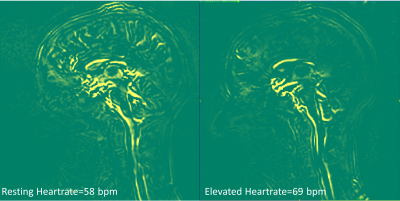 |
139 |
Changes in brain pulsatility associated with heart rate
elevation using amplified MRI and phase-contrast MRI
Haribalan Kumar1,2,3,
Haylea Rodgers3,4,
Jet Wright3,4,
Ben Bristow3,4,
Paul Condron3,5,
Taylor Emsden3,5,
Davidson Taylor3,6,
Samantha Holdsworth3,5,
Soroush Safaei2,
Gonzalo Maso Talou2,
Josh McGeown3,
Ed Maunder7,
and Eryn Kwon2,3,5
1GE Healthcare, Gisborne, New Zealand, 2Auckland Bioengineering Institute, University of Auckland, Auckland, New Zealand, 3Mātai Medical Research Institute, Gisborne, New Zealand, 4University of Otago, Dunedin, New Zealand, 5Faculty of Medical and Health Sciences & Centre for Brain Research, University of Auckland, Auckland, New Zealand, 6Ngai Tāmanuhiri, Rongowhakaata, Ngāti Porou, Tūranganui-a-Kiwa, Tūranganui-a-Kiwa, Tairāwhiti, New Zealand, 7Sports Performance Research Institute New Zealand, Auckland University of Technology, Auckland, New Zealand Keywords: Neurofluids, Neuroscience, Brain motion, image analysis Cardiac pulsatility is a key driver of brain pulsatility. However, changes in brain pulsatility in response to changes in heart rate have been sparsely studied. Using a combination of amplified MRI and phase-contrast MRI, brain parenchyma motion, blood flow, and CSF flow were measured and assessed during rhythmic hand-grip exercise to help understand the role of heart rate on brain physiology. This approach opens opportunities for probing the role of heart rate, brain fluid, and motion flow in various pathologies that affect the brain. |
|
3018.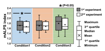 |
140 |
Effect of nighttime wakefulness on the brain's structure and
function: a study involving virtual ISMRM2021 attendees
Maho Kitagawa1,
Daisuke Sawamura2,
Yuta Urushibata3,
Hiroyuki Hamaguchi1,
Philip Kyeremeh Jnr Oppong1,
Daiki Sakamoto1,
and Khin Khin Tha1,4
1Laboratory for Biomarker Imaging Science, Hokkaido University Graduate School of Biomedical Science and Engineering, Sapporo, Japan, 2Department of Functioning and Disability, Hokkaido University Faculty of Health Sciences, Sapporo, Japan, 3Siemens Healthcare K.K., Tokyo, Japan, 4Global Center for Biomedical Science and Engineering, Hokkaido University Faculty of Medicine, Sapporo, Japan Keywords: Neurofluids, Brain, sleep, jet lag, glymphatic system, metabolite, cognition, functional connectivity Little had been reported about the effect of nighttime awakening (virtual jet lag) on the brain structure and function. In this prospective study which evaluated if short-time nighttime awakening due to virtual conference attendance affected the brain function, increased sleepiness, impaired information-processing ability, decreased mALPS index to suggest impaired glymphatic system functioning, a trend toward altered functional connectivity, were observed after nighttime awakening. The finding on mALPS index was similar to true jet lag. |
|
 Back to Meeting Home
 Back to the Program-at-a-Glance
The International Society for Magnetic Resonance in Medicine is accredited by the Accreditation Council for Continuing Medical Education to provide continuing medical education for physicians.
 Back to Meeting Home
 Back to the Program-at-a-Glance
 View Presentation Video