Digital Poster
Imaging Neurofluids: New Methods & Applications II
ISMRM & ISMRT Annual Meeting & Exhibition • 03-08 June 2023 • Toronto, ON, Canada

Digital Poster
Imaging Neurofluids: New Methods & Applications II
| Computer # | |||
|---|---|---|---|
3176.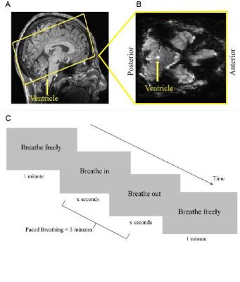 |
121 |
Respiratory Modulation of Cerebrospinal Fluid Flow During Paced
Breathing
Makaila Banks1,
Nicholas Cicero1,
Daniel Gomez2,3,
Ewa Beldzik4,
Vitaly Napadow2,3,
Jonathan Polimeni2,3,
and Laura Lewis4
1Neuroscience, Boston University, Boston, MA, United States, 2Massachusetts General Hospital, Athinoula A. Martinos Center for Biomedical Imaging, Charlestown, MA, United States, 3Department of Radiology, Harvard Medical School, Charlestown, MA, United States, 4Biomedical Engineering, Boston University, Boston, MA, United States Keywords: Neurofluids, Neurofluids, Cerebrospinal Fluid, CSF We used flow-sensitive fMRI to explore the effect of paced respiration on cerebrospinal fluid (CSF) flow. Results indicate that paced breathing tasks at rates between 0.1 Hz and 0.25 Hz increase CSF flow. All breathing paces showed a striking increase in CSF inflow-enhanced signals with the highest group-averaged amplitude being 26% during the 0.1 Hz breathing task. We conclude that diaphragmatic paced breathing is an effective modulator of CSF flow that may represent increased fluid transport relative to free breathing. |
|
3177.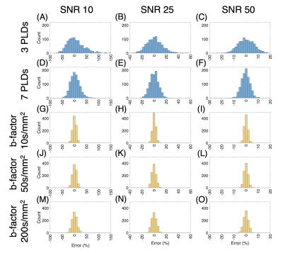 |
122 |
Effects of Noise on Blood-Brain Barrier Permeability Measurement
Error in Diffusion Weighted and Multi-Echo Arterial Spin
Labeling MRI
Yufei David Zhu1,
Donghoon Kim1,
Youngkyoo Jung1,2,
and Audrey Peiwen Fan1,3
1Biomedical Engineering, University of California, Davis, Davis, CA, United States, 2Radiology, University of California, Davis, Davis, CA, United States, 3Neurology, University of California, Davis, Davis, CA, United States Keywords: Neurofluids, Arterial spin labelling, Blood-Brain Barrier Arterial spin labeling (ASL) MRI is noninvasive and has the potential to become a sensitive, clinically useful way to measure blood-brain barrier (BBB) permeability. Here, we focused on two different ASL sequences, diffusion-weighted (DW) and multi-echo (T2), and performed simulations to compare the robustness of BBB permeability (Texch) measurement to different levels of noise. We conclude that the Texch fitting for DW ASL is more robust to noise at a physiological arterial transit time (ATT) of 1000ms at the expense of being more vulnerable to bias when the assumed ATT deviates from the true underlying ATT. |
|
3178.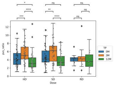 |
123 |
Perivascular Space Imaging during Therapy for Medulloblastoma
Ruitian Song1,
John O. Glass1,
Giles W. Robinson2,
Amar Gajja3,
Thomas E. Merchant4,
and Wilburn E. Reddick1
1Diagnostic Imaging, St Jude Children's Research Hospital, Memphis, TN, United States, 2Oncology, St Jude Children's Research Hospital, Memphis, TN, United States, 3Pediatric Medicine, St Jude Children's Research Hospital, Memphis, TN, United States, 4Radiation Oncology, St Jude Children's Research Hospital, Memphis, TN, United States Keywords: Neurofluids, Brain, Perivascular Space Imaging Patients with medulloblastoma treated with radio-chemotherapy (N=211) were examined by perivascular space (PVS) imaging at three time points: 0, 3, and 12 months. The patients were treated with different dose level based on risk factors. For low dose radiation, there were no significant changes in PVS between time points. A transient increase in PVS was observed for high and standard dose radiation during radiotherapy which subsequently resolved during chemotherapy. Perivascular space imaging is a potential biomarker to monitor the side effects from combined modality therapy. |
|
| 3179. | 124 |
Alterations of brain motion in idiopathic intracranial
hypertension (IIH) based on amplified MRI (aMRI)
Haribalan Kumar1,2,3,
Matthew Mcdonald3,4,
Alireza Sharifzadeh2,
Jet Wright3,
Eryn Kwon2,3,
Leigh Potter3,
Paul Condron3,5,
Taylor Emsden3,5,
Daniel Cornfeld3,
Graham Wilson3,6,
David Freschini6,
David Dubowitz7,
Miriam Scadeng7,
Helen Danesh-Meyer4,8,
Samantha Holdsworth3,7,
Sarah-Jane Guild2,9,
Soroush Safaei2,
and Gonzalo Maso Tolou2
1GE Healthcare, Gisborne, New Zealand, 2Auckland Bioengineering Institute, University of Auckland, Auckland, New Zealand, 3Mātai Medical Research Institute, Gisborne, New Zealand, 4Department of Opthalmology, University of Auckland, Auckland, New Zealand, 5Faculty of Medical and Health Sciences & Centre for Brain Research, University of Auckland, Auckland, New Zealand, 6Chelsea Hospital, Gisborne, New Zealand, 7Department of Anatomy & Medical Imaging, Faculty of Medical and Health Sciences, Centre for Brain Research, University of Auckland, Auckland, New Zealand, 8Eye Institute, Auckland, New Zealand, 9Department of Physiology, Faculty of Medical and Health Sciences, University of Auckland, Auckland, New Zealand Keywords: Neurofluids, Brain, IIH, Intracranial Pressure Heartbeat-driven brain motion has historically remained limited to the domain of research. Such motion may provide a window to characterise parenchymal and CSF pressures where direct, invasive measurements are disruptive and difficult to justify. Amplified MRI (aMRI) is a recently developed method which amplifies subtle brain motion. We show preliminary data that aMRI can be used to detect changes in brain motion associated with increased intracranial pressure in idiopathic intracranial hypertension (IIH), suggesting a potential non-invasive method for evaluating pressure levels of the subarachnoid space in patients with altered intracranial pressure. |
|
3180.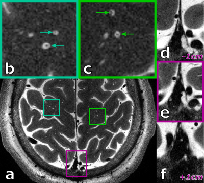 |
125 |
The potential of ultra-high resolution 7T T2-weighted TSE to
assess the glymphatic system’s structure
Hendrik Mattern1,2 and
Oliver Speck1,2,3,4
1Biomedical Magnetic Resonance, Otto von Guericke University, Magdeburg, Germany, 2German Center for Neurodegenerative Disease, Magdeburg, Germany, 3Center for Behavioral Brain Sciences, Magdeburg, Germany, 4Leibniz Institute for Neurobiology, Magdeburg, Germany Keywords: Data Acquisition, Neurofluids In this proof-of-principle study the currently untapped potential of UHF MRI is shown. Two young, healthy volunteers were scanned at 7T with 2D 0.25x0.25x1.0mm T2w TSE depicting a substantial number of PVS. At this high resolution, vessel segments inside the PVS could be studied and quantified. Further, hyperintense structures around and inside the dura and sinuses were depicted clearly. |
|
3181.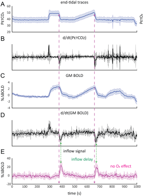 |
126 |
CO2 as an engine for neuro-fluid flow
Alex A Bhogal1,
Yunjie Tong2,
Eva van Grinsven3,
Jaco J.M. Zwanenburg1,
and Marielle E.P. Philippens3
1Center for Imaging Science, UMC Utrecht, Utrecht, Netherlands, 2Purdue University, West Lafayette, IN, United States, 3Radiotherapy, UMC Utrecht, Utrecht, Netherlands Keywords: Neurofluids, Neurofluids We use Blood Oxygen Level Dependent BOLD in combination with controlled hypercapnic stimulus to investigate the relationship between presumed cerebral blood volume changes and CSF flow in the brain. In a group of 11 subjects, we observe strong CSF inflow to the brain as a result of contracting cerebro-vasculature conforming to the Monro-Kellie Doctrine. Our results suggest that changes in arterial blood gases may provide an ‘engine’ through which to potentially enhance the clearance of waste from the brain. |
|
3182.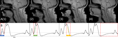 |
127 |
Effect of yawning on CSF and blood flow through the neck
Adam Martinac1,
Robert Lloyd1,
Stean Waters1,
and Lynne Bilston1
1Neuroscience Research Australia, Sydney, Australia Keywords: Neurofluids, Neurofluids, Yawning, CSF Despite being a common behaviour amongst many animals, research into the mechanics of yawning is limited. We have attempted to address this by performing sagittal real time scans and phase contrast scans to observe the flow of cerebrospinal fluid and blood through the midplane of the C3 vertebra of the spine during induced yawning. We found that over the course of a yawn venous flow and CSF flow both moved caudally during inspiration and rostrally during expiration. |
|
3183.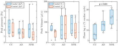 |
128 |
Fluid mechanics based distinct cerebrospinal fluid dynamics
signature in normal pressure hydrocephalus
Pragalv Karki1,
Petrice M Cogswell1,
Matthew C Murphy1,
Sandeep K Ganji1,
Jonathan Graff-Radford2,
David T Jones2,
Benjamin D Alder2,
and John Huston III1
1Department of Radiology, Mayo Clinic, Rochester, MN, United States, 2Department of Neurology, Mayo Clinic, Rochester, MN, United States Keywords: Neurofluids, Brain, Cerebrospinal fluid dynamics Normal pressure hydrocephalus (NPH) is a brain pathology with enlarged ventricles and is diagnosed as a cerebrospinal fluid (CSF) dynamics disorder based on a delayed ascent of radiotracer over the cerebral convexity and elevated flow through the cerebral aqueduct. In this study, we provide fluid dynamics-based evidence of disruption in CSF oscillation within the cerebral aqueduct in NPH patients. Particularly, we show that the peak volumetric flow is delayed with respect to peak transmantle pressure in NPH patients as compared to healthy control participants. |
|
3184. |
129 |
Characteristics of cerebral blood flow in sleep deprivation
based on arterial spin labeling
Chen Wang1,
Xiaolei Wang1,
Leilei Li1,
and Yuanqiang Zhu1
1Department of Radiology, Xijing Hospital, Air Force Medical University, Xi'an, China Keywords: Neurofluids, Nervous system ASL has been widely used in neuroimaging studies to measure the metabolic activity of local brain microstructure due to its advantages of no radiation damage, no introduction of exogenous contrast agent, high repeatability and fast scanning time.In this study, ASL perfusion imaging was used to analyze the changes of cerebral blood perfusion during normal sleep and sleep deprivation. |
|
3185.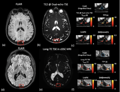 |
130 |
Concurrent measurement of perfusion parameters related to small
blood and lymphatic vessels in the human brain
Di Cao1,2,3,
Yuanqi Sun1,2,3,
Su Pan4,
Jay J. Pillai5,6,7,
Ye Qiao5,
Hanzhang Lu1,2,3,
Peter C.M. Van Zijl1,2,
Linda Knutsson1,2,8,
and Jun Hua1,2
1F.M. Kirby Research Center for Functional Brain Imaging, Kennedy Krieger Institute, Baltimore, MD, United States, 2Russell H. Morgan Department of Radiology and Radiological Science, Johns Hopkins University, Baltimore, MD, United States, 3Department of Biomedical Engineering, The Johns Hopkins University School of Medicine, Baltimore, MD, United States, 4School of Medicine, University of Maryland, Baltimore, MD, United States, 5Neuroradiology, Department of Radiology, Johns Hopkins University, Baltimore, MD, United States, 6Department of Neurosurgery, Johns Hopkins University, Baltimore, MD, United States, 7Neuroradiology, Mayo Clinic, Rochester, MN, United States, 8Department of Medical Radiation Physics, Lund University, Lund, Sweden Keywords: Neurofluids, DSC & DCE Perfusion Accumulating evidence has indicated the importance of studying the interaction between the microvascular and lymphatic systems in the brain. To date, most imaging methods can only measure blood or lymphatic vessels separately. This study proposes an MRI approach for concurrent measurement of perfusion parameters related to small blood and lymphatic vessels within one single scan. A dual-echo TSE sequence was optimized for the measurement of gadolinium(Gd)-induced blood and CSF signal changes. The proposed method showed consistent results in human brains as previous studies using separate methods. Signal changes from small blood vessels occurred faster than lymphatic vessels after intravenous Gd-injection. |
|
3186.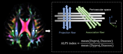 |
131 |
Abnormal glymphatic system function in patients with chronic
migraine using diffusion tensor image analysis along the
perivascular space
Xue Zhang1,2,
Wei Wang3,
Xueyan Zhang4,
Xiaoyan Bai1,2,
Ziyu Yuan3,
Ruiliang Bai5,6,7,
Bingjie Jiao6,
Yingkui Zhang2,
Zhiye Li1,2,
Peng Zhang3,
Hefei Tang3,
Yaqing Zhang3,
Xueying Yu3,
Yonggang Wang3,
and Binbin Sui2
1Department of Radiology, Beijing Tiantan hospital, Capital Medical University, Beijing, China, 2Tiantan Neuroimaging Center of Excellence, China National Clinical Research Center for Neurological Diseases, Beijing, China, 3Headache Center, Department of Neurology, Beijing Tiantan Hospital, Capital Medical University, Beijing, China, 4Department of Neurology, The First Affiliated Hospital of Zhengzhou University, Zhengzhou, China, 5Department of Physical Medicine and Rehabilitation of the Affiliated Sir Run Shumen Shaw Hospital and Interdisciplinary Institute of Neuroscience and Technology, School of Medicine, Zhejiang University, Hangzhou, China, 6key Laboratory of Biomedical Engineering of Education Ministry, College of Biomedical Engineering and Instrument Science, Zhejiang University, Hangzhou, China, 7MOE Frontier Science Center for Brain Science and Brain-machine Integration, School of Brain Science and Brain Medicine, Zhejiang University, Hangzhou, China Keywords: Neurofluids, Diffusion Tensor Imaging, Headache Chronic migraine (CM) is a common and highly disabling primary headache associated with vasomotion dysfunction. It is unclear whether glymphatic dysfunction develops due to frequent and chronic headache attacks in CM. In the current study, we evaluated the glymphatic system function in patients with CM using diffusion tensor image analysis along the perivascular space (DTI-ALPS) technique. We found a significantly higher ALPS index compared with healthy controls, and the alteration was significant only observed in the right hemisphere. Patients with CM have abnormalities in glymphatic function, and the right-prominent alterations might be a certain feature of CM. |
|
3187.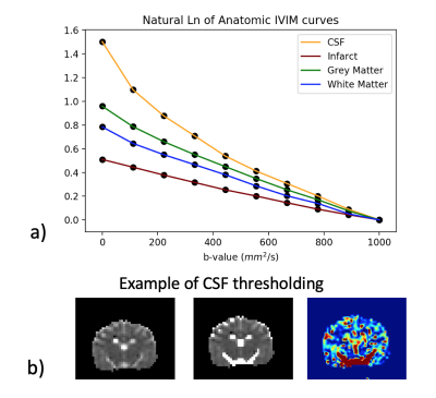 |
132 |
Machine Learning for Quantitative IVIM (qIVIM) Cerebral
Perfusion Imaging
Mira Liu1,
Julian Bertini1,
Niloufar Saadat2,
Chisondi Simba Warioba1,
Donovan Gorre1,
Timothy Carroll1,
and Gregory Christoforidis2
1University of Chicago, Chicago, IL, United States, 2Interventional Radiology, University of Chicago, Chicago, IL, United States Keywords: Neurofluids, Perfusion Intravoxel Incoherent Motion (IVIM) is a non-contrast perfusion scan that measures various speeds of molecular movement. Segmentation of IVIM signal origin, i.e. from blood, cerebrospinal fluid (CSF), or tissue water, is crucial. Simulation suggested Inversion Recovery should not be used in IVIM to remove CSF, and analysis of signal patterns suggested differing signal decay as a method of removing CSF during post-processing. A threshold determined by k-fold leave-one-out cross-validation on IVIM diffusion coefficient returned successful CSF segmentation (dice = .69), and CSF removal by supervised machine learning via linear discriminant analysis returned quantitative IVIM with strong agreement to microsphere perfusion. |
|
3188.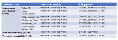 |
133 |
Cross vendors test-retest validation of diffusion tensor image
analysis along the perivascular space for evaluating glymphatic
system function
Xiaodan Liu1,2,3,
Giuseppe Barisano4,
Xingfeng Shao3,
Kay Jann3,
Hanzhang Lu5,
Konstantinos Arfanakis6,7,
Arvind Caprihan Caprihan 8,
Charles DeCarli9,
Brian T. Gold10,
Pauline Maillard9,
Clandia L. Satizabal11,
Elyas Fadaee12,
Mohamad Habes12,
Lara Stables13,
Herpreet Singh14,
Bruce Fischl15,16,17,18,
Andre van der Kouwe15,18,19,
Kristin Schwab14,
Karl G. Helmer15,18,19,
Steven M. Greenberg14,
and Danny JJ Wang3
1Radiology and Biomedical Imaging, University of California, San Francisco, San Francisco, CA, United States, 2Zilkha Neurogenetic Institue, University of Southern California, Los Angeles, CA, United States, 3Mark & Mary Stevens Neuroimaging and Informatics Institute, University of Southern California, Los Angeles, CA, United States, 4Neurosurgery, Stanford University, Palo Alto, CA, United States, 5Radiology, Johns Hopkins University School of Medicine, Baltimore, MD, United States, 6Biomedical Engineering, Illinois Institute of Technology, Chicago, IL, United States, 7Diagnostic Radiology and Nuclear Medicine, Rush University, Chicago, IL, United States, 8The Mind Research Network, Albuquerque, NM, United States, 9Neurology, University of California, Davis, Davis, Davis, CA, United States, 10Neuroscience, University of Kentucky, Lexington, KY, United States, 11Population Health Sciences and Glenn Biggs Institute for Neurodegenerative Diseases, University of Texas Health Science Center at San Antonio, San Antonio, TX, United States, 12Neuroimage Analytics Laboratory and Biggs Institute Neuroimaging Core, Glenn Biggs Institute for Neurodegenerative Diseases, University of Texas Health Science Center at San Antonio, San Antonio, TX, United States, 13Neurology, University of California, San Francisco, San Francisco, CA, United States, 14Neurology, Massachusetts General Hospital, Boston, MA, United States, 15Radiology, Harvard Medical School, Boston, MA, United States, 16Athinoula A. Martinos Center for Biomedical Imaging,Massachusetts General Hospital, Charlestown, MA, United States, 17Computer Science and AI Lab, Massachusetts Institute of Technology, Cambridge, MA, United States, 18Radiology, Massachusetts General Hospital, Boston, MA, United States, 19Athinoula A. Martinos Center for Biomedical Imaging, Massachusetts General Hospital, Charlestown, MA, United States Keywords: Neurofluids, Brain, glymphatic system The diffusion tensor image analysis along the perivascular space (DTI-ALPS) method was proposed to evaluate glymphatic system (GS) function. However, few studies validate its reliability and reproducibility. 50 participants’ DTI data from the MarkVICD consortium were recruited in this study. Two pipelines by using DSI studio and FSL software were developed for data processing and ALPS-index calculation. The mean ALPS-index was obtained by the average of bilateral ALPS-index and was used for testing the cross vendors test-retest reliability by using R studio. Mean ALPS-index demonstrated favorable inter-scanner reproducibility, inter-rater reliability and test-retest repeatability, offering a potential biomarker for GS function. |
|
3189.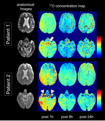 |
134 |
Visualization of Human Neurofluids Using Intrathecal 17O-Labeled
Water and MRI: A Preliminary Study
Hiroyuki Kameda1,2,
Taisuke Harada1,3,
Hiroyuki Sugimori4,
Xiawei Bai3,
Kazuyuki Minowa2,
and Kohsuke Kudo3,5
1Department of Diagnostic and Interventional Radiology, Hokkaido University Hospital, Sapporo, Japan, 2Department of Raiology, Faculty of Dental Medicine, Hokkaido University, Sapporo, Japan, 3Department of Diagnostic Imaging, Facaulty of Medicine and Graduate School of Medicine, Hokkaido University, Sapporo, Japan, 4Faculty of Health Sciences, Hokkaido University, Sapporo, Japan, 5Global Center for Biomedical Science and Engineering, Hokkaido University, Sapporo, Japan Keywords: Neurofluids, Neurofluids Imaging of neurofluids dynamics is a promising aspect in clinical imaging. Only few methods of long-term water tracking in the brain have been described for diagnostic purposes. Herein, we examined water circulation between the CSF and ISF using an intrathecal injection of 17O-labeled water and 3T-MRI in two patients with dementia. Signal changes were detected in the brain parenchyma after the intrathecal administration. This visualization technique can detect the distribution of water tracers to the CSF and brain parenchyma and could become a clinical tool for evaluating neurofluids dynamics in humans. |
|
3190.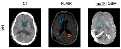 |
135 |
Intracranial Hemorrhage Patient CSF Susceptibility Variation:
Implications for Quantitative Susceptibility Mapping Reference
Selection.
Shutao Wang1,
Pascal Spincemaille2,
Magdy Selim3,
Ajith J Thomas4,
Aristotelis Filippidis5,
Yan Wen6,
Yi Wang2,
and Salil Soman1
1Radiology, Beth Israel Deaconess Medical Center, Harvard Medical School, Boston, MA, United States, 2Department of Radiology, Weill Cornell Medicine, New York, NY, United States, 3Neurology, Beth Israel Deaconess Medical Center, Harvard Medical School, Boston, MA, United States, 4Department of Neurological Surgery, Cooper University Health Care, Cooper Medical School of Rowan University, Camden, NJ, United States, 5Neurosurgery, Beth Israel Deaconess Medical Center, Harvard Medical School, Boston, MA, United States, 6GE Healthcare, New York, NY, United States Keywords: Neurofluids, Quantitative Susceptibility mapping, CSF Blood QSM is an emerging MR technique with many clinical applications, especially in the brain. CSF has been used by many researchers as the reference region to obtain quantitative QSM values for inter-subject comparison. Here, we demonstrate the QSM values at hemorrhagic CSF sites are significantly different from that at non-hemorrhagic CSF sites in the same patient with various types of intracranial hemorrhage. This finding has significant clinical implication as selection of CSF as the reference region should be made with caution in patients with intracranial hemorrhage. |
|
3191.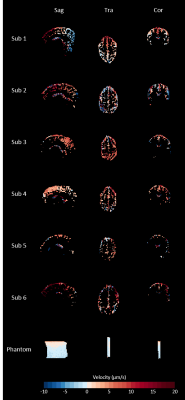 |
136 |
Assessing CSF secretion by measuring net velocity of CSF in the
human subarachnoid space using displacement encoding with
stimulated echoes at 7T.
Elisabeth C. van der Voort1,
Merlijn C.E. van der Plas1,
and Jacobus J.M. Zwanenburg1
1Department of Radiology, University Medical Center Utrecht, Utrecht, Netherlands Keywords: Neurofluids, Velocity & Flow Continuous secretion and absorption of cerebrospinal fluid (CSF) cause a net velocity that is important for cerebral clearance. However, measuring this net velocity is hampered by the cardiac and respiratory cycle which induce additional CSF motions. We show, with both simulations and in vivo measurements, that net velocity of CSF in the subarachnoid space can be measured using displacement encoding with stimulated echoes (DENSE), when contributions from heartbeat and respiration are accounted for during the analysis. Measured net velocity was 4.41±1.57 µm/s (6 subjects). Further research is needed to properly account for possible phase wraps in the measurements. |
|
3192.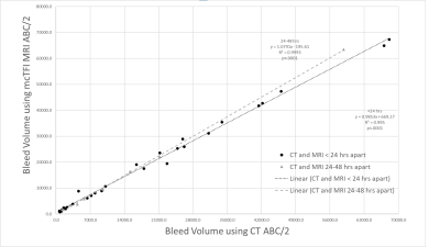 |
137 |
Application of the CT ABC/2 Method for Measuring Intracranial
Bleed Volume on mcTFI QSM MRI.
Ria Sharma1,
Geunwon Kim2,
Magdy Selim3,
Ajith J Thomas4,
Aristotelis Filippidis5,
Yan Wen6,
Pascal Spincemaille7,
Yi Wang7,
and Salil Soman1
1Radiology, Beth Israel Deaconess Medical Center, Harvard Medical School, Boston, MA, United States, 2Atrius Health, Boston, MA, United States, 3Neurology, Beth Israel Deaconess Medical Center, Harvard Medical School, Boston, MA, United States, 4Department of Neurological Surgery, Cooper University Health Care, Cooper Medical School of Rowan University, Camden, NJ, United States, 5Neurosurgery, Beth Israel Deaconess Medical Center, Harvard Medical School, Boston, MA, United States, 6GE Healthcare, New York, NY, United States, 7Department of Radiology, Weill Cornell Medicine, New York, NY, United States Keywords: Neurofluids, Brain, Intracranial Hemorrhage, ABC/2, Bleed Volume Estimation Intracranial hemorrhage (ICH) volume estimation is important for patient management. ABC/2 is a validated method of volume estimation using CT (2). Differing appearance of blood across MRI imaging techniques and blood age limits the ability of MRI to evaluate ICH volume. The QSM technique mcTFI has features well suited to bleed measurement. In this study we show that the widely used ABC/2 CT technique for ICH volume measurement applied to mcTFI QSM MRI yields ICH volumes strongly correlated with those obtained using CT. |
|
3193.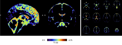 |
138 |
CSF oxygenation imaging using long TE heavily T2-weighted
Fluid-Attenuated Inversion Recovery
Manuel Taso1 and
David C Alsop1
1Division of MRI Research, department of Radiology, Beth Israel Deaconess Medical Center, Harvard Medical School, Boston, MA, United States Keywords: Neurofluids, Brain The cerebrospinal fluid plays an essential role in cerebral homeostasis, contributing to the delivery of oxygen and nutrients to brain tissue as well as clearing waste products. However, the mechanisms and functions of CSF oxygenation and their potential disturbance in pathology remain unknown. Dissolved O2 shortens T1 of water due to its paramagnetic properties. Because of its extremely long T2, selective CSF imaging can be achieved by using extremely long TE (>500ms) Spin-Echo based acquisitions. Therefore, we report here a study using ultra long TE Fluid-Attenuated Inversion Recovery to image baseline CSF oxygenation with minimal tissue contribution. |
|
 Back to Meeting Home
 Back to the Program-at-a-Glance
The International Society for Magnetic Resonance in Medicine is accredited by the Accreditation Council for Continuing Medical Education to provide continuing medical education for physicians.
 Back to Meeting Home
 Back to the Program-at-a-Glance
 View Presentation Video