Digital Poster
Inflammation Across Neurological Diseases I
ISMRM & ISMRT Annual Meeting & Exhibition • 03-08 June 2023 • Toronto, ON, Canada

Digital Poster
Inflammation Across Neurological Diseases I
| Computer # | |||
|---|---|---|---|
2608.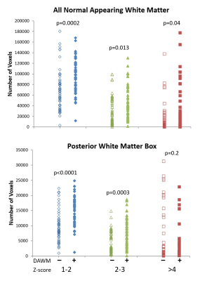 |
81 |
Geometric mean T2 of the intra/extracellular water can help
identify diffusely abnormal white matter in multiple sclerosis
Irene Margaret Vavasour1,2,
Kyle Vavasour3,
Adam Dvorak4,
Tigris Joseph2,4,
Robert Carruthers5,
Shannon Kolind1,2,4,5,
Alice Schabas5,
Ana-Luiza Sayao5,
Virginia Devonshire5,
Roger Tam1,6,
GR Wayne Moore2,5,7,
Anthony Traboulsee5,
David Li1,5,
and Cornelia Laule1,2,4,7
1Radiology, University of British Columbia, Vancouver, BC, Canada, 2International Collaboration on Repair Discoveries, University of British Columbia, Vancouver, BC, Canada, 3University of British Columbia, Vancouver, BC, Canada, 4Physics and Astronomy, University of British Columbia, Vancouver, BC, Canada, 5Medicine, University of British Columbia, Vancouver, BC, Canada, 6School of Biomedical Engineering, University of British Columbia, Vancouver, BC, Canada, 7Pathology & Laboratory Medicine, University of British Columbia, Vancouver, BC, Canada Keywords: Multiple Sclerosis, Multiple Sclerosis, diffusely abnormal white matter, T2 relaxation Diffusely abnormal white matter (DAWM) is seen on 25-50% of conventional brain MRI scans from all stages of multiple sclerosis (MS). From 8 advanced MRI metrics, geometric mean T2 of the intra/extracellular water (IET2) best separated MS participants with and without DAWM. Comparing IET2 to controls using z-scores, regions with an intermediate increase in IET2 (z-scores between 1-2) may identify DAWM voxels. |
|
2609. |
82 |
Free water alterations of deep gray matters over 1-2 years in
small vessel disease
Yawen Sun1,
Hongjiang Wei2,
and Yan Zhou1
1Department of Radiology, Ren Ji Hospital, School of Medicine, Shanghai Jiao Tong University, Shanghai, China, 2School of Biomedical Engineering, Shanghai Jiao Tong University, Shanghai, China Keywords: Dementia, Diffusion/other diffusion imaging techniques, Free water mapping This study was to evaluate the neuroinflammation-related characteristics of the deep gray matter using free-water mapping in small vessel disease over 1-2 years. Our results support the presence of elevated free water at the preclinical stage of small vessel disease, which remains persistent during the early course of the illness. The free water values changes might be the biomarkers for the mechanism of cognitive decline in the evolution of small vessel disease. |
|
2610. |
83 |
Assessment of the effects of coronary artery disease on brain
oxygen extraction fraction using quantitative susceptibility
mapping.
Ali Rezaei1,2,
Stefanie A Tremblay1,2,
Dalia Sabra1,2,3,4,
Safa Sanami1,2,
Brittany Intzandt5,
Julia Huck1,
Zacharie Potvin-Jutras1,2,
Christine Gagnon2,
Amelie Mainville-Berthiaume6,
Lindsay N Wright1,2,
Dajana Vuckovic7,
Josep Iglesies-Grau2,8,
Thomas Vincent2,
Mathieu Gayda2,
Anil Nigam2,
Louis Bherer2,8,9,
and Claudine J Gauthier1,2
1Physics Department, Concordia University, Montreal, QC, Canada, 2Centre Epic and Research Center, Montreal Heart Institute, Montreal, QC, Canada, 3Department of Biomedical Science, Faculty of Medicine, Université de Montreal, Montreal, QC, Canada, 4PERFORM Centre, Concordia University, Montreal, QC, Canada, 5BrainLab, Hurvitz Brain Sciences Program, Sunnybrook Research Institute, Toronto, ON, Canada, 6Department of Psychology, Concordia University, Montreal, QC, Canada, 7Department of Chemistry and Biochemistry, Concordia University, Montreal, QC, Canada, 8Department of Medicine, Université de Montreal, Montreal, QC, Canada, 9Research Center, Institut Universitaire de Gériatrie de Montréal, Montreal, QC, Canada Keywords: Blood vessels, Oxygenation Here we investigated the effect of coronary artery disease (CAD) on the brain oxygen extraction fraction (OEF) using quantitative susceptibility mapping (QSM). Our results, in a pilot dataset, show that OEF was significantly higher in CAD patients than in healthy controls in the caudate nucleus. This may be the result of declining cerebrovascular health in CAD patients in this region. However, since the caudate nucleus is iron-rich, this difference may also be interpreted as iron deposition as CAD has a known neuroinflammatory effect. Future work will investigate the impact of CAD on other metabolic and vascular components of brain health. |
|
2611. |
84 |
Effects of HIV-associated chronic inflammation on intracranial
vessels using semi-automatic segmentation of MRA cerebrovascular
features
Nhat Hoang1,
Henry Wang2,
Sahin Bogachan3,
Muhammad Waqas Khan4,
Abrar Faiyaz5,
Meera Singh3,
Jinjiang Pang6,
Shumin Wang6,
Li Chen7,
Chun Yuan7,
Jianhui Zhong1,
Hongmei Yang8,
Md Nasir Uddin3,
and Giovanni Schifitto3
1Physics, University of Rochester, Rochester, NY, United States, 2Radiology, University of Rochester, Rochester, NY, United States, 3Neurology, University of Rochester, Rochester, NY, United States, 4Neurology-Stroke Division, University of Rochester, Rochester, NY, United States, 5Department of Electrical and Computer Engineering, University of Rochester, Rochester, NY, United States, 6Cardiology Research, University of Rochester, Rochester, NY, United States, 7Department of Electrical and Computer Engineering, University of Washington, Seattle, WA, United States, 8Biostatistics, University of Rochester, Rochester, NY, United States Keywords: Neuroinflammation, Vessels, MRA, CSVD, HIV HIV infected individuals (HIV+) are subjected to high risks of neurological complications, including cerebrovascular disease. Quantification of vascular features may provide a tool to investigate pathomechanisms and monitor cerebrovascular disease progression. In this study we used intracranial artery feature extraction (iCafe) to compare HIV+ with age matched controls. |
|
2612.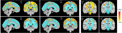 |
85 |
White matter tracts of MS patients with progression independent
of relapse activity show DTI-alterations compared to clinically
stable patients
Mario Ocampo-Pineda1,2,3,
Alessandro Cagol1,2,3,
Muhamed Barakovic1,2,3,
Po-Jui Lu1,2,3,
Jannis Müller1,2,3,
Sabine Schaedelin4,
Pascal Benkert4,
Matthias Weigel1,2,3,5,
Lester Melie-Garcia1,2,3,
Jens Kuhle2,3,
Ludwig Kappos1,2,3,
and Cristina Granziera1,2,3
1Translational Imaging in Neurology (ThINk) Basel, Department of Biomedical Engineering, Faculty of Medicine, University Hospital Basel and University of Basel, Basel, Switzerland, 2Department of Neurology, University Hospital Basel, Basel, Switzerland, 3Research Center for Clinical Neuroimmunology and Neuroscience Basel (RC2NB), University Hospital Basel and University of Basel, Basel, Switzerland, 4Clinical Trial Unit, Department of Clinical Research, University Hospital Basel, University of Basel, Basel, Switzerland, 5Division of Radiological Physics, Department of Radiology, University Hospital Basel, Basel, Switzerland Keywords: White Matter, Diffusion Tensor Imaging Progression independent of relapse activity (PIRA) has been described in patients with multiple sclerosis (MS) even in the earliest disease stages. Patients with PIRA show increased atrophy rates in multiple brain regions compared to stable patients. Here, we investigated whether patients with PIRA exhibit loss of integrity in WM tracts compared to stable patients. We studied 62 RRMS patients, 27 PIRA and 35 stable patients using a clinical DW-MRI protocol. Our results showed that PIRA patients present smaller FA values in areas of corpus callosum and along corticosprinal tract. These differences suggest neurodegeneration in major WM tracts of PIRA patients. |
|
2613. |
86 |
Microstructural alterations in gray and white matter integrity
in migraine with aura
Kyla Ann Gaudet1,
Laleh Eskandarian1,
Melanie Li2,
Susie Y Huang1,
and Katharina Eikermann-Haerter3
1Athinoula A. Martinos Center for Biomedical Imaging, Department of Radiology, Massachusetts General Hospital, Charlestown, MA, United States, 2Department of Neurology, New York University Grossman School of Medicine, New York, NY, United States, 3Division of Neuroradiology, Department of Radiology, New York University Grossman School of Medicine, New York, NY, United States Keywords: Gray Matter, Neuroinflammation Migraine is one of the most common neurological disorders. Thirty percent of migraine patients develop transient neurological symptoms, the so-called migraine aura. Migraineurs, particularly those with aura, show an increased incidence of white matter lesions (WML). To improve our understanding of WML in migraineurs, we utilized Soma and Neurite Density Image (SANDI) as an advanced diffusion magnetic resonance imaging signal model to assess in-vivo microstructure of gray matter and white matter. We found evidence for occult changes in microstructural gray and white matter integrity that point to a common underlying mechanism driving cellular inflammation and neuronal injury in migraine. |
|
2614.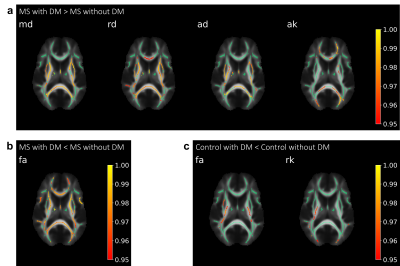 |
87 |
Elucidating the role of diabetes mellitus on multiple sclerosis
pathology using diffusion MRI
Michael Lan1,
Valentin Stepanov1,
Jenny Chen1,
Benjamin Ades-Aron1,
Dmitry S. Novikov1,
and Els Fieremans1
1Center for Biomedical Imaging, Department of Radiology, New York University Grossman School of Medicine, New York, NY, United States Keywords: Multiple Sclerosis, Diffusion/other diffusion imaging techniques Diabetes mellitus (DM) has been potentially associated with multiple sclerosis (MS), but has not been definitively shown to worsen its severity. Our study used diffusion kurtosis imaging (DKI) analyzed with tract-based spatial statistics to identify significant differences in DKI metrics in addition to measuring lesion volume in MS and Control patients with and without DM. We observed significantly increased diffusion and axial kurtosis and increased periventricular lesion volume in MS patients with DM compared to those without DM, indicating worsening inflammation and demyelination, suggesting that DM may serve as an exacerbating factor for worsening MS prognosis. |
|
2615.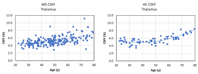 |
88 |
Change in thalamic cerebrospinal fluid fraction over the adult
lifespan and correlation with TSPO PET imaging in multiple
sclerosis patients
Thanh D. Nguyen1,
Liangdong Zhou1,
Yeona Kang2,
Emily Demmon1,
Michael Sakirsky1,
Elizabeth M. Sweeney3,
Yi Wang1,
Yi Li1,
and Susan A. Gauthier1
1Weill Cornell Medicine, New York, NY, United States, 2Howard University, Washington, DC, United States, 3University of Pennsylvania, Philadelphia, PA, United States Keywords: Multiple Sclerosis, Neurofluids We applied FAST-T2 multi-component T2 relaxometry to 200 MS patients and 66 healthy controls and found that the thalamic cerebrospinal fluid fraction (CSFF) increases with age and follows a different trajectory in MS. In 13 MS patients, we also found a strong correlation between CSFF and [11C]PK11195 uptake on PET in the thalamus and putamen, suggesting a connection between glymphatic dysfunction and microglial inflammation in the MS brain. |
|
2616.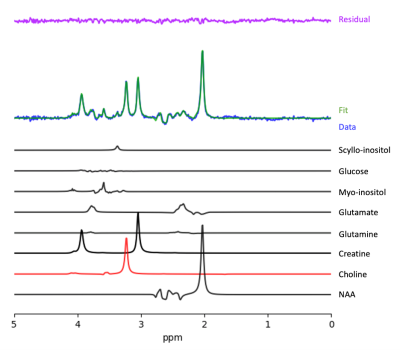 |
89 |
Dual-Task Gait Predicts Changes in Choline in the Primary Motor
Cortex of Older Individuals with Mild Cognitive Impairment
Jack TE Elkas1,
Frederico Pieruccini-Faria2,
Manuel Montero-Odasso2,
and Robert Bartha3
1Neuroscience Department, Western University, London, ON, Canada, 2Gait and Brain Lab, Parkwood Institute and Lawson Health Research Institute, London, ON, Canada, 3Department of Medical Biophysics and Robarts Research Institute, Schulich School of Medicine and Dentistry, London, ON, Canada Keywords: Dementia, Spectroscopy, Neuroinflammation, Dementia, Aging, Brain, Degenerative, Dementia, Neurodegeneration The dual-task cost on walking speed (DTC) can predict the progression of dementia in people with mild cognitive impairment (MCI). In MCI, levels of choline in the primary motor cortex, an MRS metabolite associated with inflammation, |
|
2617.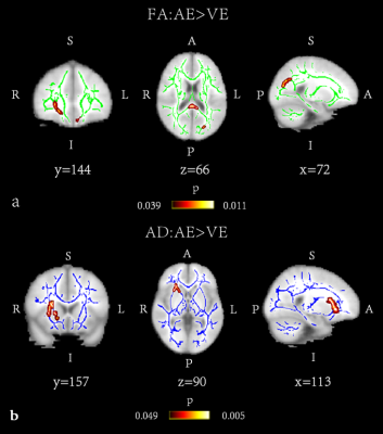 |
90 |
Diffusion tensor imaging study of white matter differences
between autoimmune encephalitis and viral encephalitis
Qingrui Li1,
Xiarong Gong2,
Qiu Bi2,
Yunzhu Wu3,
and Kunhua Wu2
1Kunming University of Science and Technology, Kunming, China, 2the Affiliated Hospital of Kunming University of Science and Technology, Kunming, China, 3MR Scientific Marketing, Siemens Healthineers, Shanghai, China Keywords: Neuroinflammation, Diffusion Tensor Imaging, encephalitis In this study, diffusion tensor imaging (DTI) analysis based on Tract-based spatial statistics (TBSS) was used to investigate the microstructural changes of white matter (WM) in viral encephalitis (VE) and autoimmune encephalitis (AE). The study found that WM injury degree in patients with VE and AE was different. The DTI parameters can separate AE, VE and healthy control group. The study results indicate that DTI could serve the function of early diagnosing and distinguishing AE from VE, and provide imaging evidence for the pathophysiological changes of WM in AE and VE patients. |
|
2618.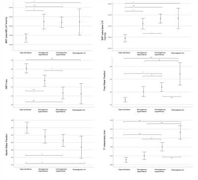 |
91 |
Deep multiple sclerosis lesion phenotyping using multimodal
quantitative MRI
Giacomo Boffa1,
Simona Schiavi1,
Francesco Tazza1,
Caterina Lapucci2,
Gian Franco Piredda3,4,5,
Domenico Zacà6,
Luca Roccatagliata7,
Tom Hilbert3,4,5,
Tobias Kober3,5,8,
Matilde Inglese1,
and Mauro Costagli1,9
1Department of Neurology, Rehabilitation, Ophthalmology, Genetics, Maternal and Child Health, University of Genoa, Genoa, Italy, 2Laboratory of Experimental Neurosciences, IRCCS Ospedale Policlinico San Martino, Genoa, Italy, 3Advanced Clinical Imaging Technology, Siemens Healthineers International AG, Lausanne, Switzerland, 4Department of Radiology, Lausanne University Hospital and University of Lausanne, Lausanne, Switzerland, 5LTS5, École Polytechnique Fédérale de Lausanne (EPFL), Lausanne, Switzerland, 6Siemens Healthcare, Milan, Italy, 7Department of Neuroradiology, Health Sciences, University of Genoa, Genoa, Italy, 8Lausanne University Hospital and University of Lausanne, Department of Radiology, Luasanne, Switzerland, 9IRCCS Stella Maris, Pisa, Italy Keywords: Multiple Sclerosis, Multi-Contrast Quantitative MRI has the potential to disentangle the heterogeneity of multiple sclerosis (MS) lesions in vivo, which can be decisive for treatment and monitoring. In this study, we distinguished different types of MS lesions based on quantitative susceptibility mapping (QSM) and characterized them using T1 relaxometry, diffusion imaging and myelin mapping. We identified four types of lesions (hypo/isointense, homogeneous hyperintense, inhomogeneous hyperintense and paramagnetic rim), which were characterized by increasing degrees of axonal and myelin disruption. Paramagnetic rim lesions were closer to the ventricular CSF, corroborating the presence of a noxious activity of CSF in MS pathology. |
|
2619.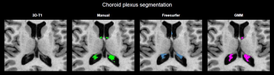 |
92 |
Enlarged choroid plexus volume in multiple sclerosis is related
with disability, cognition and brain atrophy
Samantha Noteboom1,
Jelle J. Vellema1,
Martijn D. Steenwijk1,
Helga E. de Vries2,
Frederik Barkhof3,4,
Joep Killestein5,
Eva M. M. Strijbis5,
and Menno M. Schoonheim1
1MS Center Amsterdam, Anatomy and Neurosciences, Vrije Universiteit Amsterdam, Amsterdam Neuroscience, Amsterdam UMC location VUmc, Amsterdam, Netherlands, 2Department of Molecular Cell Biology and Immunology, Vrije Universiteit Amsterdam, Amsterdam Neuroscience, Amsterdam UMC location VUmc, Amsterdam, Netherlands, 3MS Center Amsterdam, Radiology and Nuclear Medicine, Vrije Universiteit Amsterdam, Amsterdam Neuroscience, Amsterdam UMC location VUmc, Amsterdam, Netherlands, 4Institutes of Neurology and Healthcare Engineering, UCL London, London, United Kingdom, 5MS Center Amsterdam, Neurology, Vrije Universiteit Amsterdam, Amsterdam Neuroscience, Amsterdam UMC location VUmc, Amsterdam, Netherlands Keywords: Multiple Sclerosis, Neuroinflammation Enlargement of the choroid plexus (ChP) has been recently suggested in multiple sclerosis (MS), but relations with clinical and MRI outcome measures remain unclear. In this study, we compared automated segmentation approaches to assess ChP volume on 3D-T1 to manual outlines. Next, ChP volume was assessed in 327 patients with MS and 78 healthy controls. Gaussian Mixture Modelling (GMM)-based segmentation showed best agreement with manual segmentations in MS and controls. Enlargement of ChP was observed in MS compared to controls, and was associated with worse physical disability and cognitive impairment and more severe brain, cortical and thalamic atrophy. |
|
2620.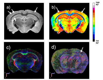 |
93 |
Examining T2* Relaxometry in Diffusion Tensor Space to Quantify
Cerebral Microbleeds in Alzheimer’s Disease Mouse Model
Emani Hunter1,
Lesley Foley2,
Yifan Zhao1,
Howard Aizenstein1,3,
Minjie Wu1,3,
and Bistra Iordanova1
1Department of Bioengineering, University of Pittsburgh, Pittsburgh, PA, United States, 2Animal Imaging Center, University of Pittsburgh, Pittsburgh, PA, United States, 3Department of Psychiatry, University of Pittsburgh, Pittsburgh, PA, United States Keywords: Alzheimer's Disease, Relaxometry, Diffusion Tensor Imaging Cerebral microbleeds are morphologic changes contributing to the progression of Alzheimer’s Disease and vascular dementias. However, the effects of microbleeds on brain connectivity across age and sex remain to be understood. In this study, we performed T2* mapping, that was registered to a DTI space to measure diffusion changes in microbleed locations. R2* and diffusion tensor indices demonstrate an inverse relationship, where diffusion is low and R2* rates are high. Our results suggest that microbleeds decrease water diffusion in the brain possibly due to increased neuroinflammation and hemosiderin accumulation, shedding light on abnormal tissue structure in Alzheimer’s disease subjects. |
|
2621.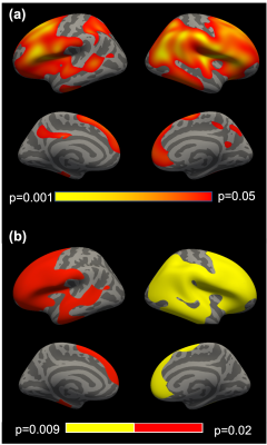 |
94 |
Cortical demyelination in chronic post-traumatic stress
disorder: A study of World Trade Center responders
Juin W. Zhou1,
Chuan Huang1,2,
Kritikos Minos3,
Sean A.P. Clouston3,
Megan K. Horton4,
Roman Kotov5,
Roberto G. Lucchini6,7,
Evelyn J. Bromet5,
and Benjamin J. Luft8,9
1Biomedical Engineering, Stony Brook University, Stony Brook, NY, United States, 2Radiology, Renaissance School of Medicine, Stony Brook, NY, United States, 3Family, Population, and Preventative Medicine, Renaissance School of Medicine, Stony Brook, NY, United States, 4Environmental Medicine and Public Health, Icahn School of Medicine at Mount Sinai, New York, NY, United States, 5Psychiatry, Renaissance School of Medicine, Stony Brook, NY, United States, 6Environmental Health Sciences, Florida International University, Miami, FL, United States, 7Medical and Surgical Specialties, Radiological Sciences, and Public Health, University of Brescia, Brescia, Italy, 8Medicine, Renaissance School of Medicine, Stony Brook, NY, United States, 9Stony Brook WTC Wellness Program, Renaissance School of Medicine, Stony Brook, NY, United States Keywords: Neurodegeneration, Psychiatric Disorders World Trade Center (WTC) responders are at mid-life, with 23% presenting with chronic post-traumatic stress disorder (PTSD). Epidemiologic studies suggest that chronic PTSD is associated with psychomotor slowing and physical functional limitations consistent with intracortical neuroinflammation. Prior neuroimaging has suggested that chronic PTSD is associated with glial activation and reduced cortical complexity, as well as neurodegeneration in the hippocampus and anterior cingulate, suggestive of PTSD-induced cortical neuropathology. Hypothesizing that these results might reduce intercortical density, we evaluated intercortical demyelination in WTC responders with chronic PTSD. We used gray-white contrast purported to measure cortical demyelination and, by extension, interneuronal health. |
|
2622.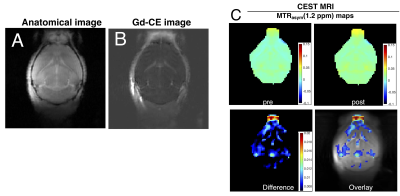 |
95 |
Dynamic dextran-enhanced CEST MRI reveals the size effect of BBB
disruption associated with neuroinflammation
Safiya Aafreen1,
Wenshen Wang2,3,
Jiadi Xu2,3,
Peter CM van Zijl2,3,
Aline Thomas2,
Jeff WM Bulte1,2,3,4,
and Guanshu Liu2,3
1Department of Biomedical Engineering, Johns Hopkins University, Baltimore, MD, United States, 2Radiology, Johns Hopkins University School of Medicine, Baltimore, MD, United States, 3Kirby Research Center, Kennedy Krieger Institute, Baltimore, MD, United States, 4Institute for Cell Engineering, Johns Hopkins University School of Medicine, Baltimore, MD, United States Keywords: Contrast Agent, Molecular Imaging, Blood-Brain Barrier; Multiple Sclerosis Prevailing imaging methods primarily focus on detecting blood-brain barrier (BBB) dysfunction utilising tracers of small molecular weight. Robust imaging methods suitable for assessing BBB permeability in the macro-molecular size range are still lacking. Our study aimed to develop and optimize a dextran-based MRI approach for detecting size-dependent BBB permeability. Demonstrated in an EAE MS mouse model, we established a dextran-based CEST MRI protocol for measuring the intracerebral distribution of non-labeled dextrans, validated the results using immunohistochemistry, and compared the CEST MRI results with conventional Gd-enhanced MRI. |
|
2623.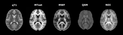 |
96 |
Longitudinal quantitative MRI changes in normal-appearing brain
tissue of patients with multiple sclerosis
Alessandro Cagol1,2,3,
Mario Ocampo-Pineda1,2,3,
Lester Melie-Garcia1,2,3,
Po-Jui Lu1,2,3,
Muhamed Barakovic1,2,3,
Matthias Weigel1,2,3,4,
Xinjie Chen1,2,3,
Antoine Lutti5,
Thanh D. Nguyen6,
Yi Wang6,
Jens Kuhle2,3,
Ludwig Kappos1,2,3,
and Cristina Granziera1,2,3
1Translational Imaging in Neurology (ThINK) Basel, Department of Biomedical Engineering, University Hospital Basel and University of Basel, Basel, Switzerland, 2Department of Neurology, University Hospital Basel, Basel, Switzerland, 3Research Center for Clinical Neuroimmunology and Neuroscience Basel (RC2NB), University Hospital Basel and University of Basel, Basel, Switzerland, 4Division of Radiological Physics, Department of Radiology, University Hospital Basel, Basel, Switzerland, 5Laboratory for Research in Neuroimaging, Department of Clinical Neuroscience, Lausanne University Hospital and University of Lausanne, Lausanne, Switzerland, 6Department of Radiology, Weill Cornell Medical College, New York, NY, United States Keywords: Multiple Sclerosis, Quantitative Imaging We explored the value of multiple longitudinal quantitative MRI (qMRI) measures in detecting microstructural changes occurring in normal-appearing tissue of patients with multiple sclerosis (PwMS). While no differences in qMRI longitudinal changes were measured between PwMS and healthy controls, progressive PwMS showed accelerated T1-relaxometry increase in normal-appearing tissue with respect to both healthy controls and relapsing-remitting PwMS, reflecting increased micro/macrostructural damage. In PwMS the rates of qMRI changes during follow-up were associated with the severity of clinical disability, with higher neurological impairment being associated with qMRI changes reflecting accelerated micro/macrostructural damage, demyelination, and axon/dendrite loss. |
|
2624.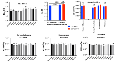 |
97 |
Preserved white matter integrity despite changes in brain
structure and metabolism in mouse with lethal irradiation and
bone marrow transplant
Min-Hui Cui1,
Roman Fleysher1,
Lidiane Torres2,
Kamalakar Ambadipudi1,
and Craig A. Branch1
1Radiology/Gruss MRRC, Albert Einstein College of Medicine, Bronx, NY, United States, 2Cell Biology, Albert Einstein College of Medicine, Bronx, NY, United States Keywords: White Matter, Transplantation, white matter, spectroscopy, irradiation, animal Adult cells from transplanted bone marrow can generate new neurons in central nervous system (CNS) in rodent and human. Here we report for the first time in live mouse that white matter integrity in lethally irradiated mouse transplanted with adult mouse bone marrow (BMTX) is preserved, reflected in increased fractional anisotropy (FA) of corpus callosum post BMTX. Stable concentration of N-acetyl-aspartate (NAA) and increased FA after BMTX suggests that donor cells along with irradiation may stimulate new neuronal proliferation. Nevertheless, decreased myo-inositol concentration may reflect its cell volume regulation function as seen in morphometric adjustments in BMTX brains. |
|
2625.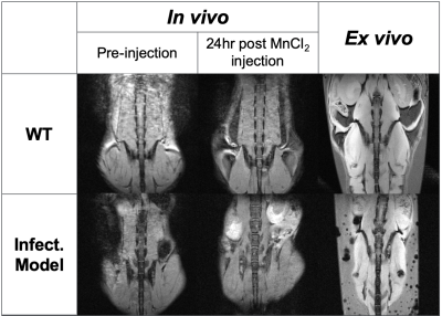 |
98 |
MRI characterization of a mouse model of Staphylococcus aureus
infection
Hannah Goldman1,
Katherine Le2,
Orlando Aristizabal1,
Matias Aristizabal1,
Mia Weissman1,
Victor Torres2,
and Youssef Zaim Wadghiri1
1Radiology, NYU Langone Health, New York, NY, United States, 2Microbiology, NYU Grossman School of Medicine, New York, NY, United States Keywords: Infectious disease, Preclinical Staphylococcus aureus is of particular concern due to the deaths associated with antimicrobial resistance and the emergence of antibiotic-resistant strains. There is not currently a vaccine as translation from preclinical models to humans has been unsuccessful. Acquiring reproducible and comparable spinal images that offer non-invasive visualization of the extent and impact of the infection are important for successful intervention or vaccine development. 3D-printed, MRI-compatible cradles enable reproducible positioning and analysis for both in vivo and ex vivo MR spinal imaging in mouse models while allowing for nondestructive analysis of the potential additional benefits to visualization associated with ex vivo MEMRI. |
|
 Back to Meeting Home
 Back to the Program-at-a-Glance
The International Society for Magnetic Resonance in Medicine is accredited by the Accreditation Council for Continuing Medical Education to provide continuing medical education for physicians.
 Back to Meeting Home
 Back to the Program-at-a-Glance
 View Presentation Video