Digital Poster
Morphology & Neuromelanin in Parkinson's Disease
ISMRM & ISMRT Annual Meeting & Exhibition • 03-08 June 2023 • Toronto, ON, Canada

Digital Poster
Morphology & Neuromelanin in Parkinson's Disease
| Computer # | |||
|---|---|---|---|
3194.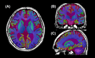 |
141 |
A Correlation Study Between Brain Sub-Region Volumes and the
Severity of Parkinson’s Disease
Zhanhao Mo1,
Lin Liu1,
Lei Zhang1,
Runyu Tang2,
Yunfei Zhang2,
Yongming Dai2,
and He Sui1
1China-Japan Union Hospital of Jilin University, Jilin, China, 2MR Collaboration, Central Research Institute, United Imaging Healthcare, Shanghai, China Keywords: Parkinson's Disease, Segmentation By applying a deep-learning based brain segmentation algorithm on T1-weighted images, the brain was segmented into 106 sub-regions and regional volumes were determined automatically. We analyzed the relationship of brain regional volumes and the severity of Parkinson’s Disease (PD) and compared regional volumes in a cohort of patients with mild and moderate PD. Our data demonstrated that regional volumes of certain brain structures had the potential ability in the characterization of PD. |
|
3195.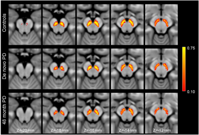 |
142 |
Reductions in nigral volume of de novo and moderate Parkinson’s
disease patients
Jason Langley1,
Kristy Hwang2,
Daniel Huddleston3,
and Xiaoping Hu1,4
1Center for Advanced Neuroimaging, University of California Riverside, Riverside, CA, United States, 22Department of Neurosciences, University of California San Diego, San Diego, CA, United States, 3Department of Neurology, Emory University, Atlanta, GA, United States, 4Department of Bioengineering, University of California Riverside, Riverside, CA, United States Keywords: Parkinson's Disease, Parkinson's Disease, substantia nigra Neuronal loss in substantia nigra pars compacta is a hallmark of Parkinson's disease. In this abstract, we examine nigral volume in a group without Parkinson's disease (non-carriers, non-manifest LRRK2 and GBA mutations), de novo Parkinson's disease, and moderate Parkinson's disease. Reductions in nigral volume are seen in the Parkinson's disease groups relative to the group without Parkinson's disease. A further reduction is seen in nigral volume of the moderate Parkinson's disease group relative to the de novo group, suggesting that nigral degeneration continues as Parkinson's disease progresses. |
|
3196.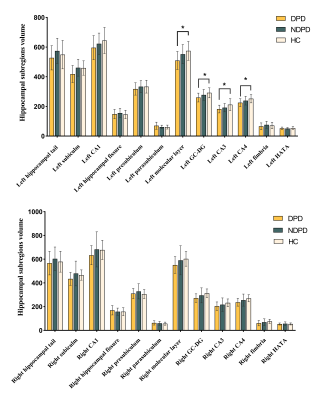 |
143 |
Altered Hippocampal Subregions Volumes Is Associated With
Emotional Impairment of Parkinson’s Disease
Mingrui Qu1,
Bingbing Gao1,
Yuhan Jiang1,
Yuan Li1,
Lizhi Xie2,
and Yanwei Miao1
1The First Affiliated Hospital of Dalian Medical University, Dalian, China, 2GE Healthcare, Shanghai, China Keywords: Parkinson's Disease, Psychiatric Disorders Depression is the main emotional manifestation associated with Parkinson's disease. The hippocampus is a core region in the limbic system, critical in regulating emotion. This study explored the subregional atrophy pattern of the hippocampus in Parkinson's disease with depression (DPD) patients and its correlation with the severity of depressive symptoms. The results of this study suggest that DPD patients exhibit a left asymmetric atrophy pattern in the hippocampus and its subregions. Partial correlation analysis showed that the left dentate gyrus, left molecular layer, left CA4 and left hippocampal volumes were negatively correlated with Hamilton depression scale scores in DPD patients. |
|
3197.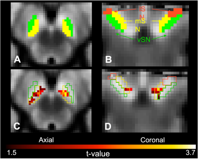 |
144 |
Association between subregions of the substantia nigra and
behavioral symptoms in patients with Parkinson disease using
neuromelanin MRI
Paula Trujillo1,
Kilian Hett1,
Kaitlyn R. Hay1,
Alexander K. Song1,
and Daniel O. Claassen1
1Neurology, Vanderbilt University Medical Center, Nashville, TN, United States Keywords: Parkinson's Disease, Neuroscience We investigated the relationship between subregions of the substantia nigra and behavioral disinhibition in patients with Parkinson disease using neuromelanin MRI. We found a significant positive relationship between disinhibition and neuromelanin MRI contrast ratio in bi-lateral clusters localized in the medial substantia nigra. These findings suggest that neuromelanin MRI can be used to assess the localization of changes in the substantia nigra and inform how dopamine pathophysiology relates to behavioral symptoms in Parkinson disease. |
|
3198.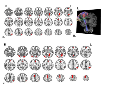 |
145 |
Baseline cerebral structural morphology predicts future freezing
of gait in early drug-naïve Parkinson’s disease
Yuting Li1,2,
Xiuhang Ruan1,
Yongzhou Xu3,
and Xinhua Wei4
1The Second Affiliated Hospital, School of Medicine, South China University of Technology, Guangzhou, China, 2Affiliated Dongguan Hospital, Southern Medical University (Dongguan People's Hospital), Dongguan, China, 3Philips Healthcare, Guangzhou, China, 4Guangzhou First People's Hospital, Guangzhou, China Keywords: Parkinson's Disease, Parkinson's Disease, drug-naïve Parkinson’s disease, Freezing of gait (FOG) We developed a model that could predict the occurrence of Freezing of gait (FOG) at the individual level using machine learning with the clinical, laboratory, and cerebral structural imaging information of early drug-naïve Parkinson’s disease (PD) patients. Data from 158 early drug-naïve PD patients at baseline were obtained from the Parkinson’s Progression Markers Initiative cohort. The predictive performance of future FOG in early PD was evaluated using elastic net-support vector machine models. T1WI morphometric markers have the potential to help predict future FOG in patients with early PD at an individual level, with improved performance when integrated with clinical variables. |
|
3199.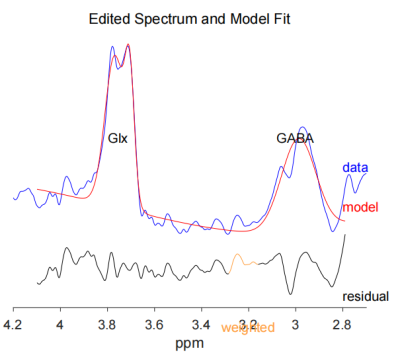 |
146 |
The evaluation of relationship between motor cortex GABA levels
and Gyrification Pattern in Patients With early-onset Parkinson
Disease
yuan Tian1,
pengfei Liu1,
and Jianxiu Lian2
1The First Affiliated Hospital of Harbin Medical University, Harbin, China, 2Philips Healthcare, Beijing, China Keywords: Parkinson's Disease, Neurodegeneration Stimulation of the motor cortex (MC) significantly reduce akinesia and bradykinesia in Parkinson Disease (PD), indicating a key role of motor cortex abnormalities in the pathogenesis of PD. Early-onset Parkinson disease (EOPD) , varying between 40 and 55 years of age, it carries additional societal and personal consequences and psychosocial disruption. Thus we aim to explore the cortical gyrification changes as well as their relationships with r-aminobutyric acid (GABA) in the motor cortex(MC) of patients with early-onset Parkinson disease (EOPD). Results showed that decreased in LGI and GABA+/Cr ratios in the motor cortex were found to be slightly coupled. |
|
3200.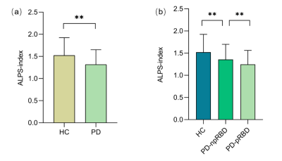 |
147 |
Glymphatic system impairment in Parkinson’s disease with rapid
movement sleep behavior disorder
Amei Chen1,
Junxiang Huang2,
Xiaofei Huang1,
Xiaofang Cheng3,
Yongzhou Xu4,
and Xinhua Wei1
1Guangzhou First People's Hospital, Guangzhou, China, 2Guangzhou Women and Children's Medical Center, Guangzhou, China, 3The Affiliated Brain Hospital of Guangzhou Medical University, Guangzhou, China, 4Philips Healthcare, Guangzhou, China Keywords: Parkinson's Disease, Diffusion Tensor Imaging, glymphatic system;rapid movement sleep behavior disorder To assess the activity of the glymphatic system in PD patients with probably rapid movement sleep behavior disorder (PD-pRBD) using diffusion tensor image analysis along the perivascular space( DTI-ALPS) index. 61 PD-pRBD, 92 PD patients with no probable RBD(PD-npRBD) and 59 healthy control (HC) were included in the study. The results showed that the ALPS index in PD-pRBD group was lower than that in PD-npRBD group. The index of ALPS in PD-pRBD was negatively correlated with REM sleep behavior disorder screening questionnaire(RBDSQ) score. We concluded that the glymphatic system function of PD with RBD is impaired. |
|
3201.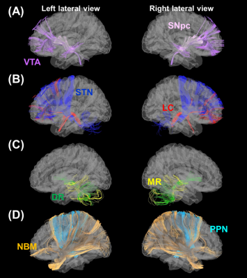 |
148 |
Axonal abnormalities in deep brain nuclear pathways across
multiple neurotransmitter systems in de novo Parkinson’s disease
patients
Yao-Chia Shih1,2,3,
Pohchoo Seow2,
Hu-Lin Christina Wang1,4,
Aeden Zi Cheng Kuek2,
Septian Hartono3,5,
Sui-Hing Yan6,
Eng King Tan3,5,
and Ling Ling Chan2,3
1Graduate Institute of Medicine, Yuan Ze University, Taoyuan City, Taiwan, 2Department of Diagnostic Radiology, Singapore General Hospital, Singapore, Singapore, 3Duke-NUS Medical School, Singapore, Singapore, 4Department of Traumatology, Far Eastern Memorial Hospital, New Taipei City, Taiwan, 5Department of Neurology, National Neuroscience Institute – SGH Campus, Singapore, Singapore, 6Department of Neurology, Far Eastern Memorial Hospital, New Taipei City, Taiwan Keywords: Parkinson's Disease, Diffusion Tensor Imaging The present study systematically and comprehensively reviewed the axonal pathways projecting from the deep brain nuclei that are emerging targets for treating Parkinson’s disease (PD). We tracked 124 deep brain nuclear pathways across different neurotransmitters systems in the high angular HCP-1065 diffusion MRI template using diffusion MRI tractography. With the trajectories of these pathways, we successfully used Parkinson's Progression Markers Initiative (PPMI) diffusion tensor imaging (DTI) data to characterize abnormal DTI changes in noradrenergic and serotoninergic pathways in relation to the severity of non-motor symptoms in de novo PD patients, suggesting a wide neural involvement of clinical PD manifestations. |
|
3202. |
149 |
Sex differences in brain structure in de novo Parkinson’s
disease: A cross-sectional and longitudinal neuroimaging study
Hui Li1,
Chen Zhang2,
Xiuqin Jia1,
and Qi Yang1
1Department of Radiology, Beijing Chaoyang Hospital, Capital Medical University, Beijing, China, 2MR Scientific Marketing, Siemens Healthineers, Beijing, China Keywords: Parkinson's Disease, Parkinson's Disease, sex difference, brain structure, cross-sectional, longitudinal The aim of this study was to investigate sex differences in brain structure at baseline and sex differences in longitudinal changes at follow-up between female and male PD patients after excluding the expected effects of age and sex. At baseline, male PD patients exhibited significantly lower brain volume and decreased cortical thickness than age-matched female PD patients. At follow-up, these sex differences remained stable and analogous progressive brain atrophy with no sex interaction were found in male and female PD patients, suggesting similar trend in disease progression between sexes over time. |
|
3203. |
150 |
Functional and Microstructural Comparative Study on Monogenic vs
Idiopathic Parkinson's Disease
Hacer Dasgin1,
Basak Soydas Turan2,
Bilge Volkan Salanci3,
Eser Lay Gun2,
Gul Yalcin Cakmakli3,
Bulent Elibol3,
and Kader K. Oguz1,4,5
1Umram, Bilkent University, Ankara, Turkey, 2Faculty of Medicine, Department of Nuclear Medicine, Hacettepe University, Ankara, Turkey, 3Faculty of Medicine, Department of Neurology, Hacettepe University, Ankara, Turkey, 4Faculty of Medicine, Department of Radiology, Hacettepe University, Ankara, Turkey, 5University of California, Davis, CA, United States Keywords: Parkinson's Disease, fMRI (resting state) Clinical presentation of monogenic PD differs from idiopathic PD, which might relate to different structural and functional alterations. Previously, we examined a small group of genetic PD patients with connectivity changes in the basal ganglia, compared with healthy control (HC)s. In this study we further examined monogenic PD patients and compared idiopathic PD group and HCs that functional and microstructural changes would be different in these groups of patients. There was no significant basal ganglia network (BGN) alteration between PD groups, but ACC activation is remarkable in both groups. Significantly decreased fractional anisotropy in mPD patients compared to iPD patients. |
|
3204. |
151 |
Reproducibility of Iron and Neuromelanin Changes of Deep Grey
Matter in 2 Large Movement Disorder Cohorts
Naying He1,
Ewart Mark Haacke1,2,
Ying Wang2,
Youmin Zhang1,
Xinhui Wang1,
Zhijia Jin1,
Yan Li1,
Peng Wu3,
and Fuhua Yan4
1Ruijin Hospital, Shanghai Jiao Tong University, Shanghai, China, 2SpinTech MRI, Bingham Farms, MI, USA 48025, Bingham Farms, MI, United States, 3Philips Healthcare, Shanghai, China, 4Ruijin Hospital, Shanghai Jiao Tong University School of Medicine, Shanghai, China Keywords: Parkinson's Disease, Neurodegeneration Diagnosing movement disorders (MDS) is a major challenge clinically. Neuromelanin and iron serve as biomarkers for MDS but few studies have evaluated a large number of cases for multiple disorders. We studied 614 patients consisting of two cohorts with different resolutions. The QSM and NM-MRI data were automatically processed using a unique template program. The results from both cohorts showed reproducible changes of iron and neuromelanin in the deep gray matter. Specifically, a loss of neuromelanin volume, an increase in iron content of the SN for both PD and MSA; increased RN iron and decreased putamen volume in MSA. |
|
3205. |
152 |
Exploring the effects of tissue iron in neuromelanin MRI
Minjun Kim1,
Sooyeon Ji1,
Joonhyeok Yoon1,
Hwihun Jeong1,
Hongjun An1,
Jiye Kim1,
Jonghyo Youn1,
Junghwa Kang2,
Eung Yeop Kim3,
Yoonho Nam2,
and Jongho Lee1
1Department of Electrical and Computer Engineering, Seoul National University, Seoul, Korea, Republic of, 2Department of Biomedical Engineering, Hankuk University of Foreign Studies, Gyeonggi-do, Korea, Republic of, 3Department of Radiology, Samsung Medical Center, Seoul, Korea, Republic of Keywords: Parkinson's Disease, Parkinson's Disease Neuromelanin MRI signal decreases in IPD patients in the substantia nigra. In this study, we investigated that the decrease is caused by not only the reduction of neuromelanin but also iron deposition. Neuromelanin reduction in IPD patients was shown by the result of the substantia nigra volume decrease. The comparison result of contrast ratio, calculated between substantia nigra and crus cerebri, of IPD patients versus healthy controls demonstrated that iron deposition also decreases the signal of NM-MRI. T2*, susceptibility, and overlapped area between substantia nigra from neuromelanin MRI and hypo-intense area from SMWI also represented the tendency of iron deposition. |
|
3206.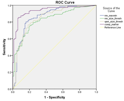 |
153 |
Quantitative Iron-Neuromelanin MRI Matches Expert Visual
Performance and Is Associated with Parkinson’s Disease Severity
Septian Hartono1,2,
Robert Chen3,
Thomas Welton1,
An Sen Tan3,
Weiling Lee3,
Peik Yen Teh3,
Celeste Chen1,
Wenlu Hou3,
Wei Ping Tham3,
Ee Wei Lim1,
Prakash Kumar1,
Yao-Chia Shih4,
Kuan Jin Lee5,
Louis Chew Seng Tan1,
Eng King Tan1,
and Ling Ling Chan3
1National Neuroscience Institute, Singapore, Singapore, 2Duke-NUS Medical School, Singapore, Singapore, 3Singapore General Hospital, Singapore, Singapore, 4Yuan Ze University, Taoyuan, Taiwan, 5Institute of Bioengineering and Bioimaging, Singapore, Singapore Keywords: Parkinson's Disease, Parkinson's Disease Nigrosome-1, a subregion of substantia nigra (SN), and neuromelanin have been identified as good biomarkers of Parkinson's disease (PD) pathology. We investigated the diagnostic accuracy of quantitative iron-neuromelanin parameters and compared these against visual analysis of nigrosome-1 and neuromelanin hyperintensity as proxies of nigral dopaminergic neurodegeneration. Susceptibility map-weighted imaging surpassed neuromelanin sensitive MRI in blinded visual PD classification; reader expertise affected performance. Composite quantitative iron-neuromelanin marker matched diagnostic accuracy of expert reader in visual evaluation of nigrosome-1 using susceptibility map-weighted imaging, potentially improving diagnostic confidence in non-experts. Quantitative susceptibility mapping showed moderate negative correlation with motor dysfunction in PD patients. |
|
3207.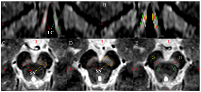 |
154 |
Differential Diagnosis Between Parkinson's Disease and Essential
Tremor with Neuromelanin Imaging of the Substantia Nigra and
Locus Coeruleus
Xinhui Wang1,
Naying He1,
Ewart Mark Haacke2,
Yu Liu1,
Peng Liu1,
Youmin Zhang1,
Zhijia Jin1,
Yan Li1,
Peng Wu3,
and Fuhua Yan1
1Radiology, Ruijin Hospital, Shanghai Jiao Tong University School of Medicine, Shanghai, China, 2Biomedical Engineering, Wayne State University, Detroit, MI, United States, 3Philips Healthcare, Shanghai, China Keywords: Parkinson's Disease, Brain, essential tremor; neuromelanin MRI; locus coeruleus;substantia nigra In this study, we used neuromelanin MRI to investigate locus coeruleus (LC) and substantia nigra changes between essential tremor (ET) and Parkinson’s disease (PD). We found the neuromelanin of LC was significantly lower in tremor-dominant PD patients than in ET patients or healthy controls, and the area under the curve of the model constructed by NM measures reached 0.92 in differentiating tremor-dominant PD from ET. These results provided new perspectives in the differential diagnosis of patients with tremor and in the investigation of the underlying pathophysiology. |
|
3208.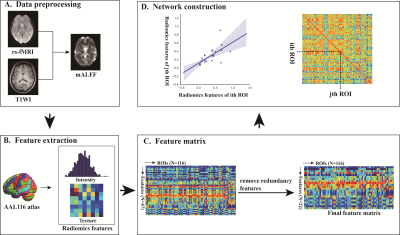 |
155 |
Amplitude of low-frequency fluctuation-based regional radiomics
similarity network: biomarker for Parkinson’s disease
Dafa Shi1,
Haoran Zhang1,
Guangsong Wang1,
and Ke Ren1
1Department of Radiology, Xiang’an Hospital of Xiamen Uneversity,School of Medicine, Xiamen University, Xiamen, China Keywords: Parkinson's Disease, Brain Connectivity Development of more clinically effective and quantifiable biomarkers to detect PD is urgently needed. It confirmed that regional radiomics similarity network (R2SN) had high reproducibility and a biological basis and provided a new perspective for understanding the human brain. Our study showed that R2SN constructed based on ALFF had good reproducibility and stability, and is biological plausibility. We found PD-related brain regions mainly located in the default-mode, sensorimotor, executive control, visual, frontoparietal network, as well as cerebellum, and striatum. ALFF-based R2SN is a novel, robust potential neuroimaging biomarker for PD and could provide new insights into connectome reorganization in PD. |
|
3209.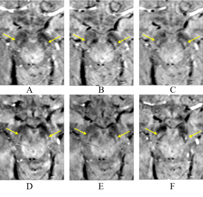 |
156 |
Imaging Nigrosome-1 using Wave-CAIPI Susceptibility Weighted
Imaging at 3T
Peng Liu1,
Naying He1,
Youmin Zhang1,
Caixia FU2,
Qing Li3,
E. Mark Haacke4,
Yu Liu1,
Wei Liu2,
Xinhui Wang1,
Daniel Polak5,
and Fuhua Yan1
1Ruijin Hospital, Shanghai Jiao Tong University School of Medicine, Shanghai, China, 2Siemens Shenzhen Magnetic Resonance Ltd., Shenzhen, China, 3Siemens Healthineers Ltd., Shanghai, China, 4Wayne State University, Detroit, MI, United States, 5Siemens Healthcare GmbH, Erlangen, Germany Keywords: Parkinson's Disease, Data Acquisition, Magnetic resonance imaging, Nigrosome-1, Susceptibility Weighted Imaging In this work, the ability of Wave-CAIPI accelerated 3D SWI for imaging the Nigrosome-1(N1) sign was evaluated. Short-TE, long-TE and conventional SWI protocols were compared for the detection of the N1 sign. Qualitative and quantitative analyses were used to evaluate the presence or absence of the N1 sign and the contrast-to-noise (CNR) of the N1 sign relative to adjacent tissues. The results showed that Wave-CAIPI SWI had no significant differences with conventional SWI for recognizing the N1 sign. Wave-CAIPI SWI can be a replacement of conventional SWI (4 min 52s) with shorter scan times of 2min 26s covering the whole brain. |
|
3210.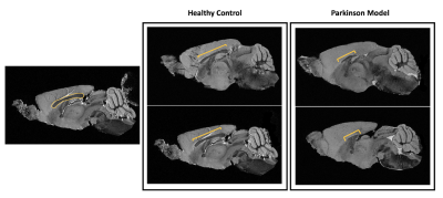 |
157 |
Major structural abnormalities in the alpha-synuclein mouse
model of Parkinson’s Disease revealed with MR Microscopy at 16.4
T
Ruxanda Lungu Baião1,
Tiago Fleming Outeiro2,
and Noam Shemesh1
1Preclinical MRI, Champalimaud Research, Champalimaud Foundation, Lisbon, Portugal, 2Department of Experimental Neurodegeneration, University of Göttingen, Goetingen, Germany Keywords: Parkinson's Disease, Neurodegeneration Parkinson's disease (PD) is associated with irreparable damage to dopaminergic neurons due to α-synuclein (aSYN) inclusions. Dopaminergic pathways include the nigrostriatal, mesolimbic, mesocortical and tuberoinfundibular systems that play vital roles. Thus, dysregulations of dopamine function may lead to brain-wide consequences. Apart from a mouse PD model of injected aSYN1, brain morphology alterations in genetic mouse models have not been described in detail using MRI. Here we explore brain morphological differences between PD models and healthy controls, crucial for further understanding of the disease. We find major morphological abnormalities in areas such as Lateral Ventricles, Corpus Callosum and the surrounding Cortex. |
|
3211.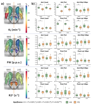 |
158 |
Multimodal assessment of nigrostriatal degeneration in de novo
Parkinson’s disease
Miguel López-Aguirre1,2,3,
Michele Matarazzo1,3,
Javier Blesa1,3,
Álvaro Sánchez-Ferro1,4,
Mariana H.G. Monje1,5,
José A. Obeso1,3,6,
and José A. Pineda-Pardo1,3,6
1HM CINAC, Hospital Universitario HM Puerta del Sur, Móstoles, Spain, 2Universidad Complutense de Madrid, Madrid, Spain, 3Center for Networked Biomedical Research on Neurodegenerative Diseases (CIBERNED), Instituto de Salud Carlos III, Madrid, Spain, 4Neurology Department, Hospital Universitario 12 de Octubre, Madrid, Spain, 5Ken and Ruth Davee Department of Neurology, Northwestern University, Chicago, IL, United States, 6Universidad San Pablo-CEU, Madrid, Spain Keywords: Parkinson's Disease, Multimodal, Dopamine, free-water DTI, Iron FDOPA uptake rate, fractional volume of free-water and R2* relaxometry are imaging biomarkers sensitive to dopaminergic decline, microstructural degeneration, and iron disruption respectively, three pathological processes developed within the nigrostriatal system in Parkinson’s disease (PD). A multimodal comprehensive characterization of these biomarkers in early PD was performed, assessing them within striatum and substantia nigra pars compacta in a de novo PD cohort. While iron disruption was not observed, PD subjects revealed dopaminergic and microstructural alterations within the nigrostriatal system, following both similar spatial patterns. These results serve to improve our understanding of nigrostriatal vulnerability and degeneration in early PD. |
|
3212.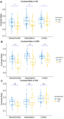 |
159 |
Neuromelanin hemispheric asymmetry in normal aging and early
Parkinson’s disease with motor symptom laterality
Xueling Liu1,
Yuxin Li1,
Puyeh Wu2,
Na Wang1,
Fengtao Liu3,
and Daoying Geng1
1Radiology Department of Huashan Hospital affiliated to Fudan University, Shanghai, China, 2GE healthcare, Beijing, China, 3Neurology Department of Huashan Hospital affiliated to Fudan University, Shanghai, China Keywords: Parkinson's Disease, Neurodegeneration, neuromelanin The bilateral neuromelanin degeneration pattern in substantia nigra in normal aging and underlying mechanisms of asymmetric motor symptoms in early Parkinson’s disease (PD) are still unknown. Here we examined the neuromelanin hemispheric asymmetry in normal elderly people and early PD patients with motor symptoms laterality by short-echo-time magnitude images derived from QSM. We demonstrated hemispheric asymmetry of neuromelanin in normal aging, as well as a left-hemispheric preferential nigral dysfunction in early-stage PDR, but a different spatial neuromelanin loss pattern in PDL. This template-based neuromelanin measurement might help to detect aging-related change and deepen our understanding of PD with motor laterality. |
|
 Back to Meeting Home
 Back to the Program-at-a-Glance
The International Society for Magnetic Resonance in Medicine is accredited by the Accreditation Council for Continuing Medical Education to provide continuing medical education for physicians.
 Back to Meeting Home
 Back to the Program-at-a-Glance
 View Presentation Video