Digital Poster
Advanced Imaging in Parkinson's Disease
ISMRM & ISMRT Annual Meeting & Exhibition • 03-08 June 2023 • Toronto, ON, Canada

Digital Poster
Advanced Imaging in Parkinson's Disease
| Computer # | |||
|---|---|---|---|
2274.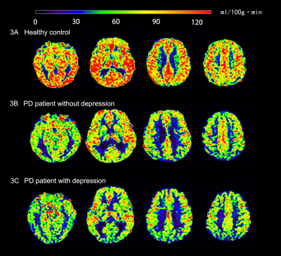 |
101 | Cerebral perfusion in Parkinson's disease with depression: an arterial spin-labeling magnetic resonance imaging study
Xinyang Li1,2, Yaotian Tian1,2, Xiaonan Wang1,2, Josef Pfeuffer3, Bénédicte Maréchal4, Kober Tobias4, Yanglei Wu5, Chunmei Li1, and Min Chen1
1Department of Radiology, Beijing Hospital, National Center of Gerontology, Beijing, China, 2Graduate School of Peking Union Medical College, Beijing, China, 3Siemens Healthcare GmhH, Erlangen, Germany, 4Siemens Healthineers International AG, Lausanne, Switzerland, 5MR Collaboration, Siemens Healthineers Ltd, Beijing, China Keywords: Parkinson's Disease, Neurodegeneration, Depression Arterial spin labeling-magnetic resonance imaging (ASL-MRI) combined with inline T1-weighted-based brain morphometry was used to evaluate regional cerebral blood flow in this study, which explored alteration of cerebral perfusion in Parkinson’s disease (PD) patients with depression and investigated its underlying neural mechanism. The results showed decreased cerebral perfusion in several brain regions in PD patients compared to healthy controls and a correlation between decreased cerebral perfusion of the right occipital white matter and right cingulate gyrus in PD patients with depression. This finding suggested that hypoperfusion of the limbic system is involved in the pathogenesis of PD with depression. |
|
2275.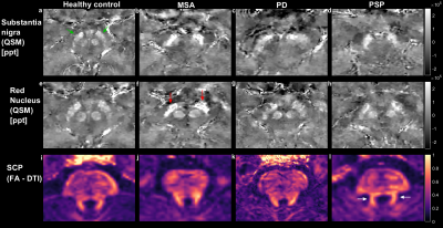 |
102 | Investigating early-stage parkinsonism using a dedicated MRI protocol and decision tree
Samy Abo Seada1, Anke W. van der Eerden1, Agnita J.W. Boon2, and Juan Antonio Hernandez-Tamames1,3
1Department of Radiology and Nuclear Medicine, Erasmus MC, Rotterdam, Netherlands, 2Department of Neurology, Erasmus MC, Rotterdam, Netherlands, 3Department of Imaging Physics, TU Delft, Delft, Netherlands Keywords: Parkinson's Disease, Multi-Contrast A dedicated MRI protocol and decision tree are presented for differentially diagnosing parkinsonisms. Decision rules are based on existing literature which identified biomarkers with a sensitivity and specificity around or above 80%. Initial in-vivo results from patients are presented, and their MRI findings are partially in agreement with existing literature. Our goal is to test our method on patients at an early-stage when their clinical diagnosis is unclear. |
|
2276.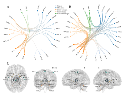 |
103 | Resting state functional connectivity alterations in motor networks of Parkinson’s disease in different frequency bands
Pengfei Zhang1, Jun Wang1, Laiyang Ma1, Yanli Jiang1, Wanjun Hu1, Guangyao Liu1, Kai Ai2, and Jing Zhang1,3
1Department of Magnetic Resonance, Lanzhou University Second Hospital, Lanzhou, China, 2Philips Healthcare, Xi'an, China, 3Gansu Province Clinical Research Center for Functional and Molecular Imaging, Lanzhou, China Keywords: Parkinson's Disease, Parkinson's Disease, Motor symptoms, Neural networks The functional alteration of the substantia nigra (SN), basal ganglia (BG), thalamus, and cortex are key hallmarks of Parkinson’s disease (PD) patients. Based on resting state functional magnetic resonance imaging, we investigated the functional connectivity (FC) of “cerebello-SN-BG-thalamo-motor cortical” network in the conventional and slow-4, slow-5 bands. PD patients demonstrated extensive FC decrease at conventional band, including inter-network connections of thalamo-cerebello-BG circuits, and connections between SN-putamen and postcentral gyrus-cerebellum. Slow-4 showed more thalamo-BG changes, while slow-5 specific FC was mainly the "cerebello-SN-BG-thalamo" circuits. The frequency specific alterations in motor related circuits may help in understanding the neuropathological mechanisms of PD. |
|
2277.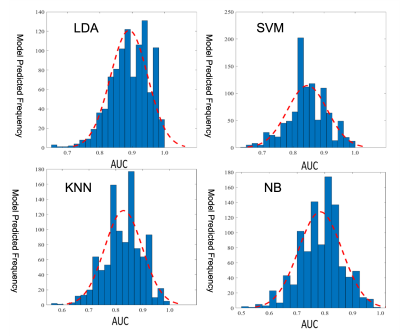 |
104 | Construction of disease progression prediction model for PD patients based on DTI data
Amei Chen1, Junxiang Huang2, Xiaofei Huang1, Xiaofang Cheng3, Yongzhou Xu4, and Xinhua Wei1
1Guangzhou First People's Hospital, Guangzhou, China, 2Guangzhou Women and Children's Medical Center, Guangzhou, China, 3The Affiliated Brain Hospital of Guangzhou Medical University, Guangzhou, China, 4Philips Healthcare, Guangzhou, China Keywords: Parkinson's Disease, Machine Learning/Artificial Intelligence, white matter connectivity, DTI, prediction model In this study, the machine learning method was used to establish a prediction model for Parkinson’s disease(PD) progression by using white matter connectivity and clinical information. A total of 123 PD patients were included. White matter network connection analysis and clinical information collection were performed for each patient. The results showed that combined with the white matter connection and clinical features, a good model of PD disease progression was established. White matter network connectivity helps predict PD progression at the individual level. |
|
2278.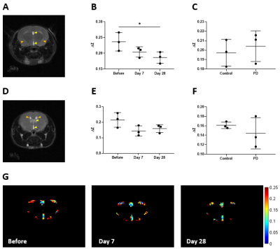 |
105 | CEST and MISL detect regional neuropathologies in a Parkinson’s disease mouse model at 3T
Joseph H. C. Lai1, Jianpan Huang1, Yang Liu1,2, Se Weon Park1,2, Jiadi Xu3, and Kannie W. Y. Chan1,2,3,4,5
1Department of Biomedical Engineering, City University of Hong Kong, Hong Kong, Hong Kong, 2Hong Kong Centre for Cerebro-Cardiovascular Health Engineering (COCHE), Hong Kong, Hong Kong, 3Russell H. Morgan Department of Radiology and Radiological Science, The Johns Hopkins University School of Medicine, Baltimore, MD, United States, 4City University of Hong Kong Shenzhen Research Institute, Shenzhen, China, 5Tung Biomedical Sciences Centre, City University of Hong Kong, Hong Kong, Hong Kong Keywords: Parkinson's Disease, CEST & MT Reveal alterations in molecules and CSF-tissue water exchange could enable interactive interventions in PD. Here we applied CEST and MISL to study the changes in a PD model at 3T. We observed significant changes (P<0.05) at 2.6ppm on day 28 and at -3.5ppm on day 7, which could indicate a substantial decrease of dopamine and lipid-related pathology at the substantia nigra, respectively. Along the nigrostriatal pathway, we observed a gradual MISL drop at the ventricles, which was significantly lower at day 28 (P<0.05) and might imply the CSF-tissue-related pathologies. Our findings could serve as potential biomarkers for early PD detection. |
|
2279. |
106 | Multimodality Neuroimaging Biomarkers in Parkinson’s Disease
Xiaomeng Zhang1, Varsha Mohan1, Dustin Wooten1, Bradley A Hooker1, Yuchuan Zhuang1, Praveen Honhar2, Sophie Holmes2,3, Mika Naganawa2, Mark Dias2, Robert Comley1, Richard E Carson2,4, Sule Tinaz5, David Matuskey2,3,5, Yanping Luo1, and Sjoerd J Finnema1
1Translational Imaging, Neuroscience, Abbvie, North Chicago, IL, United States, 2PET Center, Yale University, New Haven, CT, United States, 3Department of Psychiatry, Yale University, New Haven, CT, United States, 4Department of Biomedical Engineering, Yale University, New Haven, CT, United States, 5Department of Neurology, Yale University, New Haven, CT, United States Keywords: Parkinson's Disease, Multimodal Neuroimaging is widely used in Parkinson's disease patients to improve diagnosis, provide insights into the natural history of disease, and facilitate the development of new treatments. Different MRI sequences and contrasts have been employed to study structure and function, whilst PET have been used to image receptors, transporters, enzymes and metabolism. In this study we evaluate relationships between pathological and functional markers of neurodegeneration using a range of advanced MRI and PET methods, in the same subjects, cross sectionally and over time. To the best of our knowledge this combination of markers has yet to be reported. |
|
2280. |
107 | Diffusion and structural MRI as potential biomarkers to reflect correlation with plasma biomarkers in nondemented Parkinson’s disease
Chih-Chien Tsai1, Chun-Chao Huang2, Pei-Hao Chen3, Hsin-Fan Chiang2, Cheng‑Chih Hsieh2, Ting-Lin Chen2, Wei-Hsin Liao2, Yao-Liang Chen4, and Jiun-Jie Wang5
1Healthy Aging Research Center, Chang Gung University, Taoyuan, Taiwan, 2Department of Radiology, MacKay Memorial Hospital, Taipei, Taiwan, 3Department of Neurology, MacKay Memorial Hospital, Taipei, Taiwan, 4Department of Diagnostic Radiology, Chang Gung Memorial Hospital at Keelung, Keelung, Taiwan, 5Department of Medical Imaging and Radiological Sciences, Chang Gung University, Taoyuan, Taiwan Keywords: Parkinson's Disease, Diffusion Tensor Imaging Parkinson’s disease (PD) is a progressive neurodegenerative disease with motor dysfunction and cognitive impairments frequently. Previous studies indicated that diffusion MRI could detect white matter alteration in the brain, which can be correlated with disease severity. We use diffusion MRI to investigate the relationship between cognitive functions and these plasma biomarkers. Our study showed that the involved regions might play important roles in early cognitive decline as related to the pathological deposition of the respective plasma biomarkers. This observation might help to elucidate the early pathological change of the brain contributing to different cognitive declines in PD patients. |
|
2281.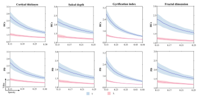 |
108 | Multiparametric Characterization of Surface-Based Morphological Brain Networks in Middle- to Late-Stage Parkinson's Disease
Jun Lu1,2, Su Yan1, Yuanhao Li1, and Wenzhen Zhu1
1Department of radiology,Tongji Hospital, Tongji Medical College, Huazhong University of Science and Technology, Wuhan, China, 2Department of CT & MRI, The First Affiliated Hospital, College of Medicine, Shihezi University, Shihezi, China Keywords: Parkinson's Disease, Microstructure As the structural basis of functional networks, the topological organization of morphological brain networks in the surface space has not been explored in more advanced stages of PD. We constructed individual morphological networks by estimating interregional similarity distribution in different cortical surface-based indices from structural MRI of PD patients and healthy controls (HCs). Graph theoretical analysis was performed to detect PD-related alterations. Compared with HCs, PD patients showed lower local efficiency and clustering coefficient for gyrification index-based morphological brain networks. These findings extend our understanding of the neurobiology of network dysfunction in middle- to late-stage PD. |
|
2282.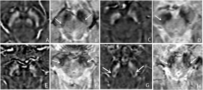 |
109 | Application of QSM and tSWI in evaluating substantia nigra "swallow tail sign" in the diagnosis of Parkinson's disease
Xiao Feng Shi1, You Min Zhang1, Na Ying He1, Zeng Hui Cheng1, Fu Hua Yan1, and Ewart Mark Haacke2
1Department of Radiology, Ruijin Hospital, Shanghai Jiao Tong University School of Medicine, shanghai, China, 2SpinTech MRI, Bingham Farms, MI, United States Keywords: Parkinson's Disease, Parkinson's Disease, swallow tail sign,substantia nigra,MRI The incidence of Parkinson's disease(PD) is high and the clinical symptoms are diverse making the diagnosis of PD difficult. Loss of the “swallow tail sign” (STS) as an imaging biomarker in the diagnosis of PD. It is of great value in the early diagnosis of PD and the improvement of prognosis. In this study, QSM and tSWI, were used to evaluate the STS of substantia nigra in PD patients. The results show tSWI can better display STS than QSM and its accuracy is higher. |
|
2283.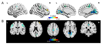 |
110 | Abnormal intrinsic neural timescale in Parkinson’s disease
Yarui Wei1, Chunyan Zhang2, Yuanyuan Peng2, Chen Chen 2, Shaoqiang Han2, Weijian Wang2, Yong Zhang2, Hong Lu3, and Jingliang Cheng2
1Department of Magnetic Resonance Imaging, The First Affiliated Hospital of Zhengzhou University, Zhengzhou, China, 2Department of Magnetic Resonance Imaging, The First Affiliated Hospital of Zhengzhou University, Zhengzhou, China, 3Department of Neurology, The First Affiliated Hospital of Zhengzhou University, Zhengzhou, China Keywords: Parkinson's Disease, fMRI (resting state) It’s unclear whether abnormal neural information stored and different temporal feature at different stages in Parkinson’s disease (PD). Here, we estimated the intrinsic timescales using the magnitude of the autocorrelation of intrinsic neural signals by resting state functional magnetic resonance imaging data in 74 PD patients, including 44 patients in the early stage and 30 patients in the late stage. Our findings suggest that PD patients exhibit abnormal intrinsic timescales in visual, sensorimotor, and cognitive systems, and at different stages, distinct patterns of intrinsic timescales in cerebral cortex, which might provide new insights for the neural substrate of PD. |
|
2284.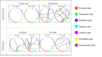 |
111 | Effect of levodopa on brain functional network connectivity in Parkinson’s disease patients with MCI.
Karthik R Sreenivasan1, Xiaowei Zhuang1, Jessica Caldwell1, Aaron Ritter2, Dietmar Cordes1, Zoltan Mari1, Natividad Stover3, Talene Yacoubian3, and Virendra Mishra1,4
1Cleveland Clinic Lou Ruvo Center for Brain Health, Las Vegas, NV, United States, 2Memory & Cognitive Disorders Program Hoag, Pickup Family Neurosciences Institute, Newport Beach, CA, United States, 3Department of Neurology, The University of Alabama at Birmingham, Birmingham, AL, United States, 4Department of Radiology, The University of Alabama at Birmingham, Birmingham, AL, United States Keywords: Parkinson's Disease, Brain Connectivity Mild cognitive impairment in PD patients (PD-MCI) is shown to be a risk factor for the development of dementia over time. However, the distinct factors that contribute to conversion from MCI to dementia are not completely understood. While earlier studies have identified altered functional connectivity (FC) it is still not clear whether PD medication affects brain FC. In the current study, we aim to quantify the impact of medication effect on FC in PD-MCI using resting-state functional MRI. Our findings suggest that altered FC was observed between the groups and with respect to the medication state in the PD-MCI group. |
|
2285.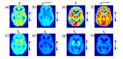 |
112 | Estimation of 11C-PE2I PET based cerebral perfusion in Parkinson’s disease patients using quantitative transport mapping network (QTMnet)
Qihao Zhang1, Dominick Romano1, Kelly McCabe Gillen2, Carly Skudin2, Shtilbans Alexander3, Thanh Nguyen2, Pascal Spincemaille2, and Yi Wang2
1Cornell University, New York, NY, United States, 2Weill Cornell Medicine, New York, NY, United States, 3Hospital for Special Surgery, New York, NY, United States Keywords: Parkinson's Disease, PET/MR 111 |
|
2286.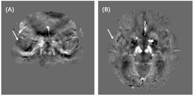 |
113 | Quantitative Susceptibility Mapping in Distinguishing Between Patients with Mild and Moderate Parkinson’s Disease
Zhanhao Mo1, Lin Liu1, Lei Zhang1, Runyu Tang2, Yunfei Zhang2, Yongming Dai2, and He Sui1
1China-Japan Union Hospital of Jilin University, Jilin, China, 2MR Collaboration, Central Research Institute, United Imaging Healthcare, Shanghai, China Keywords: Parkinson's Disease, Quantitative Susceptibility mapping Iron concentrations were associated with many significant pathological processes in the brain. The advent of Quantitative Susceptibility Mapping (QSM) enabled the assessment of magnetic susceptibility in Parkinson’s Disease (PD) brains. Our study revealed that magnetic susceptibilities of regional brain structures varied in mild and moderate PD and we investigated the possibility of early detection of PD using QSM. |
|
2287.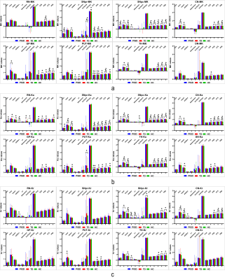 |
114 | Histogram analysis of diffusion kurtosis imaging of deep brain nuclei in Parkinson's disease with different motor subtypes
Jin Wang1, Qiu Bi2, Linyu Chen1, Shan Li3, Hongjiang Zhang2, Changxin Cheng1, Yunzhu Wu4, and Bo Wang2
1Medical school, Kunming University of Science and Technology, Kunming, China, 2Department of MRI, the First People’s Hospital of Yunnan Province, Kunming, China, 3Department of Neurology, the First People’s Hospital of Yunnan Province, Kunming, China, 4MR Scientific Marketing, Siemens Healthineers Ltd., Kunming, China Keywords: Parkinson's Disease, Gray Matter Parkinson's disease (PD) has significant clinical heterogeneity. According to its motor symptoms, PD could be divided into two principal clinical subtypes: tremor-dominant (TD) and postural instability and gait disorder (PIGD). Identifying clinical subtypes of PD could serve to better understand the underlying disease mechanisms, predict progression, and guide treatment. In this study, diffusion kurtosis imaging (DKI) combined with histogram analysis were used to evaluate the microstructural changes of the deep brain gray matter nuclei in PD patients. And it demonstrated that DKI histogram analysis was useful to diagnose and discriminate different motor subtypes of PD. |
|
2288.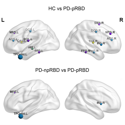 |
115 | Disrupted Brain Structural Network Connection in de novo Parkinson’s Disease with Rapid Eye Movement Sleep Behavior Disorder
Amei Chen1, Junxiang Huang2, Xiaofei Huang1, Xiaofang Cheng3, Yongzhou Xu 4, and Xinhua Wei1
1Guangzhou First People's Hospital, Guangzhou, China, 2Guangzhou Women and Children's Medical Center, Guangzhou, China, 3The Affiliated Brain Hospital of Guangzhou Medical University, Guangzhou, China, 4Philips Healthcare, Guangzhou, China Keywords: Parkinson's Disease, White Matter, White matter connectivity, rapid movement sleep behavior disorder To investigate the changes of white matter structural network connectivity in PD patients with probably rapid movement sleep behavior disorder (PD-pRBD). This study included PD-pRBD (n = 74) and PD with no probably RBD (PD-npRBD)(n = 97) and healthy contral (HC, n=73). The results showed that compared with the PD-npRBD group, the nodal efficiency (Ne) of the right insula and left middle frontal gyrus increased, while the Ne of the left temporal pole decreased. We conclude that changes in the right insula, left temporal pole and left middle frontal gyrus Ne may play key roles in the pathogenesis of PD-RBD. |
|
2289.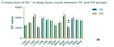 |
116 | Using R2* map to evaluate the changes of deep brain nuclei in patients with Parkinson 's disease
Man Wang1,2, Bingbing Gao1, Yanwei Miao1, and Dandan Zheng3
1the First Affiliated Hospital of Dalian Medical University, Dalian, China, 2Department of Radiology, the First Affiliated Hospital of Dalian Medical University, Dalian, China, 3Clinical and Technical Support, Philips Healthcare, Beijing, Beijing, China Keywords: Parkinson's Disease, Relaxometry, R2*,deep brain nuclei R2* is highly sensitive not only to tissue iron level, but also to the microscopic iron distribution. In this study, we first characterize the distribution of R2* within the deep brain nuclei in Parkinson's disease patients and health controls, especially the lateral difference. Secondly, we also evaluate the clinical usefulness of regional R2* for the differential diagnosis between PD and HC group. The results showed that there was no lateral difference in R2* values in the HC and PD groups. The R2* value of the right thalamus in the HC group was statistically higher than that in the PD group. |
|
2290.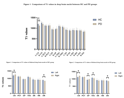 |
117 | Alterations of T1 and proton density values of deep brain nuclei based on synthetic MRI in Parkinson’s disease patients
Jinghan Zhao1, Yinghua Guo2, and Yanwei Miao1
1The First Affiliated Hospital of Dalian Medical University, Dalian, China, 2Clinical&Technical Support, Philips Healthcare, Beijing, China Keywords: Parkinson's Disease, Parkinson's Disease The objective of this study was to explore whether the alterations of T1 and proton density values could reflect the occurrence of Parkinson's disease (PD) and contribute to the prevention and diagnosis of PD. The T1 values of the left caudate nucleus (CN) and the proton density values of all nuclei were significantly reduced in the PD group compared with the control group. The T1 values of the left globus pallidus (GP) and right thalamus (THU) in the PD group were significantly reduced compared with those of the contralateral side. |
|
2291.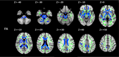 |
118 | Diffuse destruction of white matter myelin sheath integrity associated with cognitive impairment of long-term Parkinson’s disease patients
Bingbing Gao1, Liangjie Lin2, and Yanwei Miao1
1the First Affiliated hospital of Dalian Medical university, Dalian, China, 2Clinical and Technical Support, Philips Healthcare, Beijing, China Keywords: Parkinson's Disease, Dementia The incidence of Parkinson's disease patients combined with dementia increased significantly with the extension of disease course, and increased from 26.3% to 83% at twenty years after diagnosis. This study aims to use diffusion tensor imaging to detect the pattern of white matter damage in long-term PD patients with cognitive impairment, and results indicated that PD patients have diffuse FA decrease of white matter that is correlated with cognitive status. |
|
2292.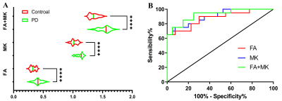 |
119 | Diagnostic efficacy of combined fractional anisotropy (FA) and mean kurtosis (MK) in detecting the Parkinson disease (PD) model rat
Yanchao Dong1, Yansheng Chen2, and Lanxiang Liu2
1Qinhuangdao Municipal No.1 Hospital, Qinhuandao, China, 2Qinhuangdao Municipal No.1 Hospital, Qinhuangdao, China Keywords: Parkinson's Disease, Parkinson's Disease, DKI Both FA and MK in DKI have value in the diagnosis of PD. We here investigated the sensitivity and specificity of FA and MK in the early diagnosis of PD rat model. PD and control group, 20 in each group. The FA, MK and FA+MK in PD group were statistically significantly different from that of the control group (P < 0.01). The Youden’s index of FA+MK value was higher than the two alone. The optimal cut-off FA+MK value was 1.404. The diagnostic efficiency of the FA+MK values were better relative to that two alone in diagnosing PD. |
|
2293.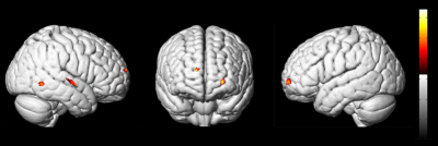 |
120 | Abnormalities of Grey Matter Volume in Parkinson’s Disease with Depression: A Voxel-Based Morphometry Study
Jiajun Cao1, Mingrui Qu1, Bingbing Gao1, Yuhan Jiang1, Yangyingqiu Liu1, Lizhi Xie2, and Yanwei Miao1
1Department of Radiology, the First Affiliated Hospital of Dalian Medical University, Dalian, China, 2GE Healthcare, Beijing, China Keywords: Parkinson's Disease, Parkinson's Disease, Voxel-Based Morphometry、Parkinson's disease with depression Depressive symptoms are common in Parkinson’s disease (PD), but the pathophysiology and neural basis underlying depression in PD is complex. To shed more light on the abnormal regions and distribution characteristics of gray matter in PD patients with depression (DPD), we conducted a Voxel-Based Morphometry (VBM) study. Compared with PD with non-depression (NDPD) patients, the gray matter volume of DPD patients decreased abnormally in the right superior temporal gyrus, right inferior temporal gyrus, left orbital superior frontal gyrus and left dorsolateral superior frontal gyrus. We speculate that changes in the brain regions are essential in the pathophysiology of DPD. |
|
 Back to Meeting Home
 Back to the Program-at-a-Glance
The International Society for Magnetic Resonance in Medicine is accredited by the Accreditation Council for Continuing Medical Education to provide continuing medical education for physicians.
 Back to Meeting Home
 Back to the Program-at-a-Glance
 View Presentation Video