Digital Poster
Imaging Aging, Dementia & Alzheimer's Disease I
ISMRM & ISMRT Annual Meeting & Exhibition • 03-08 June 2023 • Toronto, ON, Canada

Digital Poster
Imaging Aging, Dementia & Alzheimer's Disease I
| Computer # | |||
|---|---|---|---|
3311.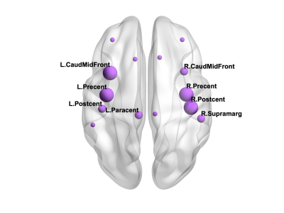 |
81 |
Network analysis reveals differences in the topology of
structural brain networks of young asymptomatic APOE-epsilon4
carriers
Eirini Messaritaki1,
Thomas M Lancaster1,2,
Katherine E Tansey3,
and Derek K Jones1
1Psychology, Cardiff University, Cardiff, United Kingdom, 2Psychology, University of Bath, Bath, United Kingdom, 3Cardiff University, Cardiff, United Kingdom Keywords: Alzheimer's Disease, Alzheimer's Disease This work explores the link between the topological properties of brain structural networks and APOE-epsilon4 in young asymptomatic adults. We investigated the sensorimotor, visual and default-mode networks. We found evidence that there are differences in the mean clustering coefficient of the sensorimotor network of carriers versus non-carriers, with the left caudal middle frontal, left precentral, right postcentral and right precentral gyri driving the differences. Interestingly, the mean clustering coefficient was higher in carriers compared to non-carriers. In contrast, no differences were found for the visual or the default-mode networks. |
|
3312.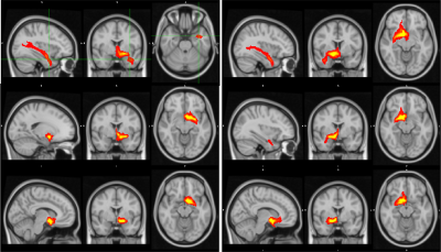 |
82 |
Mapping Connectivity of the Nucleus Basalis of Meynert in
Alzheimers using fMRI and probabilistic tractography
Sergio Adrian Becerra1,
Kaya Jordan1,
Jon Haroon1,
Kennedy Mahdavi1,
Sheldon Jordan1,2,
Rama Surya1,
and Elisabeth Rindner1
1Synaptec Network, Santa Monica, CA, United States, 2Neurology, UCLA, Los Angeles, CA, United States Keywords: Alzheimer's Disease, Alzheimer's Disease The nucleus basalis (NBM) is the major source of cholinergic innervation to the cerebral cortex, hippocampus and other subcortical structures. Focusing DTI and fMRI on the NBM can shed light on the potential state of subjects with Alzheimer’s disease. We aim to use DTI to provide insight into the white matter projections from the NBM and use fMRI to depict how it is functionally wired. We found that imaging of the NBM differentiated dementia patients from controls, and captured the level of cognitive decline. While the results are preliminary, this analysis introduces a method of observing the progression of AD. |
|
3313.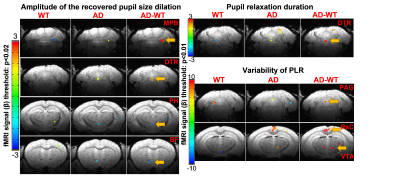 |
83 |
Pupil-fMRI correlation mapping of awake transgenic 5xFAD mice of
Alzheimer’s Disease
Xiaochen Liu1,
David Hike1,
Wenchao Yang1,
Zeping Xie1,2,
Bei Zhang1,
Andy Liu1,3,
Sang Cheon Choi1,
Biyue Zhu1,
Chongzhao Ran1,
Yuanyuan Jiang1,
and Xin Yu1
1Athinoula A. Martinos Center for Biomedical Imaging, Department of Radiology, Massachusetts General Hospital, Harvard Medical School, Charlestown, MA, United States, 2School of Traditional Medicine, Southern Medical University, Guangzhou, China, 3Department of Neuroscience, Boston University, Boston, MA, United States Keywords: Alzheimer's Disease, Alzheimer's Disease, pupil dynamics This study obtained visually stimulated high-resolution fMRI and real-time pupil dynamic signals in awake normal and AD mice. By analyzing the trial-specific pupillary responses to light (PRL), we identified unique temporal dynamic features in AD mice. By performing the pupil-fMRI event-related analysis, we detected specific subcortical functional nuclei in AD mice, showing strong correlations to the pupil dilation recovery features and the variability across different stimulation events. These results present novel function-behavioral linkage based on the pupillary responses to visual stimulation in AD mice, revealing an intriguing non-invasive and quantitative biomarker of the neurodegenerative progress of AD brains. |
|
3314.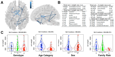 |
84 |
Identification of Brain Networks Associated with Alzheimer’s
Disease Risk
Alexandra Badea1,
Ali Mahzarnia2,
Jacques S Stout3,
Robert J Anderson2,
Hae Sol Moon4,
Zay Yar Han2,
Kate Beck5,
Jeffrey N Browndyke6,
David Dunson7,
Kim G Johnson5,
and Richard J O'Brien8
1Radiology, Neurology, BIAC, Duke Univ Medical Center, Durham, NC, United States, 2Radiology, Duke Univ Medical Center, Durham, NC, United States, 3BIAC, Duke Univ Medical Center, Durham, NC, United States, 4BME, Duke Univ Medical Center, Durham, NC, United States, 5Neurology, Duke Univ Medical Center, Durham, NC, United States, 6Psychiatry and Behavioral Sciences Department, Duke Univ Medical Center, Durham, NC, United States, 7Statistical Sciences, Duke University, Durham, NC, United States, 8Neurology, Duke University Medical Center, Durham, NC, United States Keywords: Alzheimer's Disease, Alzheimer's Disease, Aging The brain connectome helds promise to detect subtle changes in individuals at risk for Alzheimer's disease. We imaged using high resolution diffusion imaging 72 subjects enriched for the APOE4 genotype to reveal vulnerable networks associated with a composite AD risk factor including age, genotype, and sex. Sparse canonical correlation analysis (CCA) revealed a high weight associated with genotype, and subgraphs involving the cuneus, temporal, cingulate cortex, and cerebellum. Our results have identified structural brain networks and the associated weights for several risk factors for AD in preclinical stages. |
|
3315.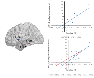 |
85 |
Predictive value of baseline functional connectivity to the
effect of transcranial magnetic stimulation on Alzheimer's
disease
Zhiwei Guo1,
Qiwen Mu1,
and Xiaoyong Zhang2
1The Second Clinicall Medical College of North Sichuan Medical College, Nanchong, China, 2Clinical Science, Philips Healthcare, Chengdu, China Keywords: Alzheimer's Disease, Alzheimer's Disease Repetitive transcranial magnetic stimulation (rTMS) may lead to a significant improvement in general cognitive function and memory function of Alzheimer's disease (AD). Several related functional connectivity may contribute to the effectiveness of rTMS. Besides, the baseline functional connectivity between the left hippocampus and left occipitotemporal cortex, left frontoinsular cortex were significantly positively correlated with the improvement of memory recognition function. The baseline functional connectivity between the left hippocampus and left occipitotemporal cortex was significantly positively correlated with the improvement of delayed recall function of memory. |
|
3316.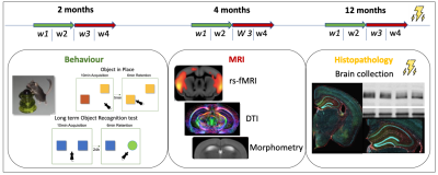 |
86 |
Sex dimorphism in early Alzheimer’s pathology: Brain
connectivity and Behavior in AppNL-F/MAPT double knock-in mice
Inès Ben Abdallah1,2,
Marion Sourty1,
Mary Mondino1,
Laetitia Degiorgis1,
Julien Lamy1,
Vincent Noblet1,
Marion Rame1,
Cristiana Pistono2,
Aminé Isik2,
Marie-Dominique Marinutti2,
Céline Héraud2,
Hiroki Sasaguri3,
Shoko Hashimoto3,
Takashi Saito3,
Takaomi Saido3,
Chantal Mathis2,
and Laura Harsan1,4
1ICube, Université de Strasbourg-CNRS, Strasbourg, France, 2LNCA, Université de Strasbourg-CNRS, Strasbourg, France, 3RIKEN Center for Brain Science, 2-1 Hirosawa, Wako-city, Saitama, Japan, 4Department of Biophysics and Nuclear Medicine, University Hospital of Strasbourg, Strasbourg, France Keywords: Alzheimer's Disease, fMRI (resting state), Preclinical MRI is a unique tool to study the complexity of functional and structural communication in the brain. To explore the brain architecture of a mouse model of Alzheimer's Disease and highlight sex-dimorphism in the emergence of AD-like signs, we used a recent mouse model, the APPNL-F/MAPT double knock-in (dKI). In preclinical imaging, we used resting-state graph theory approaches in a longitudinal study associated with behavioral evaluation. Functional connectivity of perirhinal, dorsal-hippocampus and midbrain nodes were implicated in early memory impairments in dKI female mice. Interestingly, perirhinal-cortex and dorsal-hippocampus are key regions for object-place associative memory and long-term object-recognition. |
|
3317.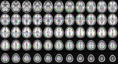 |
87 |
Network analysis of Alzheimer’s disease using group cohesive
parcellation of rsfMRI
Ajay Nemani1 and
Mark Lowe1
1Cleveland Clinic, Cleveland, OH, United States Keywords: Alzheimer's Disease, fMRI (resting state), Parcellation, Network Modelling The search for rsfMRI-based network biomarkers in Alzheimer's disease has yielded inconsistent results. These models are derived from standard anatomical and functional parcellations that may bias downstream analyses. We present a network model of Alzheimer's disease based on cohesive parcellation and compare it to traditional measures of network topology. |
|
3318.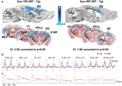 |
88 |
Hyperactive BOLD response in presymptomatic AD mice model as
potential biomarker of early AD
Taeyi You1,2,
Taekwan Lee3,
Geun Ho Im1,
Seong-gi Kim1,
Sungkwon Chung4,
and Jung Hee Lee5
1Center for Neuroscience Imaging Research, Institute of Basic Science, Suwon, Korea, Republic of, 2Biomedical Engineering, Sungkyungkwan University, Suwon, Korea, Republic of, 3Korea Brain Research Institute, Daegu, Korea, Republic of, 4Physiology, Sungkyunkwan University, Suwon, Korea, Republic of, 5Radiology, Samsung Medical Center, Seoul, Korea, Republic of Keywords: Alzheimer's Disease, fMRI (task based) Alzheimer Disease is a neurodegenerative disease that exhibits memory and cognitive deficits. Majority of research has targeted these late-stage biomarkers as treatment targets, yet all have failed in the clinical settings. Detection of early biomarkers is becoming imperative that can be easily and effectively used. fMRI is a quick and non-invasive procedure that can map whole brain activity. Here we use sensory evoked fMRI in a AD mouse model to show longitudinal changes in brain response that can be potential biomarkers for early AD. |
|
3319.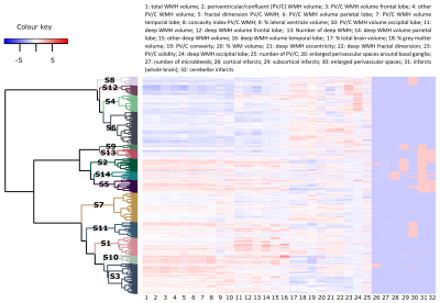 |
89 |
MRI phenotypes of the brain are related to long-term dementia
outcome in community-dwelling older adults
Jasmin A. Keller1,
Sigurdur Sigurdsson 2,
Bárbara Schmitz Abecassis 1,
Ilse M.J. Kant 3,4,
Mark A. van Buchem1,
Lenore J. Launer5,
Matthias J.P. van Osch1,
Vilmundur Gudnason2,6,
and Jeroen H.J.M. de Bresser1
1Department of Radiology, Leiden University Medical Center, Leiden, Netherlands, 2Icelandic Heart Association, Kopavogur, Iceland, 3Clinical Artificial Intelligence Implementation and Research Lab (CAIRELab) and Department of Information Technology & Digital Innovation, Leiden University Medical Center, Leiden, Netherlands, 4Department of Digital Health, University Medical Center Utrecht, Utrecht, Netherlands, 5Laboratory of Epidemiology and Population Science, National Institute on Aging, Bethesda, MD, United States, 6Faculty of Medicine, University of Iceland, Reykjavik, Iceland Keywords: Dementia, Aging, Cerebral small vessel disease Individual brain MRI markers only show at best a modest association with long-term occurrence of dementia. Therefore, it is challenging to accurately identify individuals at increased risk for dementia. We implemented a combined hierarchical clustering analysis based on neurodegenerative and neurovascular brain MRI markers and identified 14 distinct subgroups of individuals with different brain MRI phenotypes. These subgroups had a different long-term risk for dementia; especially the multi-burden brain MRI phenotype showed an increased risk (HR: 13.8 (95%-CI:4.28-44.37)). These findings may in the future be useful to determine patient prognosis and may aid in patient selection for future treatment studies. |
|
3320.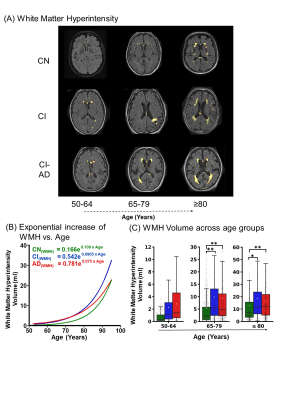 |
90 |
MRI measured Neuroanatomic volume, Cortical Thinning and WMH
load with Aging: The Early, Intermediate and Late events of
Cognitive status
Neha Yadav1,
Arkaprava Majumdar1,
and Vivek Tiwari1
1Department of Biological Sciences, Indian Institute of Science Education and Research Berhampur, Berhampur, India Keywords: Alzheimer's Disease, Aging The key to understanding brain-biomarkers depictive of normal cognition and cognitive impairment due to MCI (CI) and/or Alzheimer’s disease (CI-AD) is to identify the series of early, intermediate, and late events that encode brain health. The temporal and spatial order of events of brain health changes observed on MRI, associated with normal aging needs to be precisely delineated from the events associated with cognitive impairment. We have identified early, intermediate and late brain structural and microvascular events distinctive of CN, CI and CI-AD and developed an AI-based platform using an optimal number of MRI features distinctive of cognitive status. |
|
3321.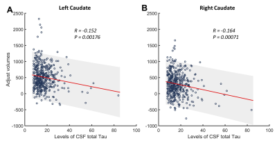 |
91 |
Associations between CSF Biomarkers of Alzheimer's Disease and
Subcortical Volume in Healthy Aging
Qixiang Lin1,
Shuai Huang2,
Aditya Bisht1,
Allan Levey1,3,
James Lah1,3,
and Deqiang Qiu2,3,4
1Department of Neurology, Emory University, Atlanta, GA, United States, 2Department of Radiology and Imaging Sciences, Emory University, Atlanta, GA, United States, 3Goizueta Alzheimer’s Disease Research Center, Emory University, Atlanta, GA, United States, 4Joint Department of BioMedical Engineering, Emory University and Georgia Institute of Technology, Atlanta, GA, United States Keywords: Alzheimer's Disease, Aging, CSF biomarker; Aβ; Tau; Subcortical; Volume This study aims to evaluate the association between subcortical volumes and cerebrospinal fluid (CSF) biomarkers of Alzheimer’s Disease (AD) in a large group of healthy aging from a single center. CSF samples were obtained and quantitative levels of amyloid-β and Tau were measured. Subcortical tissue segmentations were performed on T1-weighted MPRAGE scans and the volume of each subcortical structure were calculated. Significant correlations were found between CSF biomarkers of AD and volume of the right and the left caudate. This may indicate that the volume of caudate are associated with CSF AD biomarker in healthy aging participants. |
|
3322.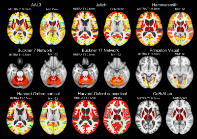 |
92 |
A comprehensive set of gray matter labels for the MIITRA atlas:
Interoperability with complementary atlases
Mohammad Rakeen Niaz1,
Yingjuan Wu1,
Abdur Raquib Ridwan2,
Shengwei Zhang2,
David A. Bennett2,
and Konstantinos Arfanakis1,2
1Biomedical Engineering, Illinois Institute of Technology, Chicago, IL, United States, 2Rush Alzheimer’s Disease Center, Rush University Medical Center, Chicago, IL, United States Keywords: Alzheimer's Disease, Aging, Aging, Atlas, Brain, Gray Matter The Multichannel Illinois Institute of Technology & Rush university Aging (MIITRA) atlas constructed using high quality MRI data on a large (N=400), diverse, community cohort of non-demented older adults, contains high resolution (0.5mm) structural and diffusion imaging templates. The present work constructed and evaluated a comprehensive set of gyral-based, cytoarchitecture-based, and functional connectivity-based gray matter labels in MIITRA space in order to enhance the functionality of the MIITRA atlas and its interoperability with complementary atlases. |
|
3323.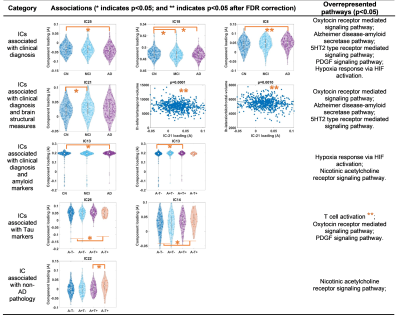 |
93 |
Multivariate data-driven approach to dissect imaging-genetic
associations in Alzheimer’s disease
Xiaowei Zhuang1,
Zhengshi Yang1,
and Dietmar Cordes1
1Lou Ruvo Center for Brain Health, Cleveland Clinic, Las Vegas, NV, United States Keywords: Alzheimer's Disease, Alzheimer's Disease, Imaging genetics To better characterize the Alzheimer’s disease pathogenesis and boost the statistical power, we applied a multivariate data-driven approach (independent component analysis (ICA)) to decompose 70000+ single nucleotide variants (SNVs) from 239 AD-associated genes into multiple functionally relevant subsets. We demonstrated that several genetic clusters identified by ICA could be specially associated with AD clinical diagnosis, AD amyloid or tau pathology, and/or MRI-derived neurodegenerative markers. This type of multivariate data-driven approach could be helpful to further delineate diagnoses-associated or neuropathology-associated genetic variants in AD. |
|
3324.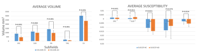 |
94 |
Hippocampal subfield volume in relation to cerebrospinal fluid
Amyloidß and cognitive assessments in early Alzheimer’s disease:
a 7T MRI study
Oluwatobi Folorunsho Adeyemi1,2,
Olivier Mougin1,
George Hutchinson1,
Penny Gowland1,
Richard Bowtell1,
and Akram Hosseini3
1University of Nottingham, Nottingham, United Kingdom, 2Physics, University of Abuja, Abuja, Nigeria, 3Nottingham University Hospital, Nottingham, United Kingdom Keywords: Alzheimer's Disease, Alzheimer's Disease, High-Field MRI, Quantitative Susceptibility Mapping, Segematation Providing a non-invasive biomarker for the diagnosis of AD has been challenging. In this study 25 participants (12 AD and 13 healthy controls) were scanned on a 7T MRI scanner. Cognitive assessments and analysis of the CSF for Amyloidb1-42 performed by trained Clinicians. The volume of the hippocampal subregion shows a linear relationship CSF- Amyloidb1-42 for all the subfield of the hippocampus, but this trend was only significant for the ERC with p=0.023. The association of the volume of ERC and CSF-amyloid-beta(1-42) suggests the potential for using high field-high resolution MRI as a biomarker for an early identification of AD. |
|
3325. |
95 |
Neuroimaging and Cognitive Testing in Healthy Aging Adults using
a Portable Low-Field MRI Scanner and Web-Based Assessment
Sean Deoni1,
Phoebe Burton2,
Jennifer Beauchemin2,
Rosa Cano-Lorente2,
Matthew De Both3,
Megan Johnson3,
Lee Ryan4,
and Mathew Huentleman3
1MNCH D&T, Bill & Melinda Gates Foundation, Seattle, WA, United States, 2Advanced Baby Imaging Lab, Providence, RI, United States, 3TGen, Pheonix, AZ, United States, 4University of Arizona, Tucson, AZ, United States Keywords: Dementia, Aging, Remote Neuroimaging In this study we sought to (1) Determine the feasibility of collecting remote MRI and cognitive data in adults and elderly individuals; and (2) Replicate previously reported population-based associations between regional brain volumes and cognitive performance with an established cognitive assessment, PAL. Overall, this initial report of at-home MRI shows that MRI data collection on a portable low-field MRI system at a participant's home is possible and offers time efficiency, convenience, and accessibility to participants who might otherwise not be able to participate. |
|
3326.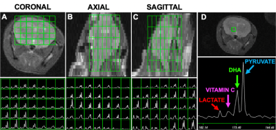 |
96 |
3D-MRSI using co-polarized HP [1-13C]pyruvate and
[1-13C]dehydroascorbate to study a mouse model of Alzheimer's
disease
Paola Porcari1,
Saket Patel1,
Elizabeth Coffee1,
Marjan Berishaj 1,
Tanya Jain2,
Kofi M. Deh1,
Nathaniel T. Kim1,
Yueming Li2,
and Kayvan Keshari1
1Radiology, Memoral Sloan Kettering Cancer Center, New York, NY, United States, 2Chemical Biology Program, Memoral Sloan Kettering Cancer Center, New York, NY, United States Keywords: Alzheimer's Disease, Alzheimer's Disease, animal model, 13C HP MRI Alzheimer’s disease (AD) is devastating and progressive disease. The non-invasive detection of early onset AD along with the mechanisms that drive its progression are still limited. Recent work has implicated metabolic reprogramming as an earlier AD potential indicator. Therefore, we investigated an AD mouse model at the early stage of development with the goal to evaluate whether co-hyperpolarized [1-13C]pyruvate and [1-13C]dehydroascorbate (DHA) along with 3D MRSI might provide insights into the brain metabolism of FAD mice at the onset of the disease. The metabolism of these translatable biomarkers would then give insight on our understanding of AD diagnosis and treatment. |
|
3327.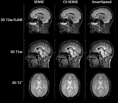 |
97 |
Accelerating 3T MRI standardized protocol for detection of
Amyloid Related Imaging Abnormality (ARIA) using Philips
SmartSpeed
Sandeep Ganji1,2,
Brian Johnson3,4,
Spencer Waddle1,
Johannes Peeters5,
Laszlo Mechtler6,
and Nandor Pinter6,7
1Philips, Rochester, MN, United States, 2Mayo Clinic, Rochester, MN, United States, 3Philips, Gainesville, FL 32608, FL, United States, 4University of Texas Southwestern Medical Center, Dallas, TX, United States, 5Philips, Eindhoven, Netherlands, 6Dent Neurologic Institute, Buffalo, NY, United States, 7Department of Neurosurgery, University at Buffalo, Buffalo, NY, United States Keywords: Alzheimer's Disease, Alzheimer's Disease Amyloid Related Imaging Abnormality (ARIA) was reported in 40% of patients treated with anti-amyloid beta drugs in phase 3 trials. It is expected to be a significant factor in the clinical application of new Alzheimer’s Disease (AD) modifying therapies and requires standardized and practical imaging. By employing Compressed-SENSE (CS-SENSE) and newer deep learning based SmartSpeed to accelerate acquisition we created a robust MRI protocol that can be completed in under ten minutes without loss of any image quality. This accelerated protocol can provide a clinically feasible strategy for scanning large populations. |
|
3328.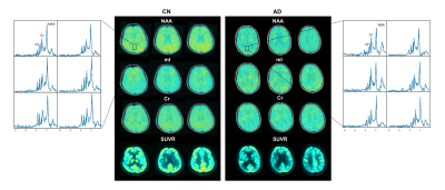 |
98 |
Brain NAA Reduction is Associated with Glucose Hypometabolism in
Functional Networks of Alzheimer’s Disease: A Hybrid
3D-MRSI/FDG-PET Study
Wenli Li1,
Miao Zhang2,
Yibo Zhao3,4,
Yudu Li3,5,
Wen Jin3,4,
Jialin Hu1,
Yaoyu Zhang1,
Danni Wang1,
Biao Li2,
Jun Liu6,
Binyin Li6,
Zhi-Pei Liang3,4,
and Yao Li1
1School of Biomedical Engineering, Shanghai Jiao Tong University, Shanghai, China, 2Department of Nuclear Medicine, Ruijin Hospital, Shanghai Jiao Tong University School of Medicine, Shanghai, China, 3Beckman Institute for Advanced Science and Technology, University of Illinois at Urbana-Champaign, Urbana, IL, United States, 4Department of Electrical and Computer Engineering, University of Illinois at Urbana-Champaign, Urbana, IL, United States, 5National Center for Supercomputing Applications, University of Illinois at Urbana-Champaign, Urbana, IL, United States, 6Department of Neurology and Institute of Neurology, Ruijin Hospital, Shanghai Jiao Tong University of Medicine, Shanghai, China Keywords: Alzheimer's Disease, Alzheimer's Disease Functional network failure has been implicated in the pathophysiology of Alzheimer’s disease (AD). FDG-PET is a well-established tool to map the glucose hypometabolism during AD progression. MRSI indexes the neuronal loss/astrogliosis noninvasively, but has been limited to single-voxel/slice techniques. Using a high-resolution 3D MRSI technique, we evaluated the neurometabolic changes in brain networks and compared them with glucose hypometabolism. Decreases in NAA and increases in mI were found in all networks. NAA reduction followed similar patterns to the hypometabolism over cognitive decline. Combined 3D MRSI and atrophy biomarkers showed comparable performance to FDG-PET in predicting cognitive decline in AD patients. |
|
3329.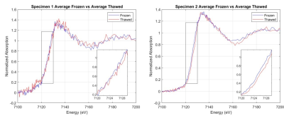 |
99 |
Interrogating the effect of thawing on Fe speciation in human
tissue using XANES
Dean Tran1,
Phillip DiGiacomo1,
Marios Georgiadis1,
Nicholas Edwards2,
Sharon Bone2,
Donald Born3,
Samuel Webb2,
and Michael Zeineh1
1Department of Radiology, Stanford University, Stanford, CA, United States, 2Stanford Synchrotron Radiation Lightsource, SLAC National Accelerator Laboratory, Menlo Park, CA, United States, 3Department of Pathology, Stanford University, Stanford, CA, United States Keywords: Alzheimer's Disease, Alzheimer's Disease, X-ray microscopy Recent studies suggest that iron and neuroinflammation are key components of AD pathology. Specifically, ferrous Fe2+ can cause oxidative stress, a possible a mechanistic link to disease progression. Correlative ex vivo MRI can detect iron-containing microglia in AD hippocampi, but whether this iron is ferrous is unknown. Synchrotron X-ray absorption near edge spectroscopy (XANES) can quantify iron oxidation state in frozen human brain samples. However, tissue thawing during the long scans might affect the oxidation state. Here, we implement the necessary hardware to interrogate that question and present preliminary evidence that, as specimens thaw, less redox-active ferrous iron is measured. |
|
3330.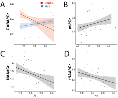 |
100 |
Neuropathology and neurometabolites in mild cognitive
impairment investigated with 11C-PiB amyloid PET and 7T MRS
Christopher William Davies-Jenkins1,2,
Kathleen E Hupfeld1,2,
Helge J Zöllner1,2,
Gwenn S Smith3,4,
and Georg Oeltzschner1,2
1The Russell H. Morgan Department of Radiology and Radiological Science, Johns Hopkins Medicine, Baltimore, MD, United States, 2F.M. Kirby Research Center for Functional Brain Imaging, Kennedy Krieger Institute, Baltimore, MD, United States, 3Department of Psychiatry and Behavioral Sciences, Johns Hopkins Medicine, Baltimore, MD, United States, 4Division of Nuclear Medicine and Molecular Imaging, The Russell H. Morgan Department of Radiology and Radiological Science, Johns Hopkins Medicine, Baltimore, MD, United States Keywords: Alzheimer's Disease, Alzheimer's Disease, MRS, PET, Amyloid, 7T, MCI Mild cognitive impairment (MCI), a prodromal stage of Alzheimer’s disease, is increasingly studied by multi-modal beta-amyloid (Aβ) PET and MRS. Most studies have targeted the PCC and a limited number of metabolites. In this study, we analyzed Aβ PET and 7T 1H-MRS data from the ACC and PCC of MCI patients and healthy controls, using a voxel-specific Aβ metric. Metabolite-amyloid correlations were investigated using multiple regression analysis. We report a novel finding of a differential GABA-amyloid relationship for MCI patients compared with controls in the ACC and support previous reports of amyloid relationships with NAA and myo-inositol. |
|
 Back to Meeting Home
 Back to the Program-at-a-Glance
The International Society for Magnetic Resonance in Medicine is accredited by the Accreditation Council for Continuing Medical Education to provide continuing medical education for physicians.
 Back to Meeting Home
 Back to the Program-at-a-Glance
 View Presentation Video