Digital Poster
Epilepsy
ISMRM & ISMRT Annual Meeting & Exhibition • 03-08 June 2023 • Toronto, ON, Canada

| Computer # | |||
|---|---|---|---|
 |
5199.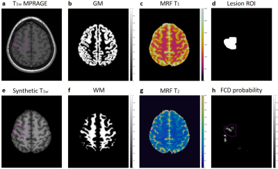 |
101 |
Combining MR Fingerprinting with Voxel-based Morphometric MRI
Analysis to Reduce False Positives for Focal Cortical Dysplasia
Detection
Zheng Ding1,2,
Siyuan Hu2,
Ting-Yu Su1,2,
Joon Yul Choi1,
Xiaofeng Wang3,
Ken Sakaie4,
Hiroatsu Murakami1,
Hans-Juergen Huppertz5,
Ingmar Blumcke1,6,
Stephen Jones4,
Imad Najm1,
Dan Ma2,
and Zhong Irene Wang1
1Epilepsy Center, Cleveland Clinic, Cleveland, OH, United States, 2Biomedical Engineering, Case Western Reserve University, Cleveland, OH, United States, 3Quantitative Health Science, Cleveland Clinic, Cleveland, OH, United States, 4Imaging Institute, Cleveland Clinic, Cleveland, OH, United States, 5Swiss Epilepsy Clinic - Klinik Lengg AG, Zurich, Switzerland, 6University of Erlangen-Nuremberg, Erlangen, Germany Keywords: Epilepsy, MR Fingerprinting We sought to improve focal cortical dysplasia (FCD) lesion detection by combining MR fingerprinting (MRF) with voxel-based morphometric MRI analysis. We acquired high-resolution MRF and T1w MPRAGE data from 29 patients and 47 age-and-gender-matched healthy controls. FCD probability maps were generated using the morphometric analysis program (MAP18). MRF T1 and T2 values in white matter were significantly higher in true-positive clusters than false-positive clusters. Using normalized MRF T1, T2, and cluster size as input, an SVM model was able to predict whether each cluster represents a true positive with high accuracy, demonstrating its potential contribution to clinical FCD detection workflow. |
5200.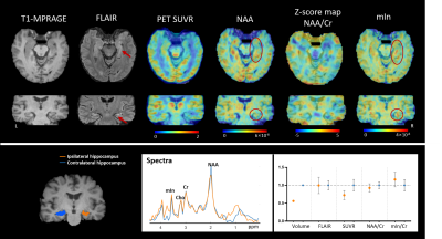 |
102 |
N-acetylaspartate and myo-inositol provide complementary
information for lateralization of mesial temporal lobe epilepsy
Hui Huang1,
Miao Zhang2,
Bingyang Cai1,
Siyu Yuan1,
Wen Jin3,4,
Yudu Li3,5,
Yibo Zhao3,4,
Zhi-Pei Liang3,4,
Yao Li1,
Biao Li2,
and Jie Luo1
1School of Biomedical Engineering, Shanghai Jiao Tong University, Shanghai, China, 2Department of Nuclear Medicine, Ruijin Hospital, Shanghai Jiao Tong University School of Medicine, Shanghai, China, 3Beckman Institute for Advanced Sciences and Technology, University of Illinois at Urbana Champaign, Urbana, IL, United States, 4Department of Electrical and Computer Engineering, University of Illinois at Urbana Champaign, Urbana, IL, United States, 5National Center for Supercomputing Applications, University of Illinois at Urbana Champaign, Urbana, IL, United States Keywords: Epilepsy, Metabolism Lateralization of drug refractory mesial temporal lobe epilepsy can be challenging for routine MR scans at 3.0T. Exogenous and radioactive tracer 18F-FDG has been widely reported to aid the lateralization of MR unidentifiable epileptic hippocampus. 1H-MRSI holds promise to provide endogenous metabolic information of the epileptogenic zone. This study demonstrated the feasibility of fast high-resolution mapping of NAA/Cr, mIn/Cr and mIn/NAA, and investigated their associations with the FDG uptake. Our experimental results showed that NAA and mIn were independently sensitive to metabolic changes in hippocampal sclerosis, providing complementary information for temporal lobe epilepsy lateralization. |
|
5201.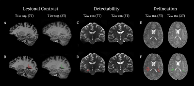 |
103 |
Quantification of Lesion Volume Differences in Structural
Epilepsy in 7T and 3T
Stefanie Chambers1,2,
Lukas Haider3,
Philipp Lazen1,
Gregor Kasprian3,
Johannes Koren4,
Robert Diehm5,
Katharina Moser5,
Matthias Tomschik1,
Jonathan Wais1,
Fabian Winter1,
Vitalij Zeiser1,
Stephan Gruber2,6,
Susanne Aull-Watschinger7,
Tatjana Traub-Weidinger8,
Christoph Baumgartner 4,
Martha Feucht5,
Christian Dorfer1,
Wolfgang Bogner2,6,
Siegfried Trattnig2,6,
Ekaterina Pataraia7,
Karl Rössler1,6,
and Gilbert Hangel1,2,6
1Department of Neurosurgery, Medical University of Vienna, Vienna, Austria, 2High-field MR Center, Department of Biomedical Imaging and Image-guided Therapy, Medical University of Vienna, Vienna, Austria, 3Division of Neuroradiology and Musculoskeletal Radiology, Department of Biomedical Imaging and Image-guided Therapy, Medical University of Vienna, Vienna, Austria, 4Department of Neurology, Hietzing Hospital, Vienna, Austria, 5Center for Rare and Complex Childhood Onset Epilepsies, Member of ERN EpiCARE, Department of Pediatrics and Adolescent Medicine, Medical University of Vienna, Vienna, Austria, 6Christian Doppler Laboratory for MR Imaging Biomarkers, Vienna, Austria, 7Department of Neurology, Medical University of Vienna, Vienna, Austria, 8Division of Nuclear Medicine, Department of Biomedical Imaging and Image-guided Therapy, Medical University of Vienna, Vienna, Austria Keywords: Epilepsy, High-Field MRI In this study, structural lesions in 3T and 7T of 12 patients suffering from intractable focal epilepsy were co-registered and manually segmented using ITK-SNAP. The rendered volumes were compared with reference to 3T lesional size in FLAIR, T1 and T2-weighted sequences. Additionally, a WMS sequence was compared to 3T FLAIR as well as 7T FLAIR. Our findings illustrate that on average, 7T showed larger lesion volume in all sequences except WMS. |
|
5202.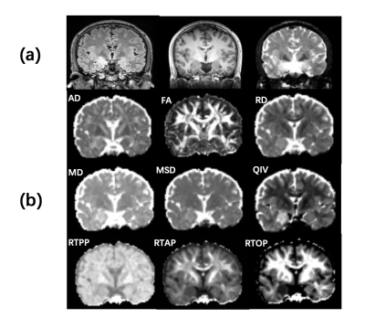 |
104 |
The diagnostic value of mean apparent propagator (MAP) MRI in
temporal lobe epilepsy (TLE) with unilateral amygdala
enlargement: A pilot study
Lu Zu1,2,
Peng Zhao2,
Mengxiao Liu3,
Xiaoli Li4,
Wei Zhang5,
Yani Cheng1,2,
and Xiangtao Lin2
1Shandong University, Jinan, China, 2Department of Radiology, Shandong Provincial Hospital, Jinan, China, 3MR scientific Marketing, Diagnostic Imaging, Siemens Healthineers Ltd, Shanghai, China, 4Department of Radiology, The Second People’s Hospital of Kunming, Kunming, China, 5The People's Hospital of Laoling, Laoling, China Keywords: Epilepsy, Diffusion/other diffusion imaging techniques, mean apparent propagator (MAP) For patients with routine MRI-negative epilepsy, noninvasive imaging is important for differential diagnosis. AE might be a subtype of MRI-negative TLE. The aim of this study was to evaluate the diagnostic efficacy of MAP-MRI and traditional DTI between TLE-AE patients and healthy controls. Results showed that MSD, QIV, RTAP, RTOP from MAP-MRI and FA from DTI in amygdala can be used as an imaging indicator to identify TLE-AE(P<0.05). Furthermore, MSD, RTAP, RTOP had a significant correlation with seizure types. MAP-MRI outperformed DTI in the diagnosis of TLE-AE, and MAP-MRI is expected to contribute in probing MRI-negative epileptogenic lesions. |
|
5203.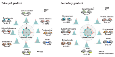 |
105 |
Connectome gradient dysfunction in benign childhood epilepsy
with centrotemporal spikes: Initial discovery
Guiqin Chen1,
Jie Hu1,
Haifeng Ran1,
Heng Liu1,
and Tijiang Zhang1
1Department of Radiology, The Affiliated Hospital of Zunyi Medical University, Zunyi, China Keywords: Epilepsy, Brain Large-scale brain network abnormalities and cognitive impairment in patients with benign childhood epilepsy with centrotemporal spikes (BECTS). And cognitive function needs hierarchical interaction support between brain-collateral systems. Whether there are changes in the interaction and functional arrangement between different network systems in patients with BECTS still ambiguous. The purpose of this study is to use the method of gradient connection to investigate the changes of macro-network function hierarchy of BECTS and its potential contribution to cognitive function in BECTS children . |
|
5204.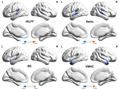 |
106 |
Static and Dynamic Alteration of Intrinsic Brain Activity might
improve MRI-negative Temporal Lobe Epilepsy Diagnosis and
Lateralization
song cheng ru1 and
cheng jing liang1
1MRI, the first affiliated hospital of zhengzhou university, zhengzhou, China Keywords: Epilepsy, fMRI (resting state), intrinsic brain activity, dynamic, cognition We comprehensively explored the potential intrinsic brain activity (IBA) abnormalities affected by MRI-negative temporal lobe epilepsy (TLE) based on six temporal dynamic indicators (dALFF, dfALFF, dReHo, dDC, dGSCorr, dVMHC) and their corresponding static indicators, the results revealed that the abnormally activated brain regions overlap markedly, including ①decreased fALFF, Reho, DC, VMHC, dfALFF, dReHo in the temporal neocortex with ipsilateral superiority. ②decreased dGSCorr and dVMHC in the occipital lobe. Moreover, many IBA indicators significantly correlated with the epilepsy duration or cognitive scale scores. The dDC, fALFF and DC showed significant discrimination ability. The ReHo and fALFF demonstrated lateralization significance. |
|
5205.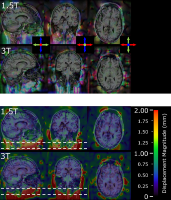 |
107 |
Spatial Distortion from Leksell G-frame for Stereotactic
Neurosurgery at 1.5T and 3T
Kiran K Seunarine1,
Martin M Tisdall2,
and Enrico De Vita1
1Physics Group, Department of Radiology, Great Ormond Street Hospital NHS Foundation Trust, London, United Kingdom, 2Department of Neurosurgery, Great Ormond Street Hospital NHS Foundation Trust, London, United Kingdom Keywords: Epilepsy, Precision & Accuracy, Stereotactic Stereo electroencephalography (sEEG) is a valuable tool for localising seizure-onset zones in focal epilepsy. Pre-surgical planning typically combines CT images, which have little spatial distortion, with MRI images, which have improved cerebral tissue contrast. However, the use of CT results in a potentially unnecessary radiation dose to the patient and additional demand on radiology services. In this work, we assess the additional distortion caused by the stereotactic Leksell G-frame in a single healthy volunteer at both 1.5T and 3T. We show that distortion is <1mm in 99.6% of the brain volume at 3T. |
|
5206.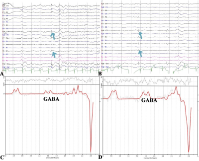 |
108 |
In vivo γ-aminobutyric acid alterations as a biomarker of the
therapeutic effect of MRI-negative temporal lobe epilepsy
Shuohua Wu1,2,
Qianqi Wang2,
Huige Zhai2,
Yiwen Zhang2,
Pu-yeh Wu3,
Gen Yan2,
and Renhua Wu1
1The Second Affiliated Hospital, Medical College of Shantou University, Shantou, China, 2The Second Affiliated Hospital of Xiamen Medical College, Xiamen, China, 3GE Healthcare, Beijing, Beijing, China Keywords: Epilepsy, Molecular Imaging, therapeutic effect Current diagnosis of MRI-negative TLE relies on clinical history and EEG or IEEG. However, IEEG is invasive and fails to monitor therapeutic effects dynamically. It is necessary to screen for effective biomarkers. We used MEGA-PRESS technique to investigate role of GABA and other metabolic alterations in TLE. We demonstrated that GABA, NAA, NAA+NAAG, and Glu levels in MTL were significantly different between epileptic and contralateral sides, and increasing index of GABA values is associated with increasing index of seizure frequency. These findings suggested that GABA is an effective biomarker for lateralization and therapeutic effect monitoring in patients with MRI-negative TLE. |
|
5207.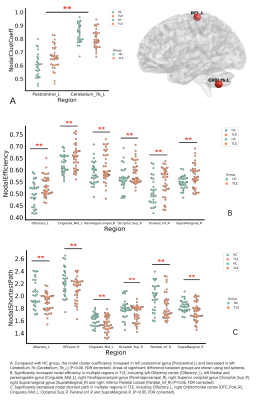 |
109 |
Diffusion spectrum imaging study of cognitive and emotional
related structural network in patients with temporal lobe
epilepsy
jiachen Li1,
jing Zhang1,
wanjun Hu1,
guangyao Liu1,
liang Zhou1,
and kai Ai2
1Department of Magnetic Resonance, Lanzhou University Second Hospital, Lanzhou, China, 2Philips Healthcare, Xi'an, China Keywords: Epilepsy, Diffusion/other diffusion imaging techniques To investigate the relationship between changes in brain networks and changes in emotion and cognition in patients with temporal lobe epilepsy. We used Diffusion spectrum imaging to construct structural brain networks. Then graph-theoretical analysis was applied to estimate structural connectivity and network properties in temporal lobe epilepsy (TLE) patients. Our study found that the brain network connectivity is altered and this alteration is associated with cognitive decline in TLE patients. This result offers a complementary understanding of neuropathology mechanism of TLE and its related cognition alteration, which may be helpful for clinical diagnosis and prognosis evaluation. |
|
5208.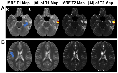 |
110 |
Detection of Lesions in Focal Epilepsy by Asymmetry Analysis of
MR Fingerprinting
Yuting Li1,
Leiyu Geng2,
Hong Ge2,
Wenbo Zhang3,
Siyuan Wu3,
and Shenghong Ju2
1Radiology, Zhongda Hospital, Medical School of Southeast University, Nanjing, China, 2Zhongda Hospital, Medical School of Southeast University, Nanjing, China, 3Medical School of Southeast University, Nanjing, China Keywords: Epilepsy, MR Fingerprinting Conventional MRI is limited in diagnosis of subtle epileptic lesions. Therefore, this study aimed to localize epileptic lesions by using MRF technology. Twenty epilepsy patients were included which underwent T1, T2 and MRF examinations. The MRI were spatially normalized to the symmetric ICBM-152 template, followed by transformation of T1 and T2 MRF maps. Absolute asymmetric index (|AI|) analysis was then performed on T1 and T2 MRF maps to detect epileptic lesions. Results showed that the |AI| analysis can detect epileptogenic foci in 75% of patients. In conclusion, the asymmetry analysis could help improve epileptic focus localization in clinical practice. |
|
5209.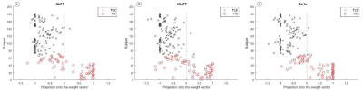 |
111 |
Diagnostic value of rs-fMRI indicators based on SVM
classification in adult patients with MRI-negative temporal lobe
epilepsy
Yang Fan1,
JIA Wen xiao1,
HANJIAERBIEKE KUKUN1,
WANG Shao yu2,
and WANG Yun ling1
1Department of Radiology, The First Affiliated Hospital of Xinjiang Medical University, Urumqi, China, 2MR Scientific Marketing MR Scientific Marketing, Shanghai, China Keywords: Epilepsy, fMRI (resting state) Rs-fMRI technology provides a range of analytical approaches that expand the scope of epilepsy research, and these algorithms provide unique views on pathophysiological processes that complement each other in interpreting regional spontaneous brain activity. ALFF, fALFF and ReHo can effectively identify MRI negative TLE (MRIn TLE) patients based on support vector machine algorithm. We hypothesized that there were abnormal changes in DMN-related brain regions in MRIn TLE patients, and the brain function indexes of angular gyrus, precuneus, and inferior parietal angular gyrus could distinguish MRIn TLE patients from HC. |
|
5210.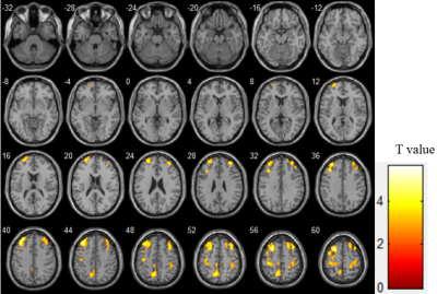 |
112 |
The potential value of PCASL in reflecting cognitive performance
for older adults with MRI-negative epilepsy
Xuyang Yin1,
Jianhong Wang2,
Yanwei Zeng1,
Zhenxu Xiao2,
Junyan Fu1,
Qianhua Zhao2,
Ding Ding2,
and Jun Zhang1
1Radiology Department, Huashan Hospital, Shanghai, China, 2Neurology Department, Huashan Hospital, Shanghai, China Keywords: Epilepsy, Arterial spin labelling Older adults with epilepsy accompanied by cognitive impairment represent a huge proportion, which calls for novel clinical techniques for early diagnosis and intervention. By applying pseudo-continuous arterial spin labeling (PCASL), we identified whole-brain voxel-based perfusion pattern of epilepsy associated with cognitive impairment, and explored correlations between cerebral blood flow (CBF) and domain-specific cognitive performance. The enrolled patients exhibited decreased CBF mostly in the bilateral frontal lobes, and there were positive associations between mean CBF and memory as well as executive function. Therefore, CBF measured by PCASL may be a useful indicator for cognitive performance in older adults with MRI-negative epilepsy. |
|
5211.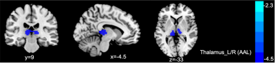 |
113 |
Structural and functional changes in patients with temporal lobe
epilepsy: a VBM and rs-fMRI study
jiachen Li1,
jing Zhang1,
wanjun Hu1,
guangyao Liu1,
liang Zhou1,
and kai Ai2
1Department of Magnetic Resonance, Lanzhou University Second Hospital, Lanzhou, China, 2Philips Healthcare, Xi'an, China Keywords: Epilepsy, fMRI (resting state) To investigate the structural and functional changes of the Temporal Lobe Epilepsy (TLE) patients, voxel-based morphometry (VBM) and resting-state functional MRI (rs-fMRI) methods were used. Our findings showed that thalamus gray matter volume decreased and performed negative correlation with self-rating anxiety scale (SAS) and self-rating depression scale (SDS). Furthermore, in rs-fMRI analysis, we found decreased functional connectivity between bilateral thalamus and right superior frontal gyrus. The results suggested that thalamus plays an important role in emotional change in TLE patients and structural and functional alterations may be related to the pathological mechanism of TLE. |
|
5212.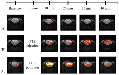 |
114 |
Focused Ultrasound Neuromodulation combined with rs-fMRI and EEG
as a preclinical tool to investigate and intervene Drug-Induced
Epilepsy
Yi-Jing Juan1,
Xiao Zhen1,
Po‑Chun Chu2,
You-Yin Chen3,
Hao-Li Liu2,
and Jyh-Horng Chen2
1Graduate Institute of Biomedical Electronics and Bioinformatics, National Taiwan University, Taipei City, Taiwan, 2Department of Electrical Engineering, National Taiwan University, Taipei City, Taiwan, 3Department of Biomedical Engineering, National Yang Ming Chiao Tung University, Hsinchu City, Taiwan Keywords: Epilepsy, fMRI (resting state), Focused Ultrasound, Neuromodulation, EEG A drug-induced epileptic animal model to investigate the feasibility of FUS neuromodulation with rs-fMRI and EEG |
|
5213.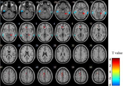 |
115 |
Mixed brain perfusion pattern and its association with epilepsy
duration: The application of arterial spin labeling in older
people with epilepsy
Xuyang Yin1,
Jianhong Wang2,
Yanwei Zeng1,
Zhenxu Xiao2,
Junyan Fu1,
Qianhua Zhao2,
Ding Ding2,
and Jun Zhang1
1Radiology Department, Huashan Hospital, Shanghai, China, 2Neurology Department, Huashan Hospital, Shanghai, China Keywords: Epilepsy, Arterial spin labelling The early identification, evaluation and intervention are important for older people with epilepsy, which calls for techniques of high convenience, repeatability and practicality in clinical application. By applying arterial spin labeling (ASL), we detected perfusion differences between older people with epilepsy and healthy controls on whole-brain voxel-based level, and conducted linear regression between cerebral blood flow (CBF) and duration of epilepsy. Our findings revealed a mixed perfusion pattern for older people with epilepsy, and the significant correlation between CBF and duration of epilepsy. Therefore, ASL has potential value in early diagnosis and evaluation of seizure activity and severity for epilepsy. |
|
5214. |
116 |
T2 mapping with GRAPPATINI in temporal lobe epilepsy improves
epileptogenic zone characterization: a pilot study
Maria Celeste Bonacci1,
Maria Eugenia Caligiuri1,
Ilaria Sammarra2,
Tobias Kober3,
Domenico Zacà4,
Francesco Fortunato2,
and Antonio Gambardella2
1Neuroscience Research Center, Università degli Studi Magna Graecia di Catanzaro, Catanzaro, Italy, 2Institute of Neurology, Università degli Studi Magna Graecia di Catanzaro, Catanzaro, Italy, 3Advanced Clinical Imaging Technology, Siemens Healthcare AG, Lausanne, Switzerland, 4Siemens Healthcare, Milan, Italy Keywords: Epilepsy, Quantitative Imaging The use of qMRI techniques in the routine imaging assessment of epilepsy patients may improve sensitivity in detecting subtle MRI abnormalities which can further aid lateralization and localization of the epileptogenic zone. T2 relaxometry might be a promising tool for a better and non-invasive lateralization of the epileptogenic zone, as well as for identifying lesions that are not visible on standard MRI. |
|
5215.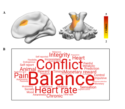 |
117 |
Structural and functional changes in drug-naïve Rolandic
epilepsy and their associated gene expression profiles
Yu Yin1 and
Heng Liu1
1the Affiliated Hospital of Zunyi Medical University, Zunyi, China Keywords: Epilepsy, fMRI (resting state) Combining structural and functional neuroimaging analyses with brain transcriptional data, the present study investigated GMV and fALFF changes in children with RE as well as their underlying gene transcriptional profiles. VBM and fALFF analyses showed altered GMV and fALFF in brain regions associated with behavioral and cognitive regulation. Transcription-neuroimaging spatial correlation analyses further identified genes correlated with GMV and fALFF changes in RE, respectively. Moreover, functional enrichment analysis demonstrated that RE-related genes were enriched for the regulation of biological process. These findings may provide us with important knowledge for understanding the pathophysiological basis of this disease. |
|
5216.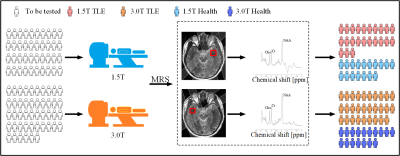 |
118 |
Quantitative Comparison of 1.5T and 3.0T 1H Magnetic Resonance
Spectroscopy for Temporal Lobe Epilepsy
Biao Qu1,
Hejuan Tan2,
Min Xiao2,
Dongbao Liu3,
Shijin Wang4,
Yiwen Zhang5,
Runhan Chen6,
Gaofeng Zheng1,
Yonggui Yang7,
Gen Yan7,
and Xiaobo Qu3
1Department of Instrumental and Electrical Engineering, Xiamen University, Xiamen, China, 2Institute of Artificial Intelligence, Xiamen University, Xiamen, China, 3Biomedical Intelligent Cloud R&D Center, Fujian Provincial Key Laboratory of Plasma and Magnetic Resonance, Department of Electronic Science, Xiamen University, Xiamen, China, 4Department of Information & Computational Mathematics, Xiamen University, Xiamen, China, 5Department of Neurology, The Second Affiliated Hospital of Xiamen Medical College, Xiamen, China, 6National Institute for Data Science in Health and Medicine, Xiamen University, Xiamen, China, 7Department of Radiology, The Second Affiliated Hospital of Xiamen Medical College, Xiamen, China Keywords: Epilepsy, Brain The study explored the diagnostic utility of different field strengths for the temporal lobe epilepsy (TLE) with 1H magnetic resonance spectroscopy (1H-MRS) which could be utilized to examine the concentrations of related metabolites. Four ratios of brain metabolites, including NAA/Cr, NAA/Cho, NAA/(Cho+Cr) and Cho/Cr were introduced to four control experiments with the Mann-Whitney U Test, the power analysis and the Paired T-Test adopted. Results suggested that 1.5T and 3.0T scanners might have comparable potential in distinguishing TLEs from HCs when 1H-MRS was used to identify patients with TLE. |
|
5217.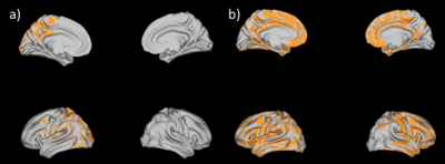 |
119 |
Neurite orientation and dispersion density imaging (NODDI) to
detect PCDH19 related microstructural anomalies.
Antonio Napolitano1,
Chiara Parrillo1,
Giulia Baldassari1,
Daniela Longo1,
Maria Camilla Rossi Espagnet1,
and Lorenzo Figa' Talamanca1
1Bambino Gesù Children's Hospital, Rome, Italy Keywords: Epilepsy, Diffusion/other diffusion imaging techniques, NODDI Protocadherin 19 (PCDH19)-clustering epilepsy (PCDH19-CE) is a genetic form of epilepsy caused by a mutation of PCDH19-CE gene that begins in the first year of life. Patients with this disorder may present intellectual disability, behavioral problems and motor and language delay. Using advanced Magnetic Resonance Imaging (MRI) methods we were able to highlight microstructural changes of brain in PCDH patients. These results shows that Neurite orientation dispersion and density imaging (NODDI) techniques might be useful tools to investigate PCDH19 anomalous arborization. |
|
5218.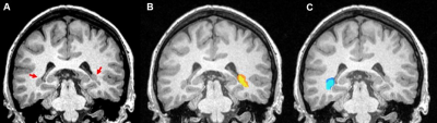 |
120 |
Epileptogenic periventricular nodular heterotopia BOLD
fluctuations are anti-correlated to non-epileptogenic nodular
heterotopias
Mark J. Lowe1,
Jian Lin1,
Ajay Nemani1,
Spencer Morris2,
Balu Krishnan2,
David Martinez2,
Juan Bulacio2,
Stephen Jones1,
Imad Najm2,
and Irene Wang2
1Imaging Institute, Cleveland Clinic, Cleveland, OH, United States, 2Neurologic Institute, Cleveland Clinic, Cleveland, OH, United States Keywords: Epilepsy, fMRI (resting state) In this study, using a limited sample size, we establish with 95% confidence that epileptogenic and non-epileptogenic periventricular nodular heterotopias can be distinguished based on their relative resting state connectivity |
|
 Back to Meeting Home
 Back to the Program-at-a-Glance
The International Society for Magnetic Resonance in Medicine is accredited by the Accreditation Council for Continuing Medical Education to provide continuing medical education for physicians.
 Back to Meeting Home
 Back to the Program-at-a-Glance
 View Presentation Video