Digital Poster
Perfusion, Blood Flow & Blood Volume II
ISMRM & ISMRT Annual Meeting & Exhibition • 03-08 June 2023 • Toronto, ON, Canada

Digital Poster
Perfusion, Blood Flow & Blood Volume II
| Computer # | |||
|---|---|---|---|
3057.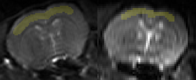 |
1 |
Cortical Blood Perfusion during Preclinical Migraine at 21.1 T
using FAIR-PASL
Dayna Leigh Richter1,2 and
Samuel Colles Grant1,2
1National High Magnetic Field Laboratory, Florida State University, Tallahassee, FL, United States, 2Chemical & Biomedical Engineering, FAMU-FSU College of Engineering, Tallahassee, FL, United States Keywords: Neurodegeneration, Arterial spin labelling, Migraine, FAIR-EPI Migraine is a neurological disorder with neurovascular implications. Blood flow changes based on brain's activity and needs, so excessive neural activation during migraine is likely to make significant alterations to cortical blood perfusion. Therefore, cortical blood perfusion was measured using FAIR-PASL to monitor the progression in preclinical migraine in female Sprague-Dawley rats. Parameters for inversion RF pulse shape and slice thickness were optimized to achieve more robust fits for monitoring cortical blood perfusion during preclinical migraine. |
|
3058.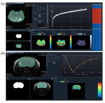 |
2 |
A python-based post-processing toolkit for rodent perfusion MRI
Jinyuan Zhang1,2,
Yishuang Yang1,2,
Rong Xue1,2,
Yan Zhuo1,2,
and Zihao Zhang1,3
1State Key Laboratory of Brain and Cognitive Science, Institute of Biophysics, Chinese Academy of Sciences, Beijing, China, 2University of Chinese Academy of Sciences, Beijing, China, 3Institute of Artificial Intelligence, Hefei Comprehensive National Science Center, Hefei, China Keywords: Software Tools, Software Tools We developed a python-based post-processing toolkit for rodent perfusion MRI with an easy-to-use graphical user interface (GUI). Until now, this toolkit provides interfaces for the post-processing of dynamic susceptibility contrast (DSC) MRI and flow-sensitive alternating inversion recovery (FAIR) pulsed arterial spin labeling (pASL). For each modality, the toolkit has function modules including image viewer, display of time series data, ROI tools, and visualization of quantitative parameter maps. This toolkit is open source and welcomes added features. It will benefit preclinical studies using perfusion MRI. |
|
3059.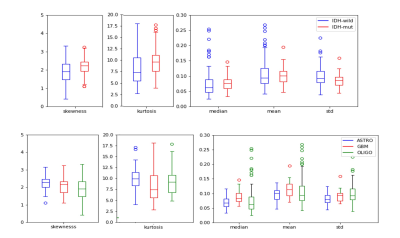 |
3 |
Relative Cerebral Blood Volume Differences Between Adult-Type
Diffuse Glioma Subgroups According to WHO 2021: A Study of 146
Gliomas
Buse Buz-Yalug1,
Ayca Ersen Danyeli2,3,
M. Cengiz Yakicier3,4,
M. Necmettin Pamir3,5,
Koray Ozduman3,5,
Alp Dincer3,6,
and Esin Ozturk-Isik1,3
1Institute of Biomedical Engineering, Bogazici University, Istanbul, Turkey, 2Department of Medical Pathology, Acibadem University, Istanbul, Turkey, 3Brain Tumor Research Group, Acibadem University, Istanbul, Turkey, 4Department of Molecular Biology and Genetics, Acibadem University, Istanbul, Turkey, 5Department of Neurosurgery, Acibadem University, Istanbul, Turkey, 6Department of Radiology, Acibadem University, Istanbul, Turkey Keywords: Machine Learning/Artificial Intelligence, Machine Learning/Artificial Intelligence The main purpose of this study is to analyze the relative cerebral blood volume (rCBV) differences of adult diffuse glioma subgroups defined in the updated WHO 2021 brain tumor classification. The IDH wildtype group had statistically significantly higher rCBV values, and IDH mutational subgroups were classified with 82.5% accuracy (precision = 82.1%, recall = 82.9%), while the accuracies of glioblastoma and oligodendroglioma classification was 81.9%, glioblastoma and astrocytoma classification was 83.3%, and astrocytoma and oligodendroglioma classification was 77.5%. |
|
3060.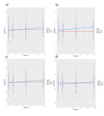 |
4 |
The impact of naproxen on Cerebral Blood Flow in preclinical
Alzheimer’s disease
Safa Sanami1,2,
Brittany N Intzandt3,
Julia Huck1,
PREVENT-AD Research Group4,
and Claudine J Gauthier2,5,6
1Physics, Concordia University, Montreal, QC, Canada, 2Centre de Recherche de l’Institut de Cardiologie de Montr´eal, Montreal, QC, Canada, 3Sunnybrook Research Institute, Toronto, ON, Canada, 4Douglas Mental Health Institute, Montreal, QC, Canada, 5Concordia University, Montreal, QC, Canada, 6PERFORM Centre, Concordia University, Montreal, QC, Canada Keywords: Data Processing, Alzheimer's Disease Alzheimer’s Disease (AD) is the most common form of dementia and displays a long preclinical phase. The use of non-steroidal anti-inflammatory drugs (NSAIDs) in this phase has a protective effect and decreases the risk of developing AD cross-sectionally. However, the effects of NSAIDs therapy long-term on cerebral hemodynamics in this early phase is unclear. This is the first study to assess whether longitudinal use of NSAIDs (i.e., naproxen) has an impact on cerebral blood flow, and whether this is associated with a change in cognition and/or cerebrospinal fluid markers. |
|
3061.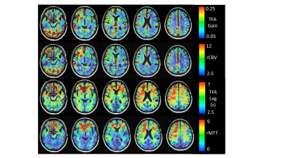 |
5 |
Assessing cerebral perfusion: transfer function analysis of the
BOLD response to hypoxia-induced changes in deoxyhemoglobin
James Duffin1,2,
Ece Su Sayin1,3,
Olivia Sobczyk2,3,
Julien Poublanc4,
Harrison T. Levine1,3,
David J. Mikulis4,
and Joseph A. Fisher1,5
1Physiology, University of Toronto, Toronto, ON, Canada, 2Department of Anaesthesiology and Pain Management, University of Toronto, Toronto, ON, Canada, 3Joint Department of Medical Imaging and the Functional Neuroimaging Lab,, University Health Network, Toronto, ON, Canada, 4Joint Department of Medical Imaging and the Functional Neuroimaging Lab, University Health Network, Toronto, ON, Canada, 5Department of Anaesthesiology and Pain Management, University Health Network, Toronto, ON, Canada Keywords: Data Analysis, Brain We used hypoxia-induced changes in deoxyhemoglobin concentration as a susceptibility contrast agent. Transfer function analysis of the resulting changes in the BOLD signal for each voxel provided cerebral perfusion measures that indicate the distribution of the strength of the signal response to the contrast agent (gain) and the lag of the response (phase or time). We relate the gain and lag to conventional resting hemodynamic measures cerebral blood volume and mean transit time, respectively. |
|
3062.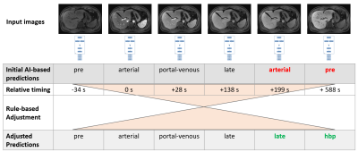 |
6 |
Deep Learning-based Automatic Perfusion Phase Identification for
Dynamic T1-weighted Liver MRI
Robert Grimm1,
Malte Müller2,
Cornelius Jacob1,
Sabine Mollus1,
Christian Tietjen3,
Moon Hyung Choi4,
Kazuki Oyama5,
Thomas Weikert6,
Andrew D Hardie7,
Jeong Hee Yoon8,
Heinrich von Busch3,
Gregor Thoermer1,
and Volker Daum2
1MR Application Predevelopment, Siemens Healthcare GmbH, Erlangen, Germany, 2Chimaera GmbH, Erlangen, Germany, 3Digital and Automation, Siemens Healthcare GmbH, Erlangen, Germany, 4Eunpyeong St. Mary’s Hospital, Catholic University of Korea, Seoul, Korea, Republic of, 5Department of Radiology, Shinshu University Hospital, Nagano, Japan, 6Department of Radiology, Universitätsspital Basel, Basel, Switzerland, 7Radiology and Radiological Science, Medical University of South Carolina, Charleston, SC, United States, 8Seoul National University Hospital and College of Medicine, Seoul, Korea, Republic of Keywords: Data Analysis, Machine Learning/Artificial Intelligence A deep learning-based approach for automatic identification of the perfusion phases in dynamic T1-weighted liver MRI is presented. First, an encoder model combined with two dense layers was trained to classify each image into pre-contrast, arterial, portal-venous, late, or hepatobiliary phase. In a second pass, classification errors are detected and adjusted, based on the expected occurrence order and relative timing to the arterial phase. The AI model reached sensitivities of 67% to 99%. Most common mis-classifications were confusions of the portal-venous or late phase with the adjacent phases. By the rule-based adjustments, the classification performance was raised to >95% accuracy. |
|
3063.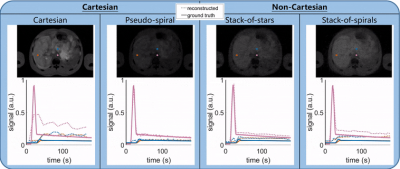 |
7 |
The ERIC phantom - Extra-dimensional respiration and inflow of
contrast: an open-source DCE digital simulation tool
Eric Schrauben1
1Radiology and Nuclear Medicine, Academic Medical Centre, Amsterdam, Netherlands Keywords: Software Tools, DSC & DCE Perfusion An open-source MATLAB-based software tool was developed for digital simulation (using the MRXCAT framework) of DCE MRI acquisition and reconstruction strategies, termed the ERIC phantom: extra-dimensional respiration and inflow of contrast. The ERIC phantom is a user-friendly graphical user interface, allowing for variable respiratory motion, acquisition parameters and trajectories, motion correction strategies, and compressed sensing reconstructions. This realistic abdominal DCE phantom can be used to investigate varying strategies for producing high quality DCE reconstructions in the presence of respiratory motion and contrast inflow. |
|
3064.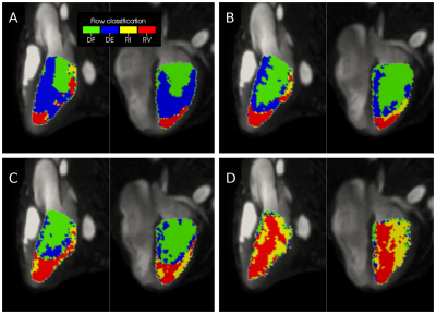 |
8 |
Normal left ventricular flow dynamics in a paediatric population
assessed by 4D Flow MRI
Fraser Maurice Callaghan1,2,
Barbara Burkhardt2,3,
Julia Geiger2,4,
Emanuela Valsangiacomo Buechel2,3,
and Christian Kellenberger2,4
1University Children's Hospital Zurich, Zurich, Switzerland, 2Children’s Research Center, University Children's Hospital Zurich, Zurich, Switzerland, 3Division of Pediatric Cardiology, University Children's Hospital Zurich, Zurich, Switzerland, 4Department of Diagnostic Imaging, University Children's Hospital Zurich, Zurich, Switzerland Keywords: Data Analysis, Cardiovascular, 4D flow 4D flow MRI provides a rich dataset that can make analysis challenging. A new technique of left ventricle (LV) registration is presented and compared with established techniques for analysis of flow dynamics in a normal paediatric population. Averaging of group flow dynamics in a common space permits additional analysis, complementary to established techniques. Our technique demonstrated flow dynamics on a common LV space and identified subtle differences in LV velocities between normal male and female subjects of a paediatric population. This represents a new tool in 4D flow data analysis for potential biomarker development. |
|
3065.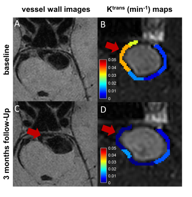 |
9 |
Intracranial Wall Permeability for Prediction of Aneurysm Growth
Using Dynamic Contrast-Enhanced MRI
Ziming Xu1,
Linggen Dong2,
Longhui Zhang2,
Yajie Wang1,
Jiaqi Dou1,
Peng Liu2,
Ming Lv2,
and Huijun Chen1
1Center for Biomedical Imaging Research, School of Medicine, Tsinghua University, Beijing, China, 2Department of Neurosurgery, Beijing Tiantan Hospital, Beijing Neurosurgical Institute, Capital Medical University, Beijing, China Keywords: Data Analysis, DSC & DCE Perfusion Size of intracranial aneurysm (IA) is considered the most important clinical factor to determine the risk of IA rupture. Accurate prediction of aneurysm growth is crucial for preventive management but remains challenging. Herein, we studied the potential predictive roles of demography and imaging characteristics in a radiological follow-up study. And wall permeability calculated by DCE-MRI was firstly observed to accurately identify the IAs with a relatively high risk of aneurysm growth (AUC = 0.875). Our study demonstrated that higher wall permeability was associated with aneurysm growth and the regions with higher wall permeability co-localized with the direction of aneurysm growth. |
|
3066.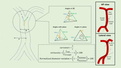 |
10 |
Association between 3D Geometry of Vertebrobasilar Arteries and
Basilar Artery Atherosclerosis: An MRI Study
Jiachen Liu1,
Rui Shen1,
Dandan Yang2,
Miaoxin Yu3,
and Xihai Zhao1
1Center for Biomedical Imaging Research, Department of Biomedical Engineering, Tsinghua University, Beijing, China, 2Department of Radiology, Beijing Geriatric Hospital, Beijing, China, 3Department of Neurology, Beijing Tiantan Hospital, Capital Medical University, Beijing, China Keywords: Data Analysis, Atherosclerosis Geometry of VBA has been proved to be associated with the presence of BA plaques, yet relative studies are lack of 3D information due to methodological difficulties. We proposed a semi-automated algorithm for quantitative measurement, which can evaluate local diameter, local curvature and angles between vessels directly in 3D space. We used the proposed algorithm to measure the 3D geometric metrics and found significant association between VBA geometry and the presence of BA plaques. Normalized diameter variation of BA and BA & PCA-R angle projected to lateral view were also associated with the presence of BA plaques independently. |
|
3067.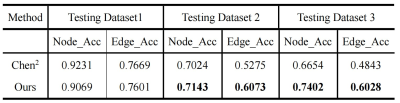 |
11 |
Automated Intracranial Artery Labeling in Patients with
Cerebrovascular Steno-occlusive Diseases
Lixin Liu1,
Yi Lv2,
Peirong Jiang3,
He Wang1,4,5,
and Zhensen Chen1,5
1Institute of Science and Technology for Brain-Inspired Intelligence,Fudan University, Shanghai, China, 2School of Compute Science and Technology, Beijing Institute of Technology, Beijing, China, 3Department of Radiology, Fujian Medical University Union Hospital, Fuzhou, China, 4Human Phenome Institute, Fudan University, Shanghai, China, 5Key Laboratory of Computational Neuroscience and Brain-Inspired Intelligence (Fudan University), Ministry of Education, Shanghai, China Keywords: Data Processing, Blood vessels Labeling of intracranial arteries is important for computer-aided diagnosis of cerebrovascular diseases and quantitative analysis of intracranial vasculature. Performance of the previously proposed automated intracranial artery labeling method based on Graph Neural Network (GNN) is limited in datasets with overt cerebrovascular steno-occlusive diseases. In this study, we improved the generalizability of the GNN-based method by using dedicated data augmentation and spatial normalization strategy. The results show that our method is more robust than the previous method in ischemic stroke patients with overt intracranial stenosis or occlusion. |
|
3068.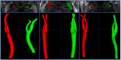 |
12 |
Automated Localization of the Extracranial Carotid Artery in
Black Blood Contrast MR Images Using a Deep Learning Approach
SeyyedKazem HashemizadehKolowri1,
Nadin Zanaty2,3,
Gador Canton2,
Niranjan Balu2,
Thomas S. Hatsukami2,
and Chun Yuan1,2
1Radiology and Imaging Sciences, University of Utah, SALT LAKE CITY, UT, United States, 2Department of Radiology, University of Washington, Seattle, WA, United States, 3Radiology, Zagazig University, Zagazig, Egypt Keywords: Machine Learning/Artificial Intelligence, Cardiovascular, Vessel Wall Imaging In this work, a deep learning approach for automated localization of carotid arteries in black blood contrast MR data is proposed. This is the first step in automated analysis of vessel wall imaging data. Carotid arteries supply oxygenated blood to the brain and are susceptible to atherosclerosis, so their vessel wall imaging is of significant importance in clinical evaluations. However, currently only qualitative assessment of VW imaging data relying on visual inspection is implemented in clinics that are not scaleable. Therefore, developing automated image processing tools to quantitatively analyze vessel wall imaging data can have a major clinical impact. |
|
3069.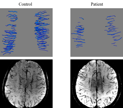 |
13 |
Automated modeling and morphologic analysis of deep medullary
veins at 7T MRI in patients with cognitive impairment
Zhixin Li1,2,3,
Jingyuan Zhang1,2,3,
Li Liang 4,
Jing An5,
Hairong Qian4,
Rong Xue1,2,3,
Yan Zhuo1,2,3,
and Zihao Zhang1,2,6
1State Key Laboratory of Brain and Cognitive Science, Institute of Biophysics, Chinese Academy of Sciences, Beijing, China, Beijing, China, 2University of Chinese Academy of Sciences, Beijing, China, Beijing, China, 3The Innovation Center of Excellence on Brain Science, Chinese Academy of Sciences, Beijing, China, Beijing, China, 4Department of Neurology, the Sixth Medical Center, Chinese PLA General Hospital, Beijing, China, 5Siemens Shenzhen Magnetic Resonance Ltd., Shenzhen, China, Shenzhen, China, 6Institute of Artificial Intelligence, Hefei Comprehensive National Science Center, Hefei, China, Hefei, China Keywords: Machine Learning/Artificial Intelligence, Modelling Deep medullary veins (DMVs) support cerebral venous drainage. They may display abnormal changes in patients with cognitive impairment. They can be visualized by multi-echo gradient echo imaging at 7T. This study proposed a segmentation and tracking method based on deep learning and shortest-path optimization. It automatically quantified the morphologic parameters of DMVs from the vascular model. These characteristics of DMVs correlated with the patients’ cognitive scores, and might reflect the pathology of vascular lesions in cognitive impairment. |
|
3070.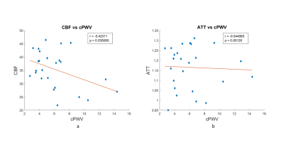 |
14 |
Carotid arterial stiffening is associated with reduced
downstream territorial perfusion of internal carotid artery in
elderly adults
Yining He1,
Jianing Tang1,2,
Tianrui Zhao1,2,
and Lirong Yan1,3
1Radiology, Northwestern University, Chicago, IL, United States, 2Biomedical Engineering, Northwestern University, Evanston, IL, United States, 3Neurology, University of Southern California, Los Angeles, CA, United States Keywords: Blood vessels, Aging Arterial stiffness is an important risk marker for poor brain aging, vascular disease, and dementia. Greater arterial stiffness leads to the transmission of excessive pulsations from the greater vessels into the downstream capillary and tissue causing microvascular dysfunction. However, previous studies have mainly focused on central or peripheral pulse wave velocity assessment. The present study has demonstrated a significant association of the PWV of the feeding arteries to the brain and its downstream territorial perfusion assessed by using two new MRI techniques, which could provide valuable insight into the neurovascular pathology of aging and brain dysfunction. |
|
3071.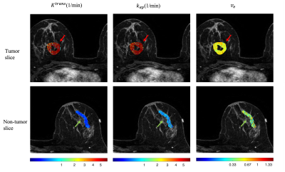 |
15 |
Retrospective quantification pharmacokinetics of clinical breast
DCE-MRI using deep learning
Chaowei Wu1,2,
Lixia Wang1,
Nan Wang1,3,
Stephen Pandol4,
Anthony G Christodoulou1,2,
Yibin Xie1,
and Debiao Li1,2
1Biomedical Imaging Research Institute, Cedars-Sinai Medical Center, Los Angeles, CA, United States, 2Department of Bioengineering, University of California, Los Angeles, Los Angeles, CA, United States, 3Radiology Department, Stanford University, Stanford, CA, United States, 4Division of Digestive and Liver Diseases, Cedars-Sinai Medical Center, Los Angeles, CA, United States Keywords: Machine Learning/Artificial Intelligence, DSC & DCE Perfusion Standard-of-care DCE-MRI suffers from a limited number of contrast phases and low temporal resolution, preventing the quantification of pharmacokinetic parameters. Quantitative DCE-MRI techniques have not yet been widely applied in the clinic due to the limited availability of specialized sequences and image reconstruction. To tackle this problem, we proposed to improve the temporal resolution of multi-phasic DCE-MRI by deep learning post-processing and demonstrated promising results in tumor delineation in the Duke-Breast-Cancer-MRI dataset. |
|
3072. |
16 |
The application of Quantitative Perfusion Analysis of GRASP for
assessing pathologic prognostic factors in rectal cancer
Mi Zhou1,
Meining Chen2,
and Yuting Wang1
1Department of Radiology, Sichuan Provincial People's Hospital, chengdu, China, 2Department of MR Scientific Marketing, Siemens Healthcare, Shanghai, China Keywords: Data Analysis, Perfusion, rectum Colorectal cancer’s pathological prognostic factors directly affect the patient's prognosis. Golden-angle RAdial Sparse Parallel (GRASP) imaging was invented to calculate the accurate perfusion parameters including influx forward volume transfer constant (Ktrans), rate constant (Kep), and plasma volume fraction (Ve). We found GRASP parameters showed significant differences in many prognostic factors, including histology type, extramural venous invasion (EMVI), lymphovascular invasion (LVI), tumor deposit (TD) and lymph node metastasis (LNM), and were independent factors for histology type, LNM, LVI and EMVI. GRASP parameters were strongly associated with preoperative prognostic factors in rectal cancer, providing useful information for treatment and follow-up protocol. |
|
3073.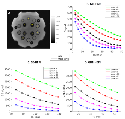 |
17 |
Evaluation of multi-echo-based Hybrid-EPI (HEPI) technique for
measuring brain oxygenation
Krishnapriya Venugopal1,
Esther A.H Warnert1,
Daniëlle van Dorth2,
Marion Smits1,
Matthias J.P van Osch2,
Dirk H.J Poot1,
and Juan Antonio Hernandez-Tamames1,3
1Radiology and Nuclear Medicine, Erasmus MC, Rotterdam, Netherlands, 2Radiology, Leiden University Medical Center, Leiden, Netherlands, 3Medical Imaging, TU Delft, Delft, Netherlands Keywords: Data Acquisition, Brain, Oxygenation The reversible component of transverse relaxation time, R2’, enables the measurement of blood oxygenation, an important biomarker in several diseases. We propose a multi-echo HEPI technique to estimate R2’. HEPI combines GRE and SE and hence provides R2* and R2, when acquired at different echo-times. The accuracy of ME-HEPI in measuring R2’ is evaluated in a phantom in this work. The sensitivity of HEPI to different oxygenation levels, attained using respiratory challenge MRI, is studied in a healthy subject. This is also investigated using a simulation tool that simulates microvasculature and MR signal for varying oxygenation in the vessels.
|
|
3074.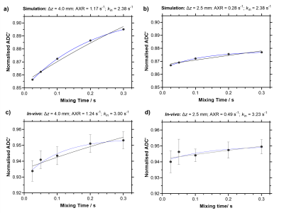 |
18 |
Reliable blood-brain barrier water exchange estimates in the rat
brain using a crusher-compensated exchange rate (CCXR) model
Yolanda Ohene1,2,
Elizabeth Powell3,
Samo Lasič4,5,
Geoff J. M. Parker3,6,
Laura M. Parkes1,2,
and Ben R. Dickie2,7
1Division of Psychology, Communication and Human Neuroscience, University of Manchester, Manchester, United Kingdom, 2Geoffrey Jefferson Brain Research Centre, University of Manchester, Manchester, United Kingdom, 3UCL, London, United Kingdom, 4Danish Research Centre for Magnetic Resonance, Copenhagen, Denmark, 5Random Walk Imaging, Åkarp, Sweden, 6Bioxydyn Limited, Manchester, United Kingdom, 7Division of Informatics, University of Manchester, Manchester, United Kingdom Keywords: Data Processing, Permeability Filter exchange imaging (FEXI) is a promising technique for measuring water exchange across the blood-brain barrier (BBB). However, the application of FEXI for the rodent brain requires thinner slices and therefore higher crusher gradients which lead to a progressive underestimation of the apparent exchange rate (AXR). Here, we implement a crusher-compensated exchange rate (CCXR) model which reduces the bias induced by the crusher gradients and allows more accurate estimates of BBB water exchange in the rat brain. |
|
3075. |
19 |
Early Prediction of Response to Neoadjuvant Systemic Therapy of
Triple Negative Breast Cancer using Radiomics on DCE-MRI
Bikash Panthi1,
Sanaz Pashapoor1,
Beatriz E. Adrada1,
Rosalind P. Candelaria1,
Mary S. Guirguis1,
Miral M. Patel1,
Rania M. Mohamed1,
Medine Boge1,
Zijian Zhou1,
Jong Bum Son1,
Ken-Pin Hwang1,
Huong T.C. Le-Petross1,
Jessica W.T. Leung1,
Marion E. Scoggins1,
Gary J. Whitman1,
Zhan Xu1,
Deanna L. Lane1,
Tanya Moseley1,
Frances Perez1,
Jason White1,
Elizabeth Ravenberg1,
Alyson Clayborn1,
Huiqin Chen1,
Jia Sun1,
Peng Wei1,
Alastair Thompson2,
Stacy Moulder1,
Anil Korkut1,
Lei Huo1,
Kelly K. Hunt1,
Jennifer K. Litton1,
Vicente Valero1,
Debu Tripathy1,
Wei Yang1,
Clinton Yam1,
Gaiane M Rauch1,
and Jingfei Ma1
1MD Anderson Cancer Center, Houston, TX, United States, 2Baylor College of Medicine, Houston, TX, United States Keywords: Radiomics, fMRI, Treatment response We developed models based on radiomic features from dynamic contrast enhanced (DCE) MR images and demonstrated that these models have potential to serve as non-invasive biomarkers for early prediction of pathologic complete response (pCR) in triple negative breast cancer (TNBC) patients undergoing neoadjuvant systemic therapy (NAST). |
|
 Back to Meeting Home
 Back to the Program-at-a-Glance
The International Society for Magnetic Resonance in Medicine is accredited by the Accreditation Council for Continuing Medical Education to provide continuing medical education for physicians.
 Back to Meeting Home
 Back to the Program-at-a-Glance
 View Presentation Video