Digital Poster
Brain Potpourri
ISMRM & ISMRT Annual Meeting & Exhibition • 03-08 June 2023 • Toronto, ON, Canada

| Computer # | |||
|---|---|---|---|
2076.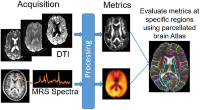 |
81 | A Machine Learning Approach for Identifying the Severity and Regional Anatomical Location of HIV Infection in the Brain
Teddy Salan1, Sulaiman Sheriff1, Sameer Vyas2, Deepika Aggarwal2, Paramjeet Singh2, and Varan Govind1
1University of Miami, Miami, FL, United States, 2Postgraduate Institute of Medical Education and Research, Chandigarh, India Keywords: Infectious disease, Machine Learning/Artificial Intelligence Identifying and monitoring viral habitats of HIV in the brain is crucial to the advancement of treatment strategies for containing the infection. However, no in-vivo methods currently exist for this purpose. In this study, we demonstrate the use of a machine learning model that integrates brain measurements from MRSI, DTI, and DKI for identifying the severity and anatomical location of microstructural and metabolic abnormalities in the brain. This information may provide important clinical and diagnostic value for the treatment of people living with HIV. |
|
2077.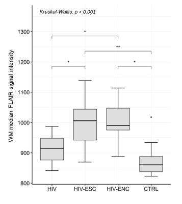 |
82 | Symptomatic cerebrospinal fluid HIV escape: a quantitative MR-based assessment
Serena Capelli1, Anna Caroli1, Giulio Pezzetti2, Francesca Ferretti3, Paola Cinque4, and Simonetta Gerevini2
1Bioengineering Department, Istituto di Ricerche Farmacologiche Mario Negri IRCCS, Ranica (BG), Italy, 2Department of Neuroradiology, ASST Papa Giovanni XXIII, Bergamo, Italy, 3Lewisham and Greenwich NHS trust, London, United Kingdom, 4Unit of Infectious Diseases, San Raffaele Scientific Institute, Milano, Italy Keywords: Infectious disease, Infectious disease, HIV This study aimed at quantitatively assessing T1-weighted, FLAIR and DWI brain alterations in 14 patients with cerebrospinal fluid HIV escape (HIV-ESC) and 7 patients with untreated HIV encephalitis (HIV-ENC), versus 11 HIV patients without neurological problems and 12 HIV-negative controls. HIV-ESC and HIV-ENC patients showed significantly higher ADC in WM and GM and increased WM FLAIR signal than the other groups. In HIV-ESC patients, the heterogeneous GM volume was negatively correlated with MRI time from infection. WM FLAIR hyperintensity in HIV-ESC and HIV-ENC patients may reflect vasogenic edema, with mass effect depending on its degree and possible underlying brain atrophy. |
|
 |
2078.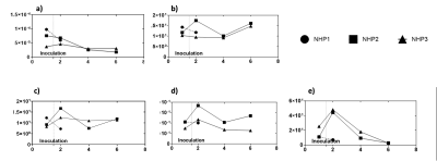 |
83 | Feasibility of textural analysis and very-low-field magnetic resonance for imaging Nipah virus infection
Kunal Aggarwal1,2, Yu Cong3, Ji Hyun Lee4, Venkatesh Mani3, Claudia Calcagno5, Michael R. Holbrook5, and Sairam Geethanath6
1Department of Electrical and Computer Engineering, Technical University Munich, Munich, Germany, 2Deptartment of Diagnostic, Molecular and Interventional Radiology, Mount Sinai Hospital, New York City, NY, United States, 3National Institute of Allergy and Infectious Diseases, Fort Detrick, MD, United States, 4Radiology and Imaging Sciences, Clinical Center, National Institutes of Health, Bethesda, MD, United States, 5National Institute of Allergy and Infectious Diseases, National Institutes of Health, Fort Detrick, MD, United States, 6Mount Sinai Hospital, New York, NY, United States Keywords: Infectious disease, Infectious disease Our study focuses on textural analysis of high field and low field MR images and its validation through simulation. We acquired MR images of three NHPs at 3T injected with NiV and simulated them into low field to perform texture analysis for different neuroanatomies. The low field simulation was done by changing contrast, resolution and SNR. The validation of low field simulation was performed by scanning phantom at 3T and 0.05T and passing the data though low field simulator. Results showed four out of 14 textural features had similar changes in 2 NHPs. |
| 2079. | 84 | Correlation of Endolymphatic Hydrops and Perilymphatic Enhancement with Clinical Features of Ménière’s Disease
Jinye Li1, Mengxiao Liu2, Linsheng Wang3, Chuanting Li3, Lixin Sun3, and Yafei Liu4
1shangdong provincial ENT hospital, Jinan, China, 2Siemens healthineers, Shanghai, China, 3Shandong Provincial ENT Hospital, Jinan, China, 4Shandong Mental Health Center, Jinan, China Keywords: Head & Neck/ENT, Head & Neck/ENT To investigate the correlation of the EH grade and PE with clinical features in MD, we evaluated their correlations by using the cubic spline models and multivariate analysis. We found that a nonlinear or linear correlation of EH grade and SIR with duration of disease-related symptoms and hearing thresholds. Additionally, the higher duration of vertigo and LFHT were associated with a higher EH grade, as well as duration of tinnitus and vestibular EH for SIR. Consequently, the grades of EH and the extent of PE in patients with MD can be evaluated by use of some clinical features and PTA. |
|
2080.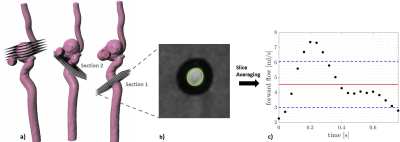 |
85 | Effect of a flow-diverter stent on flow in the vessel and aneurysm measured with MRI.
Lana Bautz1, Oluwabusayo A. Oni2, Hivnu Toraman3, Olav Jansen3, Jan-Bernd Hövener1, Naomi Larsen3, and Mariya S. Pravdivtseva1
1Department of Radiology and Neuroradiology, Section Biomedical Imaging, Molecular Imaging North Competence Center (MOIN CC), University Medical Center Schleswig-Holstein (UKSH), Kiel University, Kiel, Germany, Kiel, Germany, 2University of Florida, Gainesville, FL, United States, 3Department of Radiology and Neuroradiology, University Medical Center Schleswig-Holstein (UKSH), Kiel, Kiel, Germany Keywords: Data Analysis, Treatment, Evaluation of aneurysm treatment Flow diverter stents are effective for the treatment of intracranial aneurysms at a high risk of rupture. Still, complications may occur after stent implantation, so evaluation of aneurysm treatment is required. Treatment follow-up can be performed with 4D phase-contrast MRI. In this work, flow within the aneurysm and adjacent vessels with and without stent was measured with 4D flow. Strong metal artifacts were observed at the parent vessel which resulted in MRI signal voids and a 30 % reduction of measured flow. No artifacts were seen within an aneurysm sac, here flow was reduced up to 83 %. |
|
2081.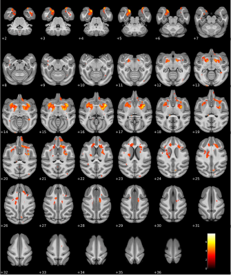 |
86 | Voxel-Based Brain Morphometry: an Optimized Image Analysis Pipeline and Proof-of-Concept in a Non-human Primate Model of Ebola Virus Infection
Byeong-Yeul Lee1, Ji Hyun Lee2, Jeffrey Solomon3, Marcelo Castro1, Venkatesh Mani1, Joseph Laux1, Winston T. Chu2, Matthew G. Lackemeyer1, Jordan K. Bohannon4, Anna N. Honko5, Ian Crozier3, Jens H. Kuhn1, and Claudia Calcagno1
1Integrated Research Facility at Fort Detrick, National Institute of Allergy and Infectious Diseases, National Institutes of Health, Frederick, MD, United States, 2Center for Infectious Disease Imaging, Radiology and Imaging Sciences, Clinical Center, National Institutes of Health, Bethesda, MD, United States, 3Clinical Monitoring Research Program Directorate, Frederick National Laboratory for Cancer Research, Frederick, MD, United States, 4National Biodefense Analysis and Countermeasures Center, Frederick, MD, United States, 5Boston University School of Medicine, Microbiology, Boston, MA, United States Keywords: Infectious disease, Infectious disease We present an optimized voxel-based morphometry magnetic resonance imaging (MRI) analysis pipeline for non-human primate brain images, and its preliminary application in Ebola virus (EBOV)-exposed rhesus monkeys. Results suggest the optimized pipeline can detect brain morphometric changes after EBOV exposure in this model. Further analyses will be required to confirm and build upon these findings, including their implications for acute and post-acute neurological findings in human survivors. Future studies may make use of this and other optimized, voxel-based pipelines to shed further light on the role of central nervous system involvement in EBOV and other infectious diseases. |
|
2082.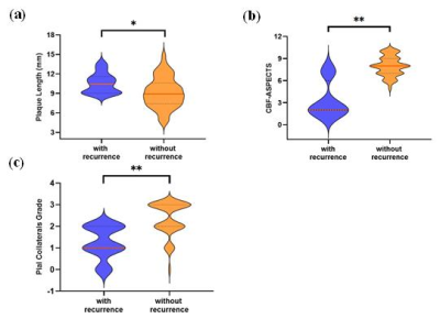 |
87 | Association of Plaque Characteristics, Pial Collaterals and Perfusion with Recurrent Ischemic Stroke in Middle Cerebral Artery Stenosis
Jia-Wei Yu1, Rui-Ying Li1, Xian-Ce Zhao2, Shan Huang2, and Deng-Ling Zhao1
1Department of Radiology, Zhongda Hospital, Medical School, Southeast University, Nanjing, China, 2Philips Healthcare, Shanghai, China Keywords: Stroke, Atherosclerosis Association between plaque characteristics, perfusion, pial collaterals (PCs) and recurrent ischemic stroke is unclear. Few analyses focus on combination of blood vessels, perfusion and PCs. In this study, 62 patients presenting with a transient ischemic attack or acute ischemic stroke caused by middle cerebral artery atherosclerosis were involved. All patients underwent the routine head MRI/MRA scan and high-resolution vessel wall imaging examinations. We performed a comprehensive assessment of plaque characteristics, perfusion and PCs. We concluded that brain perfusion and PCs are correlated with recurrence of ischemic stroke and their combination provided incremental value in identifying the recurrence of ischemic stroke. |
|
2083.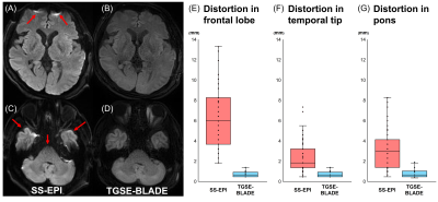 |
88 | Application of slice-accelerated one-minute TGSE-BLADE DWI in clinical practices
Sachi Okuchi1, Yasutaka Fushimi1, Satoshi Nakajima1, Akihiko Sakata1, Takuya Hinoda1, Sayo Otani1, Azusa Sakurama1, Krishna Pandu Wicaksono1, Hiroshi Tagawa1, Yang Wang1, Satoshi Ikeda1, Shuichi ito1, Miyuki Takiya1, Kun Zhou2, and Yuji Nakamoto1
1Department of Diagnostic Imaging and Nuclear Medicine, Graduate School of Medicine, Kyoto University, Kyoto, Japan, 2Siemens Shenzhen Magnetic Resonance Ltd., Shenzhen, China Keywords: Stroke, Artifacts TGSE-BLADE DWI has been reported to reduce geometric distortion and susceptibility artifacts, however the long acquisition time prevent its clinical application. We reduced acquisition time to 1 minute for TGSE-BLADE DWI using slice acceleration (1-min TGSE-BLADE DWI). We compared distortion and artifacts between SS-EPI DWI and 1-min TGSE-BLADE DWI, and evaluated diagnostic performance for acute infarctions of 1-min TGSE-BLADE DWI. The result shows that the 1-min TGSE-BLADE-DWI has better quality image in terms of distortion and artifacts, and higher diagnostic performance for acute infarctions. TGSE-BLADE DWI with slices acceleration is a promising method for evaluating lesions in acute stroke patients. |
|
2084. |
89 | Subclinical vascular damage is associated with cognitive decline in middle-to-old age community subjects with high vascular risks
Yao Zhang1, Shan Xu2, Ruiting Zhang1, and Peiyu Huang2
1Department of Radiology, The Second Affiliated Hospital, Zhejiang University School of Medicine, Hangzhou, China, 2The Second Affiliated Hospital, Zhejiang University School of Medicine, Hangzhou, China Keywords: Vessels, fMRI (resting state) Due to the insidious development of vascular degeneration, assessing subclinical vascular changes in people at high risks may aid early detection and intervention. We explored the relationship between the subclinical vascular function changes and vascular risks, vascular imaging markers, and cognition in a middle-to-old age community cohort. We found that larger vascular contractility in the intracranial arteries was related to younger age, less lacune, and better cognition. Participants with diabetes had a longer blood transit time from the ICAs to the capillary. These results suggested that subclinical vascular imaging markers were associated with brain parenchymal damage and cognitive decline. |
|
2085.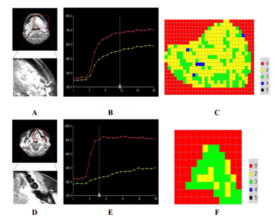 |
90 | Assessing tumor internal heterogeneity and pathological findings of oral tongue cancer using voxel-based TIC analysis
Xing Yang1, Ke Xue2, Zhen Tian1, Jingbo Wang1, Yongming Dai2, and Yingwei Wu1
1Shanghai Ninth People’s Hospital, affiliated to Shanghai Jiao Tong University, School of Medicine, Shanghai, China, 2MR Collaboration, Central Research Institute, Shanghai United Imaging Healthcare, Shanghai, China Keywords: Head & Neck/ENT, DSC & DCE Perfusion Evaluating the heterogeneous characteristics and lymph node metastasis status of the tumor would be of importance to stratify patients to have the individually tailored management. In this study, we investigated the feasibility of pixel-by-pixel TIC method in evaluating tumor heterogeneity and predicting histological tumor grade and LNM in tongue SCC. We found that the pixel-by-pixel TIC analysis approach allowed the detection of the internal heterogeneity of the whole tumor. Ratio of Type 2 TIC pattern would facilitate the distinction of SCCs with different histological grades and LNM status, implying its tremendous potential in tumors with high heterogeneity. |
|
2086.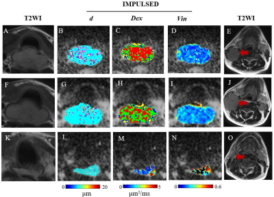 |
91 | Quantitative Microstructure Mapping of Head and Neck Squamous Cell Carcinoma Using Time-Dependent Diffusion MRI: A Preliminary Study
Hangzhi Liu1, Xinyan Wang1, Chen Zhang2, Thorsten Feiweier3, Xiaohong Chen4, and Junfang Xian1
1Department of Radiology, Beijing Tongren Hospital, Capital Medical University, Beijing, China, Beijing, China, 2MR collaborations, Siemens Healthcare, Beijing, China, Beijing, China, 3Siemens Healthcare GmbH,Neurology Applications Development, Erlangen, Germany, Erlangen, Germany, 4Department of Otolaryngology Head and Neck Surgery, Beijing Tongren Hospital, Capital Medical University, China, Beijing, China Keywords: Head & Neck/ENT, Head & Neck/ENT This study investigated the feasibility of IMPULSED(Imaging Microstructural Parameters Using Limited Spectrally Edited Diffusion) to evaluate cell microstructure of head and neck squamous cell carcinoma. The results showed that mean cell diameter, extracellular diffusion coefficient and intracellular volume fraction are 9.47±3.33 µm,1.95±1.04μm²/ms and 0.23±0.14% respectively and consistent with the previous pathological reports. Patients with cervical lymph node metastases had significantly smaller mean cell diameters than those without metastases. Mean cell diameter decreased with increasing cycles of immunotherapy. This suggests that IMPULSED can quantitatively characterize cell properties of tumor in head and neck squamous cell carcinoma patients. |
|
2087.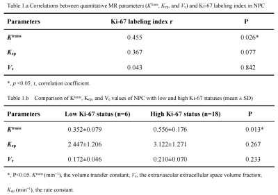 |
92 | Correlation between magnetic resonance perfusion and Ki-67 expression in nasopharyngeal carcinoma
Nan Wang 1, Lijun Wang1, Lizhi Xie2, and Ailian Liu1
1Department of Radiology, the First Affiliated Hospital of Dalian Medical University, Dalian, China, 2GE Healthcare, MR Research China, Beijing, China Keywords: Sparse & Low-Rank Models, Cancer Nasopharyngeal carcinoma (NPC) is a common malignancy in Southeast Asia. In this study, we explore the correlation between the quantitative parameters derived from dynamic contrast-enhanced magnetic resonance (DCE-MRI) of NPC with Ki-67 index. Results showed that the Ktrans mean was positively correlated with Ki-67. The Ktrans mean value was significantly higher in cases with a high Ki-67 status than in those with a low Ki-67 status (AUC 0.8519), and the cutoff value, sensitivity, and specificity was 0.479, 77.78%, and 100%, respectively, suggesting the Ktrans parameter of DCE-MRI has the most utility in distinguishing between high and low Ki-67 levels. |
|
2088. |
93 | DWI Imaging for the Discrimination of Cytotoxic Lesions and Acute Ischemic Infarction of the Corpus Callosum: Utility of Relative ADC.
Xing Tang1 and Anqi Chen1
1Department of Radiology, Xijing Hospital, Fourth Military Medical University, Xi'an, China Keywords: Head & Neck/ENT, Diffusion Tensor Imaging Cytotoxic lesions of the corpus callosum (CLOCCs) is a rare clinical-imaging syndrome, which is difficult to differentiate from acute ischemic infarction of the splenium of the corpus callosum (AII-SCC) by conventional MRI, DWI and ADC value. In this study, a novel relative ADC (rADC) value was proposed. Results showed that the rADC value of CLOCCs is significantly lower than that of AII-SCC, which Indicated that rADC value may have the potential to distinguish these two diseases. |
|
2089.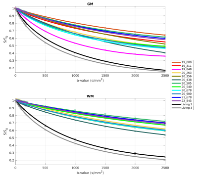 |
94 | Comparison of post mortem in situ and in vivo intravoxel incoherent motion of the human brain
Melanie Bauer1,2, Celine Berger1,2, Eva Scheurer1,2, and Claudia Lenz1,2
1Institute of Forensic Medicine of the University of Basel, Department of Biomedical Engineering, Basel, Switzerland, 2Institute of Forensic Medicine of the University of Basel, Health Department Basel-Stadt, Basel, Switzerland Keywords: Head & Neck/ENT, Diffusion/other diffusion imaging techniques Performing intravoxel incoherent motion (IVIM) magnetic resonance imaging offers the possibility to differentiate various diffusion processes according to their varying molecule speeds. In this study, the IVIM parameters perfusion fraction, diffusion and pseudo-diffusion were determined in the human brain for 12 post mortem in situ and 2 in vivo cases. Our results show that the IVIM parameters decrease after death and that they are higher in gray matter than in white matter. Besides, the age at death, the core temperature of the subjects and the post mortem interval have an effect on the post mortem IVIM parameters. |
|
2090.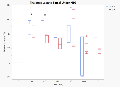 |
95 | Estrogen Deprivation has a Negligible Impact on Thalamic Metabolism under Central Sensitization
Dayna L. Richter1,2, Samuel Holder1,2, Harrison D Craythorne2, and Samuel Colles Grant1,2
1National High Magnetic Field Laboratory, Florida State University, Tallahassee, FL, United States, 2Chemical & Biomedical Engineering, FAMU-FSU College of Engineering, Tallahassee, FL, United States Keywords: Neuroinflammation, Spectroscopy, Relaxation-enhanced spectroscopy, lactate, total creatine Previous examinations of the nitroglycerin (NTG)-based central sensitization, a chemical model of migraine, in the Sprague-Dawley model revealed increases in thalamic lactate signal. This held across sexes, albeit at different strengths. To investigate the potential source of any sex differences, estradiol deprivation is examined using 1H relaxation-enhanced spectroscopy localized to the thalamus. While the same energetic deprivation is seen, estradiol deprivation had negligible impact on the thalamic metabolic profile under NTG. Further examinations under alternate modalities are needed. |
|
2091.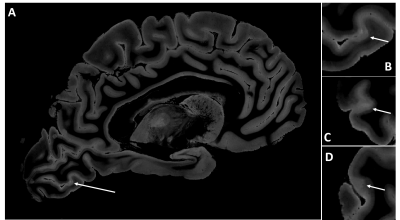 |
96 | Ultra High Spatial Resolution MRI of Intact Ex Vivo Multiple Sclerosis Brain
Ken Sakaie1, Jagjit Sidhu1, Kedar Mahajan2,3, Kunio Nakamura4, Daniel Ontaneda2, Bruce Trapp3, Mark J. Lowe1, Stephen Jones1, and Emmanuel Obusez1
1Imaging Institute, The Cleveland Clinic, Cleveland, OH, United States, 2Mellen Center, The Cleveland Clinic, Cleveland, OH, United States, 3Neurosciences, The Cleveland Clinic, Cleveland, OH, United States, 4Biomedical Engineering, The Cleveland Clinic, Cleveland, OH, United States Keywords: Multiple Sclerosis, Multiple Sclerosis Treatment options for progressive multiple sclerosis (MS) are limited and the lack of sensitive and specific MRI biomarkers is a major hurdle in therapeutic development. A number of pathophysiological processes are repeatedly found on histology but are difficult to identify accurately on MRI. Ultra-high spatial resolution MRI of intact ex vivo brain promises to help bridge the gap between MRI and pathology. We demonstrate progress towards visualizing potential biomarkers for progressive MS. |
|
2092.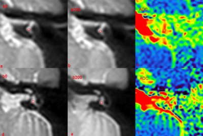 |
97 | The diagnostic value of TSE-DWI in patient with Benign paroxysmal positional vertigo (BPPV): a feasibility study
Cuilin Yin1, Jingjing Bai1, Ningning Ding1, Kai Ai2, and Jian Yang1
1The First Affiliated Hospital of Xi'an Jiaotong University, Xi'an, China, 2Philips Healthcare, Xi'an, China Keywords: Head & Neck/ENT, Diffusion/other diffusion imaging techniques, Benign paroxysmal positional vertigo, turbo spin-echo diffusion weighted imaging This study explored the feasibility of TSE-DWI in inner ear and determined the diagnostic accuracy and interobserver performance of TSE-DWI in BPPV. The method is to evaluate the difference of ADC values between benign paroxysmal positional vertigo (BPPV) patients and healthy controls. Our research shows that The ADC values of semicircular canal in the BPPV patients (affected side) were higher than that of the contralateral ear. Moderate diagnostic accuracy of DWI was seen in the diagnosis of BPPV. Therefore, we consider that the TSE-DWI derived ADC value may represent promising novel imaging markers of inner ear disease. |
|
2093.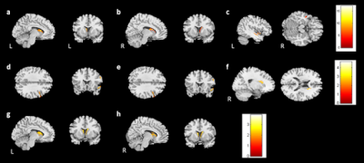 |
98 | Association of longitudinal changes in cerebral microstructure with cognitive function in breast cancer survivors after adjuvant chemotherapy
Yi-Fang Wu1, Vincent Chin-Hung Chen2,3, Yuan-Hsiung Tsai2,4, and Jun-Cheng Weng1,3,5
1Department of Medical Imaging and Radiological Sciences, and Department of Artificial Intelligence, Chang Gung University, Taoyuan, Taiwan, 2School of Medicine, Chang Gung University, Taoyuan, Taiwan, 3Department of Psychiatry, Chang Gung Memorial Hospital, Chiayi, Taiwan, 4Department of Diagnostic Radiology, Chang Gung Memorial Hospital, Chiayi, Taiwan, 5Medical Imaging Research Center, Institute for Radiological Research, Chang Gung University and Chang Gung Memorial Hospital at Linkou, Taoyuan, Taiwan Keywords: Neuroinflammation, Cancer Adjuvant chemotherapy for breast cancer might impact cognitive function and brain structure. In this study, we investigated the cerebral microstructural changes in breast cancer survivors after adjuvant chemotherapy and the correlation with cognitive function with longitudinal study designs. The results showed brain volume reduction in thefrontal and temporal regions were also observed in patients from baseline to postchemotherapy. An association between brain volume and cognitive performance was also found in the limbic system. According to our findings, the study was useful in developing a prediction model as well as a guide for cancer treatment. |
|
2094.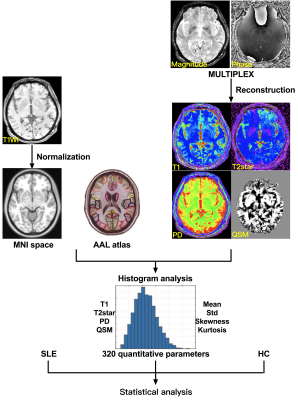 |
99 | Brain abnormalities in systemic lupus erythematosus using an advanced multi-parametric MR imaging method of MULTIPLEX: a feasibility study
Jiaying Mo1, Hai Lin2, Yongming Dai2, Xiangliang Tan1, and Yikai Xu1
1Department of Medical Imaging Center, Nanfang Hospital, Southern Medical University, Guangzhou, China, 2MR Collaboration, Central Research Institute, United Imaging Healthcare, Shanghai, China Keywords: Head & Neck/ENT, Brain, Systemic lupus erythematosus Quantitative MRI can measure a variety of physiological tissue parameters, such as longitudinal T1 value, transverse T2 value and proton density, iron content and fat content. We applied an advanced multi-parametric MR imaging technique of MULTIPLEX that provides the maps of T1, T2*, proton density and quantitative susceptibility mapping, to investigate the brain abnormalities in systemic lupus erythematosus (SLE). We found that the MULTIPLEX could provide comprehensive and quantitative assessment of SLE-related regions captured in multiple MRI parameters, which might assist in our understanding of the neuropathological mechanism of SLE. |
|
 Back to Meeting Home
 Back to the Program-at-a-Glance
The International Society for Magnetic Resonance in Medicine is accredited by the Accreditation Council for Continuing Medical Education to provide continuing medical education for physicians.
 Back to Meeting Home
 Back to the Program-at-a-Glance
 View Presentation Video