Digital Poster
Neurodegeneration I
ISMRM & ISMRT Annual Meeting & Exhibition • 03-08 June 2023 • Toronto, ON, Canada

| Computer # | |||
|---|---|---|---|
1526.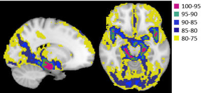 |
61 | Assessing pulsatility ratio in different sized cerebral arterial vessels across varying levels of blood pressure
Tae Kim1 and Peter J Gianaros2
1Radiology&Bioengineering, University of Pittsburgh, Pittsburgh, PA, United States, 2Psychology, University of Pittsburgh, Pittsburgh, PA, United States Keywords: Neurodegeneration, Hypertension Pulsatility derived from the BOLD signal was used to assess the relationship of pulsatility damping in different sized cerebral arterial vessels across levels of blood pressure. Individuals with higher blood pressure exhibit reduced cerebrovascular compliance, and less damping of BOLD pulsatility among these individuals may be a potential indicator of cerebrovascular pathogenesis. |
|
1527.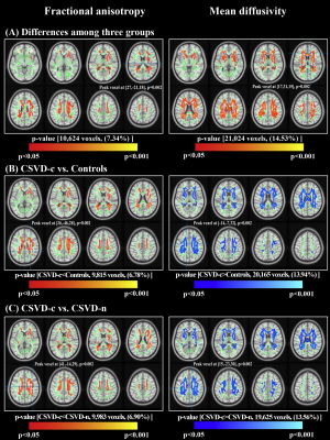 |
62 | Characterization of white matter microstructural abnormalities in cerebral small vessel disease with cerebral microbleeds
Chaofan Sui1, Hongwei Wen2, Shengpei Wang3, Mengmeng Feng4, Haotian Xin4, Yian Gao1, Jing Li5, Lingfei Guo1, and Changhu Liang1
1Department of Radiology, Shandong Provincial Hospital Affiliated to Shandong First Medical University, Jinan, China, 2Key Laboratory of Cognition and Personality (Ministry of Education); School of Psychology, Southwest University, Chongqing, China, 3Research Center for Brain-inspired Intelligence Institute of Automation, Chinese Academy of Sciences, Beijing, China, 4Department of Radiology, Shandong Provincial Hospital, Shandong University, Jinan, China, 5Department of Radiology, Beijing Friendship Hospital, Capital Medical University, Bijing, China Keywords: Neurodegeneration, White Matter To characterize white matter (WM) microstructural abnormalities in patients with cerebral small vessel disease (CSVD) coexisting with cerebral microbleeds (CMBs) and to further investigate the exact mechanism by which CMBs influence cognitive decline in patients with CSVD at the group and individual levels. Fractional anisotropy (FA), mean diffusivity (MD), axial diffusivity (AD) and radial diffusivity (RD) images from 49 CSVD patients with CMBs (CSVD-c), 114 CSVD patients without CMBs (CSVD-n), and 83 controls were analyzed using DTI-derived tract-based spatial statistics to detect WM diffusion changes among groups. |
|
1528.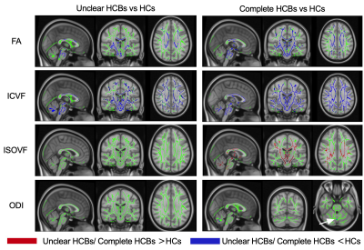 |
63 | “Hot cross bun” Sign Grades Means Different White Matter Changes in MSA-C: A Neurite Orientation Dispersion and Density Imaging Study
Chuanying Shi1, Peng Wu2, Jixin Luan3, and Xiance Zhao2
1Radiology, Liaocheng People's Hospital, Liaocheng, China, 2Philips Healthcare, Shang Hai, China, 3China-Japan Friendship Hospital, Chinese Academy of Medical Sciences & Peking Union Medical College, Bei Jing, China Keywords: Neurodegeneration, Diffusion/other diffusion imaging techniques, voxel-wise tract-based spatial statistics White matter (WM) changes play an important role in the progression of multiple system atrophy cerebellar type (MSA-C), and doctors need an imaging marker to grade the WM alterations. We observed different WM alterations between MSA-C patients with unclear “hot cross bun” sign (HCBs) and patients with complete HCBs, using NODDI, analyzed by TBSS. Compared with healthy controls, patients with complete HCBs showed broader different regions of intracellular volume fraction (ICVF) than ones with unclear HCBs, and meanwhile, difference of orientation dispersion index (ODI) and isotropic volume fraction (ISOVF) were only shown in complete HCBs ones. Difference of ICVF between patients with unclear and complete HCBs was also obvious. All of these findings indicated HCBs might be an imaging marker to assess the WM changes in MSA patients. |
|
1529.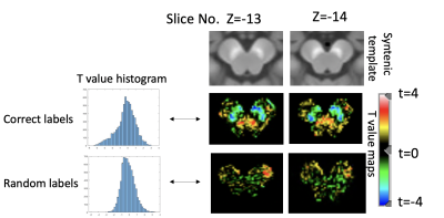 |
64 | Mapping neuromelanin loss in clinically uncertain parkinsonism differentiates neurodegenerative from non-neurodegenerative
Yue Xing1,2,3, Stefan Pszczolkowski1, Saadnah Naidu1,2,3, Tayyib Hayat1,2,3,4, Jonathan Evans4, Christophe R Tench1,3, Schwarz T Stefan1,2,5, and Dorothee P Auer1,2,3
1Mental Health and Clinical Neuroscience Unit, University of Nottingham, Nottingham, United Kingdom, 2Sir Peter Mansfield Imaging Centre, University of Nottingham, Nottingham, United Kingdom, 3NIHR Nottingham Biomedical Research Centre, University of Nottingham, Nottingham, United Kingdom, 4Nottingham University Hospitals, Nottingham, United Kingdom, 5Department of Radiology, Cardiff and Vale University, Cardiff, United Kingdom Keywords: Neurodegeneration, Neurodegeneration, clinical uncertain Parkinsonism, NM-MRI Brain dopamine transporter SPECT imaging is routinely used to assess striatal dopaminergic deficit in the differentiation of essential tremor from degenerative parkinsonism in clinically uncertain parkinsonism (CUP). Neuromelanin (NM)-MRI detects nigral depigmentation with good diagnostic accuracy in confirmed Parkinson’s Disease; but its diagnostic value in CUP is unclear. Using voxel-based analysis following optimised automatic registration, we investigated the topography of NM loss in CUP, and show that voxels that best discriminate between non-neurodegenerative and neurodegenerative CUP are co-located in the dorsolateral substantia nigra. |
|
1530.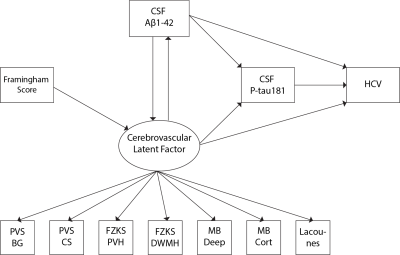 |
65 | Vascular contribution to the preclinical stages of Alzheimer’s disease: Insights from the EPAD cohort
Luigi Lorenzini1, Alessio Maranzano2, Lyduine E Collij1, Carole H Sudre3, Robin Wolz4, Sven Haller5, Kaj Blennow6, Giovanni B Frisoni7, Pierre Payoux8, Pablo Lage-Martinez9, Michael Ewers 10, Gael Chatelat11, Adam Waldman 12, Joanna Wardlaw 12, Nick Fox 13, Craig Ritchie 14, Philip Scheltens 15, Pieter Jelle Visser 15, Alle Meije Wink 1, Henk JMM Mutsaerts 1,
Juan Domingo Gispert 16, Silvia Ingala 1, and Frederik Barkhof1
1Amsterdam UMC, Amsterdam, Netherlands, 2Department of Neurology-Stroke Unit and Laboratory of Neuroscience, Istituto Auxologico Italiano IRCCS - Department of Pathophysiology and Transplantation, “Dino Ferrari” Center, Università degli Studi di Milano, Milan, Italy, Milan, Italy, 3MRC Unit for Lifelong Health and Ageing at UCL, London, UK, London, United Kingdom, 4IXICO, London, United Kingdom, 5CIRD Centre d’Imagerie Rive Droite, Geneva, Switzerland, Geneva, Switzerland, 6Department of Psychiatry and Neurochemistry, Institute of Neuroscience and Physiology, the Sahlgrenska Academy at the University of Gothenburg, Sweden, Gothenburg, Sweden, 7University Hospitals and University of Geneva, Geneva, Switzerland, 8Department of Nuclear Medicine, Toulouse CHU, Purpan University Hospital, Toulouse, France, 9Centro de Investigación y Terapias Avanzadas, Neurología, CITA‐Alzheimer Foundation, San Sebastian, Spain, 10German Center for Neurodegenerative Diseases (DZNE), Feodor-Lynen-Strasse 17, Munich, Germany, 11Université de Normandie, Unicaen, Inserm, U1237, PhIND "Physiopathology and Imaging of Neurological Disorders", Institut Blood-and-Brain @ Caen-Normandie, Cyceron, 14000, Caen, France, 12Centre for Clinical Brain Sciences, The University of Edinburgh, Edinburgh, Scotland, 13Dementia Research Centre, Department of Neurodegenerative Disease, UCL Queen Square Institute of Neurology, London, United Kingdom, 14Centre for Dementia Prevention, The University of Edinburgh, Edinburgh, Scotland, 15Department of Neurology, Alzheimer Center Amsterdam, Amsterdam Neuroscience, Vrije Universiteit Amsterdam, Amsterdam UMC, Amsterdam, Netherlands, 16Barcelonaβeta Brain Research Center (BBRC), Pasqual Maragall Foundation, Barcelona, Spain Keywords: Neurodegeneration, Alzheimer's Disease, Cerebrovascular, radiological Evidence suggests that cardiovascular risk factors and cerebrovascular disease contribute to the Alzheimer’s disease (AD) pathophysiology. It is still unclear to what extent vascular and amyloid pathology have a synergistic or independent influence in preclinical AD stages. We used structural equation models in a cohort of cognitively unimpaired individuals to investigate if cerebrovascular pathology mediates the relationship between cardiovascular risk factors and CSF Aβ1-42, and show that cerebrovascular pathology accelerates downstream AD markers (p-Tau181 and hippocampal volume) by increasing amyloid pathology. |
|
1531.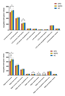 |
66 | Depression symptoms in Parkinson's disease correlate with amygdala subregions atrophy
Mingrui Qu1, Bingbing Gao1, Yuhan Jiang1, Yuan Li1, Lizhi Xie2, and Yanwei Miao1
1The First Affiliated Hospital of Dalian Medical University, Dalian, China, 2GE Healthcare, Shanghai, China Keywords: Neurodegeneration, Psychiatric Disorders Depression is the most frequent psychiatric disorder in Parkinson's disease (PD). Amygdala pathology has been suggested to contribute to some clinical features of PD, including deficits of olfaction and mood disorders. We aimed to more accurately measure alterations in the volume of each amygdala nucleus in Parkinson's disease with depression (DPD) patients. Then the volume of each specific amygdala nucleus would be associated with the severity of depressive symptoms. This study showed that DPD patients had multiple amygdala subregions atrophy. The bilateral lateral amygdala and left accessory basal nucleus were negatively correlated with the severity of depression in DPD patients. |
|
1532. |
67 | Increase of Magnetic Transfer Contrast in Middle Cerebellum Peduncles in Patients with Multiple System Atrophy
Haiying Lyu1, Qing Li2, Yufei Huang1, and Yong Lu3
1Radiology, Ruijin Hospital Affiliated to Shanghai Jiao Tong University School of Medicine, Shanghai, China, 2MR Collaborations, Siemens Healthineers Ltd., Shanghai, China, 3Ruijin Hospital / Luwan Branch Affiliated to Shanghai Jiao Tong University School of Medicine, Shanghai, China Keywords: Neurodegeneration, Magnetization transfer, Multiple system atrophy The magnetic transfer (MT) MRI imaging has long been used for quantifying neuromelanin containing nucleus, but its ability of quantifying the magnetization exchange between free water and macromolecules has also allowed its potential usage in detecting white matter changes. Patients with multiple system atrophy has unique pathological changes in pontocerebellar regions. In this study, we have not only found significant increase of MT contrast in MCP regions of MSA, but also proved significant correlation between this alteration and brainstem or cerebellar atrophy, which might offer new insights for future studies. |
|
1533.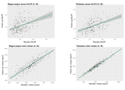 |
68 | Examining the consistency of brain MRI structural and functional readouts using repeat imaging from the longitudinal UK Biobank MRI Study.
Guocheng Jiang1, Jennifer S Rabin2,3,4,5, Walter Swardfager2,6, Hugo Cogo-Moreira7, and Bradley J MacIntosh1,8
1Department of Medical Biophysics, University of Toronto, Toronto, ON, Canada, 2Hurvitz Brain Sciences Research Program, Sunnybrook Research Institute, Toronto, ON, Canada, 3Harquail Centre for Neuromodulation, Sunnybrook Health Sciences Centre, Toronto, ON, Canada, 4Department of Medicine Division of Neurology, University of Toronto, Canada, Toronto, ON, Canada, 5Graduate Department of Rehabilitation Science, University of Toronto, Toronto, ON, Canada, 6Department of pharmacology and toxicology, University of Toronto, Toronto, ON, Canada, 7Department of Education, ICT and Learning, Østfold University College, Halden, Norway, 8Department of Radiology and Nuclear Medicine, Oslo University Hospital, Oslo, Norway Keywords: Neurodegeneration, fMRI, Structural MRI We aim to study structural and functional changes over time with a focus on the thalamus and hippocampus regions of interest. Longitudinal analysis was performed in N=274 UK Biobank participants. We find strong correlations between the baseline and follow-up MRI readouts. Namely, it is possible to predict thalamus and hippocampus volume changes across a mean of 2.25 years using baseline volume and resting state functional activation estimates. This work helps characterizing the longitudinal consistencies within both hippocampal and thalamic MR readouts and helps setting up prediction models as the next step. |
|
1534.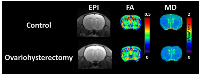 |
69 | Brain alterations in ovariohysterectomized rats revealed by diffusion tensor imaging
Shin-Lei Peng1, Sheng-Min Huang2, and Chun-Chieh Chan1
1Department of Biomedical Imaging and Radiological Science, China Medical University, Taichung, Taiwan, 2Institute of Biomedical Engineering and Nanomedicine, National Health Research Institutes, Miaoli, Taiwan Keywords: Neurodegeneration, Diffusion Tensor Imaging Women undergoing hysterectomy with oophorectomy have an increased risk of Alzheimer’s disease. However, postoperative neuroimaging data on pathogenic processes in the brain are limited. This study was aimed to investigate the potential effect of ovariohysterectomy on brain integrity in the rat model using diffusion tensor imaging technique. Compared to the control group, the ovariohysterectomy group showed significantly lower fractional anisotropy in the corpus callosum, bilateral striatum, and bilateral cortex, suggesting neuronal injury in ovariohysterectomized rats. Therefore, neuroimaging should be performed to monitor brain alterations in women after hysterectomy with bilateral oophorectomy in clinical settings. |
|
1535.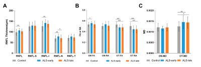 |
70 | Evaluation of the anterior visual pathway in amyotrophic lateral sclerosis with optical coherence tomography and diffusion tensor imaging
Jing Zhang1, Yali Zhao1, Yuan Yang2, and Hongyu Wu1
1Radiology, Tongji Hospital, Tongji Medical College, Huazhong University of Science and Technology, Wuhan, China, 2Neurology, The Central Hospital of Enshi Tujia and Miao Autonomous Prefecture, Enshi, China Keywords: Neurodegeneration, Diffusion Tensor Imaging Involvement of the visual pathway in amyotrophic lateral sclerosis (ALS) has been demonstrated. However, the exact damage mechanisms such as how the changes in the distinct parts of the anterior visual pathway (AVP) are still poorly understood and remain highly controversial. We observed altered RNFL, fractional anisotropy, mean diffusivity and relative anisotropy of AVP by the multimodal use of optical coherence tomography and DTI in ALS individuals compared to the controls. Our findings shed light on the dynamic pathophysiological effects on the morphology of the AVP and the potential association between microstructural damage to the AVP and executive function. |
|
1536.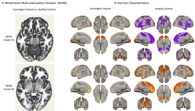 |
71 | Disruptions in Functional Connectivity in Pre-manifest Huntington Disease: A Data-Driven, Whole-Brain fMRI Study
Simon Laganiere1, Mark A Halko2, Luis Sierra3, Clementina Ullman3, Karen Hildebrand3, Magdaline Mwangi3, Julia Dierker3, Samuel Frank1, Kaitlin Toal3, and Sheeba Anteraper4
1Beth Israel Deaconess Medical Center and Harvard Medical School, Boston, MA, United States, 2McLean Hospital, Belmont, MA, United States, 3Beth Israel Deaconess Medical Center, Boston, MA, United States, 4Stephens Family Clinical Research Institute, Carle Foundation Hospital, Urbana, IL, United States Keywords: Neurodegeneration, fMRI (resting state), Huntington disease, striatum, data-driven multi-voxel pattern analysis Huntington disease (HD) is a progressive, autosomal dominant disease caused by a pathological expansion of CAG repeats in the HTT gene1,2. A clinical diagnosis of HD is made at the appearance of unequivocal motor signs. However, in the “premanifest” stage, due to slowly progressive neurodegenerative changes1,2, subtle motor, psychiatric and cognitive decline occurs many years prior to diagnosis3. The sequelae of neural circuit dysfunction remains unclear. Improving our mechanistic understanding of functional brain connectivity alterations prior to disease manifestation will help identify sensitive biomarkers of disease progression1,4–6. Data-driven analysis of high-quality functional magnetic resonance imaging data will guide such efforts. |
|
1537.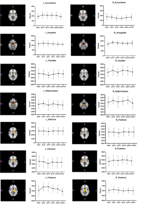 |
72 | Pretherapeutic specific gray matter volume for predicting MRgFUS thalamotomy-mediated tremor response in Parkinson's disease
Xiaoyu Wang1 and Xin Lou1
1Department of Radiology, Chinese PLA General Hospital, Beijing, China Keywords: Neurodegeneration, MR-Guided Interventions, MRgFUS This is the first study to assess magnetic resonance-guided focused ultrasound (MRgFUS) VIM thalamotomy on gray matter (GM) volume in tremor-dominant Parkinson’s disease (PD). MRgFUS has good efficacy and safety in the treatment of PD. We used three methods to extract specific GM volumes of nine PD patients before and after treatment for calculating their difference and correlation analysis. We found that the specific GM regions may predict tremor responses in PD after thalamotomy, and the results help to better understand the distant effect of MRgFUS thalamotomy and the involvement of GM in tremor control in PD. |
|
1538.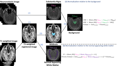 |
73 | Neuromelanin-MRI Assessment in Dementia with Lewy Bodies
Rotem Iris Orad1,2,3, Moran Artzi1,2,4, Noa Bregman2,3, Anat Mirelman2,4,5, Avner Thaler2,4,5, Amgad Droby2,4,5, Orly Goldstein6, Mali Gana-Weisz6, Netanell Avisdris1, Avi Orr-Urterger2,4,6, Nir Giladi2,3,4,5,6,7, Tamara Shiner2,3,4,7, and Dafna Ben Bashat1,2,4
1Sagol Brain Institute, Tel Aviv Sourasky Medical Center, Tel Aviv, Israel, 2Sackler School of Medicine, Tel Aviv University, Tel Aviv, Israel, 3Center for Cognitive Neurology Unit, Neurological Institute, Tel Aviv Sourasky Medical Center, Tel Aviv, Israel, 4Sagol School of Neuroscience, Tel Aviv University, Tel Aviv, Israel, 5Laboratory of Early Markers of Neurodegeneration, Center for the Study of Movement, Cognition, and Mobility, Neurological Institute, Tel Aviv Sourasky Medical Center, Tel Aviv, Israel, 6The Genomic Research Laboratory for Neurodegeneration, Neurological Institute, Tel Aviv Sourasky Medical Center, Tel Aviv, Israel, 7Movement Disorders Unit, Neurological Institute, Tel Aviv Sourasky Medical Center, Tel Aviv, Israel Keywords: Neurodegeneration, Genetics, Dementia with Lewy Bodies Reduced neuromelanin-MRI signal and volume are known in Parkinson's disease (PD). However, changes in neuromelanin-MRI with Dementia with Lewy bodies (DLB) and whether there are genotype-related differences are unknown. In this study, we showed reduced neuromelanin signal and volume in patients with DLB compared to healthy controls. Yet, no differences were detected between genotype groups: DLB with and without GBA mutation. Results may suggest the same pathomechanism underlying DLB, PD and other neurodegenerative parkinsonian syndromes, with depletion of neuromelanin within the substantia nigra. |
|
1539.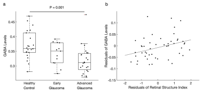 |
74 | Reduction of GABA in the visual cortex of glaucoma patients is linked to decreased neural specificity.
Ji Won Bang1, Carlos Parra1, Kevin Yu1, Gadi Wollstein1,2,3, Joel S Schuman1,2,3,4, and Kevin C Chan1,2,3,4,5
1Department of Ophthalmology, NYU Grossman School of Medicine, NYU Langone Health, New York University, New York, NY, United States, 2Center for Neural Science, College of Arts and Science, New York University, New York, NY, United States, 3Department of Biomedical Engineering, Tandon School of Engineering, New York University, New York, NY, United States, 4Neuroscience Institute, NYU Grossman School of Medicine, NYU Langone Health, New York University, New York, NY, United States, 5Department of Radiology, NYU Grossman School of Medicine, NYU Langone Health, New York University, New York, NY, United States Keywords: Neurodegeneration, Brain Glaucoma is an age-related neurodegenerative disease of the visual system. Although increasing number of studies indicated its widespread involvements of the eye and the brain, very little is known about the underlying metabolic mechanisms. Thus, here we investigated the GABAergic and glutamatergic systems in the visual cortex of glaucoma patients, as well as neural specificity. Our study demonstrated that glaucoma is accompanied by the reduction of GABA and glutamate in the visual cortex. Further, the reduction of GABA but not glutamate predicted neural specificity. This suggests that GABA loss in the visual cortex degrades the neural specificity in glaucoma. |
|
1540.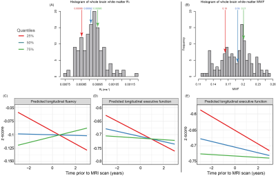 |
75 | Evidence of association between lower cerebral white matter myelin content and rapid cognitive decline in cognitively unimpaired individuals
Zhaoyuan Gong1, Murat Bilgel2, Matthew Kiely1, Curtis Triebswetter1, Luigi Ferrucci3, Susan M. Resnick2, Richard G. Spencer1, and Mustapha Bouhrara1
1Laboratory of Clinical Investigation, National Institute on Aging, Baltimore, MD, United States, 2Laboratory of Behavioral Neuroscience, National Institute on Aging, Baltimore, MD, United States, 3Translational Gerontology Branch, National Institute on Aging, Baltimore, MD, United States Keywords: Neurodegeneration, Aging, Myelin Myelin plays an essential role in the normal functioning of the central nervous system. However, how myelination influences longitudinal changes in cognitive performance, especially in cognitively normal (CN) individuals, remains unclear. Using a linear mixed-effects regression analysis, we examined the association between myelin content and changes in cognitive domain scores obtained over several years prior to the time of the MRI scan. We demonstrated strong and statistically significant relationships between myelin content and the rates of change in cognitive performance in several white matter regions. These findings highlight the importance of white matter, specifically myelin integrity, in cognitive functioning. |
|
1541.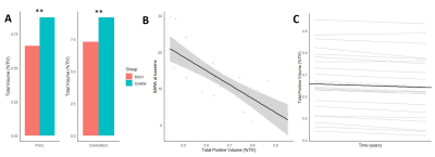 |
76 | Altered neurochemical profiles and volume loss in the cerebellum and brainstem as biomarkers in spinocerebellar ataxia type 1
Kirsten Kapteijns1, Teije van Prooije1, Jack JA van Asten2, Marcel Verbeek1, Bart van de Warrenburg1, and Tom WJ Scheenen2
1Neurology, Radboudumc, Nijmegen, Netherlands, 2Medical Imaging, Radboudumc, Nijmegen, Netherlands Keywords: Neurodegeneration, Spectroscopy, Single Voxel Spectroscopy Spinocerebellar ataxia type 1 is a rare, progressive movement disorder primarily affecting the cerebellum and closely connected brain regions. We assessed the dynamics of MR biomarkers and their utility as progression and/or predictive biomarkers in SCA1. SCA1 patients showed an altered neurochemical profile when compared to controls. Additionally, we measured a lower volume of affected regions in patients, clearly distinguishing them from controls. Altered volume correlated strongest with clinical scores, confirming the potential to use MR-markers to monitor SCA1 progression. Furthermore, MRS markers correlated with consequent change in volumetric measures, showing preliminary evidence for their predictive value in SCA1. |
|
1542. |
77 | Cortical thickness alterations in patients with T2DM and its correlation with cerebral small vessel diseases and cognitive function
Jinghan Zhao1, YangYingqiu Liu1, wei Du1, Yuhan Jiang1, Peng Sun2, Weiwei Wang1, and Yanwei Miao1
1The First Affiliated Hospital of Dalian Medical University, Dalian, China, 2Philips Healthcare, Beijing, China Keywords: Neurodegeneration, Diabetes, small vessel diseases (CSVD)、cognition、freesurfer The objective of this study was to explore the changes of cortical thickness in patients with type 2 diabetes mellitus, and the correlation between the thinning cerebral regions and cerebral small vessel diseases (CSVD) burden. The thickness of the left medial orbitofrontal cortex was negatively correlated with the centrum semi-ovale enlarged perivascular space (CSO-EPVS) score and CSVD total burden score. The thickness of the left lateral occipital cortex was negatively correlated with CSO-EPVS score, basal ganglia enlarged perivascular space (BG-EPVS) score, and CSVD total burden score, and positively correlated with mini-mental state examination (MMSE) delayed memory score. |
|
1543.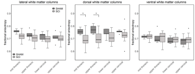 |
78 | Remote neurodegeneration in a rat contusion spinal cord injury model: a combined MRI and histological study
Gergely David1, Alice Motovylyak2, Felix Schlegel3, Zsofia Kovacs3, Matthew Budde4, Christian Kündig1, Jan Klohs3,5, and Patrick Freund1,6,7
1Spinal Cord Injury Center, Balgrist University Hospital, University of Zurich, Zurich, Switzerland, 2Department of Biomedical Engineering, Marquette University and Medical College of Wisconsin, Milwaukee, WI, United States, 3Institute for Biomedical Engineering, University of Zurich and ETH Zurich, Zurich, Switzerland, 4Department of Neurosurgery, Medical College of Wisconsin, Clement J Zablocki Veterans Affairs Medical Center, Milwaukee, WI, United States, 5Neuroscience Center Zurich, University of Zurich and ETH Zurich, Zurich, Switzerland, 6Department of Neurophysics, Max Planck Institute for Human Cognitive and Brain Sciences, Leipzig, Germany, 7Wellcome Trust Centre for Human Neuroimaging, UCL Queen Square Institute of Neurology, University College London, London, United Kingdom Keywords: Neurodegeneration, Trauma MRI has been widely used to investigate the structural damage after traumatic spinal cord injury (SCI). While most small animal studies have focused on the injury site, remote SCI-related damage along the neuraxis has received less attention. Here, we demonstrate that ex vivo diffusion MRI and cross-sectional area measurements are sensitive to remote neurodegeneration in a rat contusion SCI model, showing gray matter and dorsal column atrophy alongside decreased fractional anisotropy in the dorsal columns several spinal levels rostral to the injury epicenter. Imaging findings were consistent with SMI32 immunohistochemistry with axonal degeneration mostly concentrated in the dorsal column. |
|
1544. |
79 | Detecting longitudinal alterations of cerebral white matter associated with breast cancer and chemotherapy using GQI
Wei Chuang1, Vincent Chin-Hung Chen2,3, Yuan-Hsiung Tsai2,4, and Jun-Cheng Weng1,3,5
1Department of Medical Imaging and Radiological Sciences, and Department of Artificial Intelligence, Chang Gung University, Taoyuan, Taiwan, 2School of Medicine, Chang Gung University, Taoyuan, Taiwan, 3Department of Psychiatry, Chang Gung Memorial Hospital, Chiayi, Taiwan, 4Department of Diagnostic Radiology, Chang Gung Memorial Hospital, Chiayi, Taiwan, 5Medical Imaging Research Center, Institute for Radiological Research, Chang Gung University and Chang Gung Memorial Hospital at Linkou, Taoyuan, Taiwan Keywords: Brain Connectivity, Diffusion/other diffusion imaging techniques In 2020, breast cancer is the most prevalent type of cancer and has the highest incident rate in women worldwide. The extensively used adjuvant chemotherapy might have detrimental effect on human brain and results in chemotherapy-related cognitive impairment (CICI) in breast cancer patients. The present study performed longitudinal design, aiming to investigate the microstructural and macroscale white matter alterations by generalized q-sampling imaging (GQI). Our results suggested that the patients had changes in local white matter integrity and network performance in DAN before treatment and frontal lobe connection after treatment. |
|
 Back to Meeting Home
 Back to the Program-at-a-Glance
The International Society for Magnetic Resonance in Medicine is accredited by the Accreditation Council for Continuing Medical Education to provide continuing medical education for physicians.
 Back to Meeting Home
 Back to the Program-at-a-Glance
 View Presentation Video