Digital Poster
Brain Connectivity in Health II
ISMRM & ISMRT Annual Meeting & Exhibition • 03-08 June 2023 • Toronto, ON, Canada

| Computer # | |||
|---|---|---|---|
3488.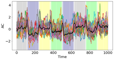 |
81 |
Autocorrelation of the fMRI signal dynamically changes during
different brain states
Ali M Golestani1,
Nichole R Bouffard2,
Morris Moscovitch2,3,
and Morgan D Barense2,3
1University of Calgary, Calgary, AB, Canada, 2Department of Psychology, University of Toronto, Toronto, ON, Canada, 3Rotman Research Institute at Baucrest, Toronto, ON, Canada Keywords: Brain Connectivity, fMRI The autocorrelation (AC) of fMRI signal is affected by brain function and state. We used fMRI data acquired during working memory, mathematical computation, visual attention, and rest, and showed that the AC signal of gray matter dynamically changes across different task blocks. The dynamic AC (dAC) can be used to estimate brain-state changes with high accuracy. Classifier accuracy is correlated with the task performance; Subjects with better task performance have better accuracy in the dAC-based brain state classification. This study shows that the AC of the fMRI signal over time dynamically changes by the cognitive processes. |
|
3489.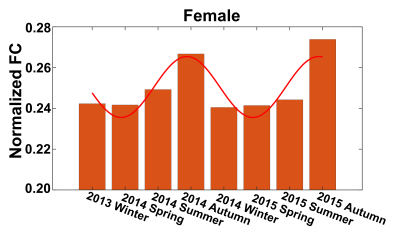 |
82 |
Seasonal variations of functional connectivity of human brains
Lyuan Xu1,2,
Soyoung Choi1,
Yu Zhao1,3,
Muwei Li1,3,
Baxter P. Rogers1,3,4,5,
John C. gore1,3,4,
Yurui Gao1,4,
and Zhaohua Ding1,2,4
1Vanderbilt University Institute of Imaging Science, Vanderbilt University Medical Center, Nashville, TN, United States, 2Department of Electrical and Computer Engineering, Vanderbilt University, Nashville, TN, United States, 3Department of Radiology and Radiological Sciences, Vanderbilt University Medical Center, Nashville, TN, United States, 4Department of Biomedical Engineering, Vanderbilt University, Nashville, TN, United States, 5Department of Psychiatry and Behavioral Sciences, Vanderbilt University Medical Center, Nashville, TN, United States Keywords: Brain Connectivity, Brain Connectivity, Seasonality Seasonal variations have been observed in various aspects of human behavior. While studies have explored the seasonality effects in cognition and mood, possible underlying seasonal variations of human brain activity have not gained wide attention. Functional magnetic resonance imaging (fMRI) based on blood-oxygenation-level-dependent (BOLD) effects can detect and map functional activity and thus provides opportunities to characterize seasonal variations in brain functions. This work used fMRI data from the Human Connectome Project (HCP) to quantify seasonal patterns of brain connectivity. Knowledge of seasonality effects in brain activity offers the potential of advancing our understanding of seasonal variations in human behavior. |
|
3490.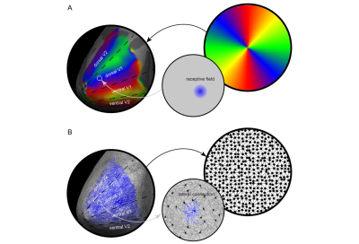 |
83 |
Topology of visual cortical connections shapes spatial structure
of perception
Chunxiang Jiang1,2,3,
Alun Metcalf1,
Jack Briggs1,
Lijuan Zhang2,3,
and Chen Song1
1Cardiff University Brain Research Imaging Centre, Cardiff University, Cardiff, United Kingdom, 2Shenzhen Institute of Advanced Technology, Chinese Academy of Sciences, Shenzhen, China, 3University of Chinese Academy of Sciences, Beijing, China Keywords: Brain Connectivity, fMRI Visual experience is highly spatial. Much of what we perceive, we perceive in space. However, the spatial structure of visual experience is often taken for granted and its neural mechanism is rarely studied. Here we tested a novel theoretical framework that proposes lateral connections in primary visual cortex as the neural basis underlying the spatial structure of visual experience. We found that the inhomogeneity in spatial localization ability across visual field correlated with the variability in lateral connection length across primary visual cortex, suggesting a mapping between the topography of visual cortical connectivity and the spatial structure of visual perception. |
|
3491.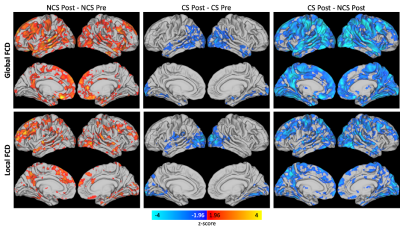 |
84 |
Functional connectivity density changes in adolescent contact
and non-contact sport athletes
Alexander D. Cohen1,
Benjamin L. Brett2,
Milan D. Patel1,
Kelly D. Ristow1,
Michael A. McCrea2,
and Yang Wang1
1Radiology, Medical College of Wisconsin, Milwaukee, WI, United States, 2Neurosurgery, Medical College of Wisconsin, Milwaukee, WI, United States Keywords: Brain Connectivity, Traumatic brain injury, Functional connectivity density Repetitive head impact exposure (RHIE) during contact sports (CS) might have potentially long-term neurological effects. We evaluated the effect of sport, time, and their interaction on local and global functional connectivity density (FCD) as measured using an advanced multiband multi-echo EPI sequence in CS and non-contact sport (NCS) middle school and high school athletes. Significant effects of sport and the interaction between sport and time on both local and global FCD were detected, which might suggest functional connections alterations due to contact sport participation. |
|
3492.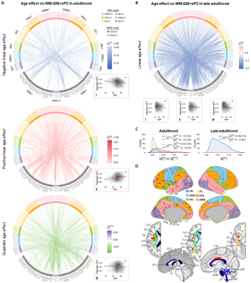 |
85 |
Reorganization of Functional Connectivity between White and Grey
Matters during Normal Aging
Yurui Gao1,2,
Yu Zhao2,3,
Muwei Li2,3,
Dylan R Lawless2,4,
Kurt G Schilling3,
Lyuan Xu2,4,
Andrea T Shafer5,
Lori L Beason-Held5,
Susan M Resnick5,
Baxter P Rogers2,3,
Zhaohua Ding2,4,
Adam W Anderson1,2,
Bennett A Landman1,2,3,4,
and John C Gore1,2,3
1Biomedical Engineering, Vanderbilt University, Nashville, TN, United States, 2VUIIS, Vanderbilt University Medical Center, Nashville, TN, United States, 3Radiology, Vanderbilt University Medical Center, Nashville, TN, United States, 4Electrical and Computer Engineering, Vanderbilt University, Nashville, TN, United States, 5Laboratory of Behavioral Neuroscience, National Institute on Aging, Baltimore, MD, United States Keywords: Brain Connectivity, Aging Resting state BOLD signals in white matter (WM) bundles have been found to be partially synchronized with signals in gray matter (GM) volumes, suggesting WM-GM functional connectivity (FC). However, little is known about whether or how these relationships change during normal aging, and traditional graph models are inappropriate. We introduced a novel graph model and applied it to assess WM-GM network properties in 1,462 healthy subjects (22–96years) and their age effects. Results show heterogenous alterations in WM-GM rsFC over adulthood with decreases mainly during late adulthood. Our results demonstrate there is substantial reorganization of WM-GM correlations during normal aging. |
|
3493. |
86 |
Comparing left vs right handed normal subjects using high
spatial-resolution resting-state fMRI at 7T
Stephen Jones1,
Ajay Nemani1,
Xuemei Huang1,
and Mark Lowe1
1Cleveland Clinic, Cleveland, OH, United States Keywords: Brain Connectivity, Brain Connectivity We present data from a series of resting-state data obtained at 7T using 1 mm isotropic resolution in separate concatenated groups of right handed (RH) and left handed (LH) normal control subjects. Whole brain connectivity maps are obtained from a large number of different seed parcels, many derived from FreeSurfer. We demonstrate the ability of this unique data to recapitulate known connectivity maps at the large scale and discern new findings at small scales. |
|
3494.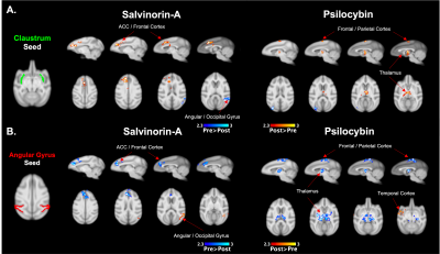 |
87 |
Acute Effects of Psilocybin and Salvinorin-A on Functional
Connectivity
Frederick Andrew Bagdasarian1,
Hanne Hansen1,2,
Chi-Hyeon Yoo1,
Michael Placzek1,
Jacob Hooker1,
and Hsiao-Ying Wey1
1Radiology, Athinoula A. Martinos Center for Biomedical Imaging, Massachusetts General Hospital, Harvard Medical School, Charlestown, MA, United States, 2Neurobiology Research Unit, Institute of Clinical Medicine, University of Copenhagen, Copenhagen, Netherlands Keywords: Brain Connectivity, fMRI (resting state) This work utilized fMRI to assess the influence of the psychedelics, Psilocybin, a serotonergic agonist, and Salvinorin-A, a kappa-opioid receptor (KOR) agonist, on functional connectivity (FC) in non-human primates. We used a seed-based FC analysis, probing regions of interest associated with psychedelic hallucinogens. Our findings highlight the overlapping and differing influence of these substances on FC relative to the subcomponents of the default mode network and the claustrum. This work may provide insight on the mechanisms of action of psychedelics that target differing receptor systems. Key Words: Functional connectivity; Claustrum; DMN; Psychedelics. |
|
3495.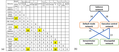 |
88 |
Effect of acute severe hypoxic environment on brain network
connectivity
Daehun Kang1,
Koji Uchida2,
Clifton Haider3,
Erin Gray1,
Myung-Ho In1,
Joshua Trzasko1,
Norbert Campeau1,
Kirk Welker1,
Jeffrey Gunter1,
Yunhong Shu1,
Matt A Bernstein1,
Max Trenerry4,
David III Holmes3,
Michael Joyner2,
Timothy Curry2,
and John III Huston 1
1Radiology, Mayo Clinic, Rochester, MN, United States, 2Anesthesiology and Perioperative Medicine, Mayo Clinic, Rochester, MN, United States, 3Physiology and Biomedical Engineering, Mayo Clinic, Rochester, MN, United States, 4Psychiatry and Psychology, Mayo Clinic, Rochester, MN, United States Keywords: Brain Connectivity, fMRI, Hypoxia Acute exposure to a severely low oxygen environment (<10%) can cause temporary deterioration of cognitive performance. To better understand the mechanism of the hypoxic-related temporary cognitive impairment in the human brain, we examined functional connectivity changes in brain networks during acute severe hypoxia while subjects performed a cognitive task. The acute severe hypoxic environment temporarily increases functional connectivity among salience (SN), default mode (DMN), executive central (ECN), sensorimotor, and visuospatial networks. We observed that increased connectivity of SN to DMN and to ECN during acute severe hypoxia is related with the behavioral cognitive deterioration. |
|
3496.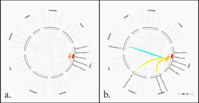 |
89 |
Functional Connectivity in Smokeless Tobacco Dependent Users in
a Craving ‘Primed’ State
Vasishta Polisetty1,
S Senthil Kumaran2,
Priyanka Bhat3,
Pankaj .2,
Anju Dhawan1,
and Sonali Jhanjhee1
1Dept of Psychiatry, All India Institute of Medical Sciences, New Delhi, New Delhi, India, 2Dept of NMR, All India Institute of Medical Sciences, New Delhi, New Delhi, India, 3Biomedical Engineering, Indian Institute of Technology, Delhi, New Delhi, India Keywords: Psychiatric Disorders, Brain Connectivity, Addiction Tobacco is a leading cause of preventable cancer and death and is consumed in various forms. There is a paucity of literature regarding cue-induced craving in smokeless tobacco, commonly found in the South East Asian Region. This study aimed to investigate the functional connectivity in a ‘primed’ state after cue-induced craving. We found facilitatory connectivity within salience network (SN) and inhibitory connectivity between default mode network (DMN) and anterior cingulate cortex (ACC) in smokeless tobacco users. |
|
3497.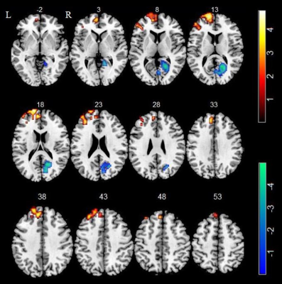 |
90 |
Abnormal neural activity in male chronic smokers revealed by
resting-state functional MRI
Xiaoyu Niu1,
Xinyu Gao1,
Mengzhe Zhang1,
and Yong Zhang1
1Department of MRI, First Affiliated Hospital of Zhengzhou University, Zhengzhou, China Keywords: Brain Connectivity, Brain, chronic smokers, amplitude of low frequency fluctuations, resting state functional connectivity, functional magnetic resonance imaging We combined two methods of resting-state functional magnetic resonance imaging (rs-fMRI) to explore the abnormal neural activity in male chronic smokers. The amplitude of low frequency fluctuation (ALFF) was first calculated and brain regions with significant differences in ALFF between two groups were used as seeds for further resting-state functional connectivity (rs-FC) analysis. Our findings revealed increased spontaneous regional activity in the superior frontal gyrus (SFG) with reduced functional connectivity to visual attention areas and cerebellar subregions in smokers compared with controls, which may play an important role in the pathophysiology of smoking. |
|
3498.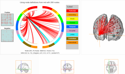 |
91 |
Mood repair in long-term meditators is predicted by rsfMRI
functional connectivity of the insula and correlates with FA of
the uncinate fasciculus
Quentin Dessain1,
Laurence Dricot2,
Nicolas Delinte1,
Manon Daussort1,
Benoit Macq1,
Phillipe de Timary2,
and Ron Kupers2
1Institute of Information and Communication Technologies, Electronics and Applied Mathematics, Université Catholique de Louvain, Louvain La Neuve, Belgium, 2Institute Of NeuroScience, Université Catholique de Louvain, Bruxelles, Belgium Keywords: Brain Connectivity, Brain We used rsfMRI and diffusion imaging to study functional and structural brain changes underlying the psychological trait mood repair in long-term meditators. Results of connectome-based predictive modeling showed that measured “mood repair” scores in meditators could be successfully predicted from rsfMRI data during meditation. The highest degree node in the underlying network was in the anterior ventral insula. Diffusion imaging data further revealed a role of the uncinate fasciculus in the mood repair trait in meditators. The uncinate fasciculus is part of the limbic network, connecting anterior temporal lobe with the inferior prefrontal cortex. |
|
3499.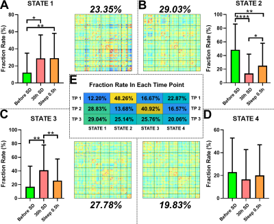 |
92 |
The Restoration Ability of Short Nap after Sleep Deprivation on
the Brain Cognitive Function: A Dynamic Functional Connectivity
Analysis
Ziliang Xu1,
Minwen Zheng1,
and Yuanqiang Zhu1
1Department of Radiology, Xijing Hospital, Fourth Military Medical University, Xi'an, China Keywords: Brain Connectivity, fMRI (resting state) As we known, consequences caused by Sleep deprivation (SD) are temporary and can be fully reversed with sufficient sleep. However, in many cases, long-duration recovery sleep is not feasible. Inspire by recent study, a short nap may be sufficient for rapid reversal of SD-induced brain function deficits. Based on dynamic functional connectivity and psychomotor vigilance task, our results showed that temporary cognitive impairment cause by SD could reversed to some extent by a nap. Additionally, dominant DFC states differed before and after SD, and their relative proportions affected the degree of cognitive impairment after SD and recovery after a nap. |
|
3500.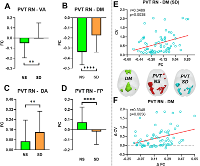 |
93 |
Effects of Abnormal Brain Network Connectivity after Sleep
Deprivation Based on Intra-Individual Variance
Ziliang Xu1,
Minwen Zheng1,
and Yuanqiang Zhu1
1Department of Radiology, Xijing Hospital, Fourth Military Medical University, Xi'an, China Keywords: Brain Connectivity, fMRI (task based) Sleep deprivation (SD) can cause task performance and negative correlation strength between default mode (DM) and task related network decreasing, but the detailed mechanism is still unclear. Intra-individual variance (IIV) is thought to associated with the negative correlation strength between DM and task active network. Thus, in this study, IIV was used to evaluate the relationship between the changing of this negative correlation strength and the task performance after SD. Results showed that SD may increase the healthy subjects’ IIV through reducing the competition relationship between DM and task active network, which resulted in the temporal impairments of cognitive function. |
|
3501. |
94 |
Risk taking tendency correlate with imbalance functional link
between brain networks following 36 hours total sleep
deprivation
Jiyuan Li1,
Yunlong Yue1,
Yunlong Song2,
and Yanfang Jin1
1Department of MRI, Beijing Shijitan Hospital, Capital Medical University, Beijing, China, 2Department of CT and MRI, The General Hospital of the Air Force People’s Liberation Army, Beijing, China Keywords: Brain Connectivity, fMRI (resting state) 36 hours sleep deprivation produces a significant deficit in vmPFC functional connectivity and default mode networks (DMN), along with the enhanced functional link between vmPFC and executive control networks (ECN). Furthermore, the negative correlation between vmPFC-DMN and vmPFC-ECN coupling during RW were diminished after TSD. We also find that TSD induced the significantly negative correlation between vmPFC-ECN networks and risk-taking behavior. These results demonstrate that an absence of sleep substantially impaired the balance of large scale brain networks and which in turn predicts risk-taking behavior following 36 hours of TSD. |
|
3502.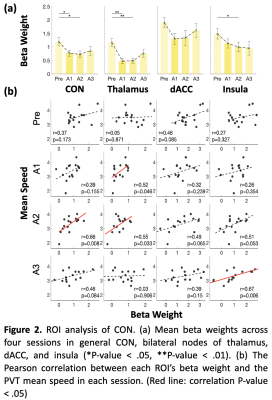 |
95 |
Dynamic associations between tonic alertness and brain activity
during sleep inertia
Yi-Chia Kung1,2,3,
Shou Chen4,
Fan-Chi Hsiao5,
Wei-Chou Chang1,3,
Changwei Wesley Wu6,7,
and Ching-Po Lin2
1Department of Radiology, Tri-Service General Hospital, Taipei, Taiwan, 2Institute of Neuroscience, National Yang Ming Chiao Tung University, Taipei, Taiwan, 3National Defense Medical Center, Taipei, Taiwan, 4McConnell Brain Imaging Center, Montreal Neurological Institute, McGill University, Montréal, QC, Canada, 5Department of Counseling and Industrial/Organizational Psychology, Ming Chuan University, Taoyuan, Taiwan, 6Graduate Institute of Mind, Brain and Consciousness, Taipei Medical University, Taipei, Taiwan, 7Brain and Consciousness Research Center, Taipei Medical University-Shuang Ho Hospital, New Taipei, Taiwan Keywords: Brain Connectivity, fMRI (task based) Sleep inertia (SI) occurs during the transition from sleep to waking, accompanied by temporary hypoalertness and decreased tonic alertness around half an hour. We used the psychomotor vigilance task (PVT) to detect the dynamic alteration of tonic alertness during SI by monitoring the Cingulo-opercular network activity through simultaneous EEG-fMRI scanning. The alteration was greater when comparing the Pre PVT session to early wakefulness than to the late wakefulness, and was easily detectable under the task demands. A dynamic of tonic alertness could be observed during SI under the demands of the task. |
|
3503.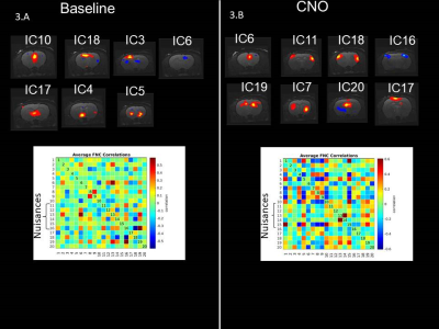 |
96 |
Preliminary evaluation of brain neural networks following
chemogenetically-induced activation of the motor cortex in rats
Mathias Lysholt Mathiasen1,
Sascha Gude1,
Henrik Lundell1,
Hartwig Roman Siebner1,2,3,
and Nathalie Just1
1DRCMR, Copenhagen University Hospital Amager and Hvidovre, Hvidovre, Denmark, 2Department of Neurology, Copenhagen University Hospital Bispebjerg and Frederiksberg, Copenhagen, Denmark, 3Department of Clinical Medicine, Faculty of Health and Medical Sciences, University of Copenhagen, Copenhagen, Denmark Keywords: Brain Connectivity, fMRI (resting state), Chemogenetics Genetic and viral tools coupled with functional magnetic resonance imaging (fMRI) techniques provide unique opportunities to investigate the functional neural networks in the rodent brain. Chemogenetic tools can be used to selectively activate or inhibit specific neural populations and their projections, and in combination with fMRI, can reveal the underlying mechanisms of brain activity. We present early resting-state and pharmacological fMRI results regarding CNO-mediated activation of the hM3Dq DREADD in the motor cortex of rats. |
|
3504.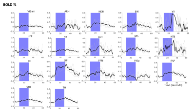 |
97 |
Functional relevance of cholinergic projections in the
ChAT-IRES-Cre mouse brain using optogenetic functional MRI
Thajunnisa Ashraf Sajitha1,
Russell W Chan1,
Patryk Filipiak2,
Muneeb Faiq1,
Royce P Lee1,
Steven H Baete2,
and Kevin C Chan1,3,4
1Department of Ophthalmology, NYU Langone Health, New York University Grossman School of Medicine, New York, NY, USA 10017, New York, NY, United States, 2Center for Advanced Imaging Innovation and Research (CAI2R), Department of Radiology, NYU Langone Health, New York University Grossman School of Medicine, New York, NY, USA 10017, New York, NY, United States, 3Department of Radiology, New York University Grossman School of Medicine, New York, NY, USA 10017, New York, NY, United States, 4Center for Neural Science, NYU College of Arts and Sciences, New York, NY, 10003, New York, NY, United States Keywords: Brain Connectivity, fMRI (task based) Recent studies have mapped the whole-brain cholinergic projections in the mouse brain. However, their functional relevance remains unclear. In this study, we used optogenetic functional MRI to observe the functional brain responses upon stimulating the cholinergic neurons in the basal forebrain. We observed significant BOLD percentage change in 14 out of 19 brain regions evaluated, indicating their functional relevance to the cholinergic system. |
|
3505.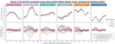 |
98 |
Associations between reading ability and white matter tract
integrity in adolescents
Pierre Nedelec1,
Samuel Lashof-Regas1,
Leo Sugrue1,
and Andreas Rauschecker1
1Radiology & Biomedical Imaging, University of California, San Francisco, San Francisco, CA, United States Keywords: Brain Connectivity, Adolescents Across a demographically diverse sample of over 7000 US adolescents, the relationship between white matter tracts and reading skills was evaluated using automated tractometry. Fractional Anisotropy (FA) values within several white matter tracts demonstrated strong positive associations with reading ability. Higher FA values were associated with higher reading scores, as assessed by the NIH Toolbox Oral Reading Recognition Test (TORRT). These relationships persisted even after correction for several potential confounding variables. |
|
3506. |
99 |
Age-dependent co-variation networks in myelin-related images
Nan-Hao Chen1,2,
Li-Ping Chen3,
Chia-Wei Hsu3,
Chin-Hua Yang1,3,4,
and Hsu-Hsia Peng1
1Department of Biomedical Engineering and Environmental Sciences, National Tsing Hua University, Hsinchu, Taiwan, 2Institute of Biomedical Engineering and Nanomedicine, National Health Research Institutes, Miaoli, Taiwan, 3Department of Medical Imaging, National Taiwan University Hospital Hsinchu Branch, Hsinchu, Taiwan, 4Department of Radiology, Taoyuan General Hospital, Taoyuan, Taiwan Keywords: Brain Connectivity, Aging, Myelin co-variation network Myelin co-variation networks was used to investigate the myelinic alteration in brain degradation. However, a systematic investigation of age-related co-variation networks of multiple myelin-related images remains deficient. We aimed to investigate the age-related effect on myelin co-variation networks in gray matter and white matter. Our results revealed that different myelin-related indices presented different age-dependent evolution of co-variation networks. The age-dependencies of strength of co-variation and topological attributes were mostly in complex polynomial pattern. The investigation of multiple age-related myelin co-variation networks might comprehend the synchronous changes between brain regions and demonstrated its usefulness in predicting the alteration of myelinic tissue. |
|
3507.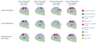 |
100 |
Action execution and observation network: the structural
connectome supporting variable force perception
Fulvia Palesi1,
Letizia Casiraghi2,
Giovanni Savini3,
Sonia Paternò1,
Egidio D'Angelo1,4,
and Claudia AM Gandini Wheeler-Kingshott1,4,5
1Dept. Brain and Behavioural Sciences, University of Pavia, Pavia, Italy, 2Dept. Mental Health and Dependence, ASST of Pavia, Pavia, Italy, 3Dept. Neuroradiology, IRCCS Humanitas Research Hospital, Milan, Italy, 4Brain Connectivity Center, IRCCS Mondino Foundation, Pavia, Italy, 5NMR Research Unit, Department of Neuroinflammation, Queen Square Multiple Sclerosis Centre, UCL Queen Square Institute of Neurology, University College London (UCL), London, United Kingdom Keywords: Brain Connectivity, Tractography & Fibre Modelling, Action execution observation network Inter-individual interaction is at the basis of social life making it crucial to characterize the functional networks subtending action execution (AE) and action observation (AO). We investigated the integrative properties of previously defined AE and AO functional networks in relation to force perception in terms of their underlying structural brain connectivity. We found that both networks show a strong connectivity between areas involved in sensory/force perception and in motor planning. The cerebellum was engaged both in executing the force-related task and in simulating motor responses, supporting the existence of specific brain networks involving the cerebellum in mirroring and mentalizing mechanisms. |
|
 Back to Meeting Home
 Back to the Program-at-a-Glance
The International Society for Magnetic Resonance in Medicine is accredited by the Accreditation Council for Continuing Medical Education to provide continuing medical education for physicians.
 Back to Meeting Home
 Back to the Program-at-a-Glance
 View Presentation Video