Digital Poster
Spinal Cord Imaging
ISMRM & ISMRT Annual Meeting & Exhibition • 03-08 June 2023 • Toronto, ON, Canada

| Computer # | |||
|---|---|---|---|
1545.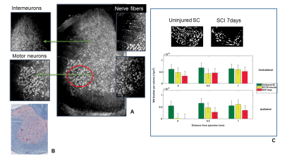 |
81 | Study of mouse models of lesion by using a multi technique approach.
Laura Maugeri1,2, Charles Nicaise3, Aleksandar Jankovski4,5, Emil Malucelli6, Mauro DiNuzzo2,7, Alessia Cedola8, Federico Giove2,7, and Michela Fratini2,8
1Institute of Nanotechnology Lecce Unit & Rome Unit, CNR, LECCE, Italy, 2Laboratory of Neurophysics and Neuroimaging (NaN), IRCCS Santa Lucia Foundation, Rome, Italy, 3URPhyM – NARILIS, Université de Namur, Namur, Belgium, 4Institute of NeuroScience (IoNS), NEUR division, Université catholique de Louvain (UCLouvain), Brussels, Belgium, 5Department of Neurosurgery, Université catholique de Louvain (UCLouvain), CHU UCL Namur, Yvoir, Belgium, 6Department of Pharmacy and Biotechnology, University of Bologna, Bologna, Italy, 7Museo Storico della Fisica e Centro Studi e Ricerche Enrico Fermi, Rome, Italy, 8Institute of Nanotechnology, CNR, ROME, Italy Keywords: Spinal Cord, Data Processing, SPINAL CORD INJURY Innovative biomarkers as well as new modalities to integrate structural information at higher level should be tuned up in order to understand the mechanisms underlying the pathology evolution. Here, we show some results obtained by integrating morphological information from X-ray phase contrast microtomography with histology combined with immunohistochemistry. Thanks to this approach, we demonstrated the possibility to use the cell number variation as a biomarker for pathological conditions. |
|
1546.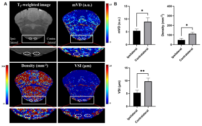 |
82 | In vivo MR vessel size imaging of brain vascular plasticity after experimental spinal cord injury
junchao qian1, ke zhou1, Chen Zhou2, yu tian3, Yanan Zhang2, Tianwei Song2, and Junchao Qian2
11.Institute of Health and Medical Technology, Hefei Institutes of Physical Science, Hefei Cancer Hospital, Chinese Academy of Sciences, Hefei 230031, P. R. China, Hefei, China, 2Institute of Health and Medical Technology, Hefei Institutes of Physical Science, Hefei Cancer Hospital, Chinese Academy of Sciences, Hefei, China, 31.Institute of Health and Medical Technology, Hefei Institutes of Physical Science, Hefei Cancer Hospital, Chinese Academy of Sciences, Hefei 230031, P. R. China, Hefei,China, China Keywords: Blood vessels, Blood vessels Spinal cord injury (SCI) leads to neuronal cell death, axonal damage and demyelination. Brain undergo anatomical changes following SCI. Recently MR vessel size imaging has shown promising application in visualizing neovascularization. In this study we explored the possibility to vascular morphology changes and angiogenesis in the regions along the cranial corticospinal tract (CST) in SCI using vessel size imaging. The results showed increased microvascular density (Density), mean vessel diameter (mVD) and vessel size index (VSI) values in contralateral pyramids four weeks post-injury compared to pre-injury levels. Thus, vessel size imaging could provide valuable information of neovascularization in brain after SCI. |
|
1547. |
83 | Contribution of diffusion MRI to neuropathic pain in the SNL rat model: correlation between gliosis and diffusion parameters
Sang-Jin Im1, Seokha Jin1, and HyungJoon Cho1
1Department of Biomedical Engineering, Ulsan National Institute of Science and Technology (UNIST), Ulsan, Korea, Republic of Keywords: Spinal Cord, Diffusion Tensor Imaging In this study, magnetic resonance imaging is used for anatomical and pathological studies of the spinal cord, investigating the mechanisms of neuralgia through diffusion magnetic resonance imaging techniques. However, analysis of ganglion changes in the spinal cord in multi-shell diffuse-weighted MRI signals remains challenging due to the ambiguous relationship between sympathetic and MRI signals. To study these relationships, we construct a neuropathic pain model using rats by performing in ex-vivo MRI experiments and neuropathic cell staining to provide insight into the treatment of neuropathic pain. |
|
1548.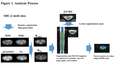 |
84 | Anisotropic and Isotropic Kurtosis Estimation of Spinal Cord Microstructure in Multiple Sclerosis and Neuromyelitis Optica Spectrum Disorder
Masaaki Hori1,2, Akifumi Hagiwara2, Kazumasa Yokoyama3, Issei Fukunaga4, Katsuhiro Sano2, Koji Kamagata2, Katsutoshi Murata5, Shohei Fujita2,6, Christina Andica7, Akihiko Wada2, Kouhei Kamiya1,2, Julien Cohen-Adad8, and Shigeki Aoki2,7
1Toho University Omori Medical Center, Tokyo, Japan, 2Radiology, Faculty of Medicine, Juntendo University, Tokyo, Japan, 3Neurology, Faculty of Medicine, Juntendo University, Tokyo, Japan, 4Radiological Technology, Faculty of Health Science, Juntendo University, Tokyo, Japan, 5Siemens Japan K.K., Tokyo, Japan, 6Radiology, The University of Tokyo, Tokyo, Japan, 7Faculty of Health Data Science, Juntendo University, Chiba, Japan, 8NeuroPoly Lab, Polytechnique Montreal, Montreal, QC, Canada Keywords: Spinal Cord, Spinal Cord We investigated the microstructural changes in the spinal cords of patients with multiple sclerosis (MS) and neuromyelitis optica spectrum disorder (NMOSD) using anisotropic kurtosis and isotropic kurtosis (Kiso) derived from 2-shell single diffusion encoding (SDE) MRI data with spherical mean techniques (SMT) and mean signal diffusion kurtosis imaging. There was a significant difference in Kiso between MS and NMOSD at the level of C4 (P=0.032, Mann-Whitney U test with Bonferroni correction). Therefore, Kiso derived from 2-shell SDE data might potentially be useful for evaluating the spinal cord microstructure in MS and NMOSD patients. |
|
1549.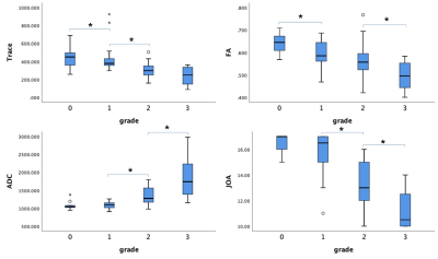 |
85 | Feasibility of MUSE DTI in differentiation of spinal cord injury severity in cervical spondylotic myelopathy
Haoyue Shao1, Xiangyu Tang1, Qiufeng liu1, and Weiyin Vivian Liu2
1Tongji Hospital Affiliated to Tongji Medical College of Huazhong University of Science and Technology, Wuhan, China, 2GE Healthcare, Beijing, China Keywords: Spinal Cord, Diffusion/other diffusion imaging techniques, cervical spondylotic myelopathy MUSE (Multiplexed Sensitivity-Encoding) is a novel diffusion weighted imaging with 2 to 3-excitations, phase acquisition step reduction, to achieve high-resolution diffusion imaging, higher signal-to-noise ratio, fewer motion artifacts and magnetic field inhomogeneities. In this study, we applied MUSE-based diffusion tensor imaging to investigate the compression-caused microstructure changes in the spinal cord of each subject. Our results suggested that MUSE-DTI computed parameters (Trace, FA, ADC) have dependable diagnostic values in detecting CSM (cervical spondylotic myelopathy) and that the ADC value is the best indicator of spinal cord compression. |
|
1550.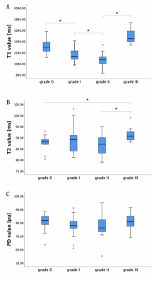 |
86 | Quantitative evaluation of the spinal cord compression in patients with cervical spondylotic myelopathy using synthetic MRI
Xiangyu Tang1, Haoyue Shao1, Weiyin Vivian Liu2, Qiufeng Liu1, and Wenzhen Zhu1
1Radiology department, Tongji hospital, Tongji medical college, Huazhong University of science and technology, Wuhan, China, 2MR Research, GE Healthcare, Beijing, China Keywords: Spinal Cord, Quantitative Imaging, Synthetic MRI, Cervical spondylotic myelopathy The present study for the first time applied synthetic MRI in diagnosis of patients with cervical spondylotic myelopathy. Multiple relaxation maps (T1, T2 and PD maps) and contrast-weighted images were obtained in a single scan of synthetic MRI. Our study demonstrated that T1 and T2 relaxation times of spinal cord at maximal compression level (MCL) changed with a grade dependent difference and T1MCL value could sensitively reflect the microstructural alteration of compressive spinal cord and even MCL at grade I, Moreover, T1MCL and T2MCL value was related to clinical scores and diameter values of the spinal cord at MCL. |
|
1551.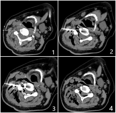 |
87 | CT-Guided Cervical Spinal Cord and Nerve Root Adhesiolysis for Intractable Hiccup:Based on the evidence of 3.0 T magnetic resonance neuroimaging
YANG MaoJiang1, Liu JianHao2, ANUP Bhetuwal1, QIONG Xian3, ZHANG HanWen1, YANG HanFeng1, Chen Meining4, and XU XiaoXue1
1Affiliated Hospital of North Sichuan Medical College, Nanchong 637000, Nanchong, China, 2The Fifth People's Hospital of Chengdu, Chengdu, China, 3Second Affiliated Hospital of North Sichuan Medical College,Nanchong 637100, Nanchong, China, 4MR Scientific Marketing, Siemens Healthcare, Shanghai, China, Shanghai, China Keywords: Head & Neck/ENT, Head & Neck/ENT The preoperative neuroimaging examination can clearly reflect the abnormalities of cervical intervertebral disc, cervical plexus and brachial plexus, help us understand the potential pathogenesis of intractable hiccup, and improve the efficacy. |
|
1552.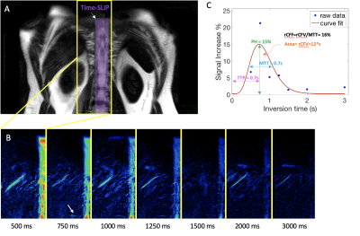 |
88 | Does spinal CSF perfuse out of the spine?
Diana Vucevic1, Vadim Malis1, Won Bae1, and Mitsue Miyazaki1
1Radiology, UCSD, San Diego, CA, United States Keywords: Spinal Cord, Visualization Cerebrospinal fluid (CSF) perfusion in the spinal canal is one of the greatest mysteries in the scientific community. CSF can be imaged without the use of gadolinium-based contrast agents (GBCA) using our novel spin-labelling MRI technique at 3 Tesla. Perfusion of CSF in the spine can be seen in the thoraco-lumber region from the nerve roots. This CSF outflow may be absorbed by the venous plexus along the dorsal nerve roots. The second pathway It may be from the nerve roots, recirculated to lymphatic tissue and into the thoracic duct. |
|
1553.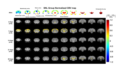 |
89 | Effects of neuropathic pain on cerebral hemodynamics in spinal nerve ligation model
SEOKHA JIN1 and Hyung Joon Cho1
1UNIST, Ulsan, Korea, Republic of Keywords: Neuroinflammation, Neuroinflammation, Neuropathic pain In this study, the effects of neuropathic pain on cerebral hemodynamics was investigated. For the neuropathic pain model, a spinal nerve ligation model was operated and validated by Von Frey test. To estimate the cerebral hemodynamics, DSC-MRI was performed up to 28 days after surgery. As a result, it is possible to specify when or where the neuropathic pain strongly affects cerebral perfusion. |
|
1554.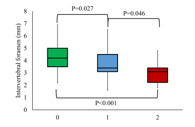 |
90 | Nodular change of the cervical nerve root on quantitative double-echo steady-state in cervical spondylosis
Keisuke Nitta1, Hajime Yokota2, Takayuki Sada1, Ryuuna Kurosawa1, Koji Matsumoto1, Takashi Namiki3, Masami Yoneyama3, Yoshitada Masuda1, and Takashi Uno2
1Radiology, Chiba University Hospital, Chiba-shi, Japan, 2Diagnostic Radiology and Radiation Oncology, Chiba University Hospital, Chiba-shi, Japan, 3Philips Japan, Tokyo, Japan Keywords: Spinal Cord, Neuro Evaluation of intervertebral foramen stenosis is essential in the diagnosis of cervical spondylosis. However, that is not easy because the intervertebral foramen is thin. We focused on the nodular change of the cervical nerve root, which is often visualized in patients with cervical spondylosis. We investigated the relationship between nodular changes and intervertebral foramen stenosis and confirmed the correlation between them. Thus, the identification of the nodular change may be helpful in the diagnosis of cervical spondylosis. |
|
1555.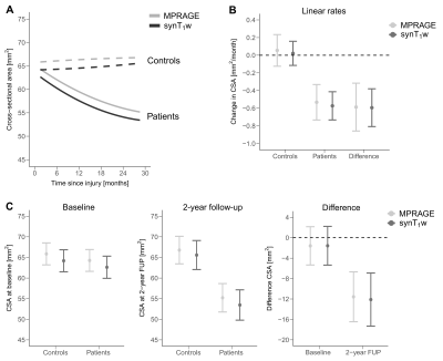 |
91 | Reliability of spinal cord measures based on Synthetic T1-weighted MRI derived from multi-parametric mapping (MPM)
Simon Schading1, Maryam Seif1,2, Tobias Leutritz2, Markus Hupp1, Armin Curt1, Nikolaus Weiskopf2,3, and Patrick Freund1,2,4
1Spinal Cord Injury Center, Balgrist University Hospital, Zürich, Switzerland, 2Department of Neurophysics, Max Planck Institute for Human Cognitive and Brain Sciences, Leipzig, Germany, 3Felix Bloch Institute for Solid State Physics, Leipzig University, Leipzig, Germany, 4Wellcome Trust Centre for Neuroimaging, UCL Queen Square Institute of Neurology, London, United Kingdom Keywords: Spinal Cord, Quantitative Imaging MRI acquisition time is a main factor for patient's comfort and motion artifacts. To avoid additional time consuming T1-w MPRAGE imaging for measuring spinal cord atrophy, when quantitative multi-parameter maps (MPM) are available, we reconstructed and validated a synthetic T1-w (synT1-w) based on MPMs. Test-retest repeatability was assessed and synT1-w images were compared with T1-w MPRAGE in a longitudinal study following spinal cord injury. Measures derived from synT1-w demonstrated high intra- and inter-site repeatability with only small difference when compared to MPRAGE. The estimated effects of spinal cord atrophy agreed with those obtained from MPRAGE. |
|
1556.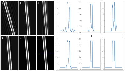 |
92 | Controlled Modeling of Cerebrospinal Fluid Flow Artifacts with a Simple Digital Spine Phantom
Daniel V Litwiller1, R Scott Hinks2, Ajeetkumar Gaddipati3, Trevor Kolupar3, and Suchandrima Banerjee4
1GE Healthcare, Denver, CO, United States, 2GE Healthcare, Albuquerque, NM, United States, 3GE Healthcare, Waukesha, WI, United States, 4GE Healthcare, Menlo Park, CA, United States Keywords: Spinal Cord, Artifacts Robust spine imaging with conventional fast spin echo remains a clinical challenge due to motion artifacts from multiple sources. Flow artifacts from cerebrospinal fluid can be especially problematic for assessments of the spinal cord itself. In addition, judging the effectiveness of various protocol modifications can be challenging in vivo due to the stochastic nature of the relationship between image timing and the cardiac cycle. In this work, we present results of a simple digital spine phantom that can be used in the controlled evaluation of strategies to mitigate artifacts from CSF flow. |
|
1557.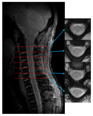 |
93 | High Resolution Spinal Cord Imaging at 7T with Rosette Trajectory and Compressed Sensing.
Sultan Zaman Mahmud1,2, Seyedeh Nasim Adnani1,2, Thomas S. Denney1,2, and Adil Bashir1,2
1Department of Electrical and Computer Engineering, Auburn University, Auburn, AL, United States, 2Auburn University MRI Research Center, Auburn University, Auburn, AL, United States Keywords: Spinal Cord, New Trajectories & Spatial Encoding Methods, Rosette Imaging, Compressed Sensing. MRI is very useful to investigate spinal cord pathologies non-invasively. However, signal to noise ratio (SNR), spatial resolution and motion artifacts are some of the main challenges in spinal cord MRI. Ultra high field MRI such as 7T, can improve the SNR enabling high spatial resolution. The inherent property of low susceptibility to motion of non-Cartesian imaging technique such as Rosette, can improve the motion related artifacts. So the goal of this study was to develop a technique for high-resolution spinal cord imaging at 7T using Rosette MRI. |
|
1558.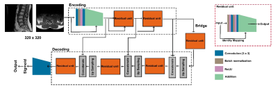 |
94 | An automatic tool for lumbar deformities and intervertebral disc degeneration using deep-learning based 3D segmentation
Yao-Wen Liang1, Chao-Hung Kuo2, Feng-Mao Chiu1, Ting-Chun Lin1, Yu-Hao Lan1, Chih-Yu Wang1, and You-Yin Chen1,3
1Department of Biomedical Engineering, National Yang Ming Chiao Tung University, Taipei, Taiwan, 2Department of Neurosurgery, Neurological Institute, Taipei Veterans General Hospital, Taipei, Taiwan, 3Ph.D. Program for Neural Regenerative Medicine, Taipei Medical University, Taipei, Taiwan Keywords: Spinal Cord, Machine Learning/Artificial Intelligence Degenerative alterations are the most common cause of lumbar spinal stenosis (LSS) in seniors. LSS is detected more often due to enhanced imaging technology and an aging population. Imaging data help individuals with chronic symptoms considering invasive therapy. This study combined the approach of assessing disc degeneration with stenosis assessment, using a deep learning-based segmentation module and a home-made autonomous measuring system. |
|
1559.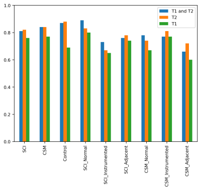 |
95 | Radiomic Analyses of the Instrumented Spinal Cord After Treatment for Degenerative Myelopathy and Acute Cord Injuries
Azadeh Sharafi1, Andrew Klein1, and Kevin M Koch1
1Radiology, Medical College of Wisconsin, Milwaukee, WI, United States Keywords: Spinal Cord, Radiomics Quantitative MRI of the spinal cord can provide insightful information about injured or degenerated cord tissue. However, the presence of instrumentation used treat such conditions can compromise the ability to perform such analyses. In this preliminary study, first and second-order radiomics features were computed across axial segmented spinal cord in subjects with 1) degenerative cervical myelopathy and 2) spinal cord injury patients treated with spinal fusion. Radiomics were computed using T1 and T2 weighted metal-artifact suppressed 3D-MSI and analyzed and successfully utilized to model local fusion status within the clinical cohorts and also against non-instrumented control subjects. |
|
1560. |
96 | Quantitative Analysis of Posterior Decompression in Chiari Malformation by Cine-PC MRI and 3D-T2w Scanning
Xinyu Wang1, Fengtan Li1, Xinli Wang1, Chen Zhang2, and Jianxun Qu3
1Tianjin Medical University General Hospital, Tianjin, China, 2MR Scientific Marketing, Siemens Healthcare, Beijing, China, 3MR Collaboration, Siemens Healthcare, Beijing, China Keywords: Spinal Cord, Spinal Cord In this study, the cine-phase contrast (cine-PC) sequence was used to compare the flow rate of cerebrospinal fluid in the midbrain aqueduct of patients with Chiari type I before and after posterior decompression and to analyze the difference in the ratio of cerebellar volume to posterior fossa volume between patients with Chiari type I and normal controls. Cine-PC magnetic resonance imaging and 3D-T2 scanning are shown to be effective for surgically evaluating patients undergoing posterior decompression surgery. |
|
1561.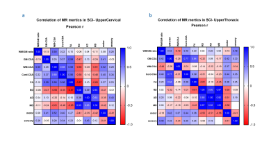 |
97 | Quantitative Magnetic Resonance Imaging to Quantify the Macro and Microstructural Changes in Normal and Pediatrics with Spinal Cord Injury
Shiva Shahrampour1, Mahdi Alizadeh1, Devon Middleton1, Benjamin De Leener2, Laura Krisa1, Adam E. Flanders3, Scott H. Faro3, Julien Cohen-Adad2, MaryJane Mulcahey1, and Feroze B. Mohamed1
1Thomas Jefferson University, Philadelphia, PA, United States, 2Polytechnique Montréal, Montreal, QC, Canada, 3Thomas Jefferson University Hospital, Philadelphia, PA, United States Keywords: Spinal Cord, Pediatric Spinal Cord Injury (SCI) in the pediatric population is relatively rare but carries significant psychological and physiological consequences. Advanced qMRI techniques has shown promising results to evaluate spinal cord integrity at a macro and microstructural level. |
|
1562.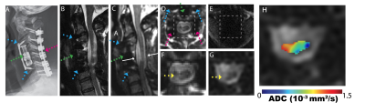 |
98 | A Cohort Study of Spinal Cord Diffusivity After Treatment of Cervical Spondylotic Myelopathy with Instrumented Spinal Fusion
Kevin Koch1 and Andrew S Nencka1
1Radiology, Medical College of Wisconsin, Milwaukee, WI, United States Keywords: Spinal Cord, Spinal Cord This study analyzed spinal cord diffusivity in CSM subjects treated with metallic spinal fusion decompression. Metal-artifact-suppressed diffusion-weighted MRI were collected on 38 CSM subjects and 25 controls. Diffusivity was analyzed as a function of cohort and level instrumentation (instrumented, non-instrumented, and adjacent segment). In addition, the impact of mJOA symptom scores and duration since the fusion procedure on diffusivity were modeled. Multi-linear mixed effects models accounted for demographic variations and multiple-measures (at different cord levels) within each subject. The results of the study identified consistent reductions in diffusivity due to the presence of fusion instrumentation. |
|
1563.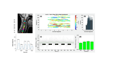 |
99 | Spinal cord fMRI to investigate the Relapsing-Remitting Multiple Sclerosis (RRMS) patients
Michela Fratini1,2, Lorenzo Giovannelli3, Laura Maugeri1,3, Mauro Di Nuzzo4, Marta Moraschi5, Maria Guidi4, Daniele Mascali4, Irene Egidi4, and Federico Giove3,4
1Nanotec, CNR, Roma, Italy, 2IRCSS Santa Lucia Foundation, Rome, Italy, 3IRCCS Santa Lucia Foundation, Roma, Italy, 4CREF, Roma, Italy, 5Radiation Oncology, Campus Bio-Medico University of Rome, Roma, Italy Keywords: Spinal Cord, fMRI (task based), BOLD signal We have studied Relapsing-Remitting Multiple Sclerosis (RRMS) patients group with spinal cord fMRI during a multilevel force grip motor task. We demostrated the possibility to use spinal cord fMRI as a biomarker for clinical application. |
|
1564.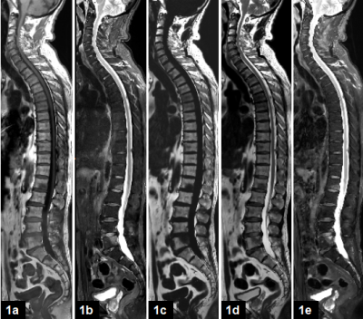 |
100 | Application of Water-Fat Imaging in Spinal Diseases: comparison with conventional T1-weighted and T2-weighted STIR Images
Nan Wang1, Guobin Li2, Shuheng Zhang2, Dan Yu3, Yongming Dai3, and Qingwei Song1
1the First Affiliated Hospital of Dalian Medical University, Dalian, China, 2Shanghai United Imaging Healthcare Co., Ltd, Shanghai, China, 3MR Collaboration, Central Research Institute, United Imaging Healthcare, Shanghai, China Keywords: Cancer, Whole Body Water-Fat Imaging (WFI) based on chemical shifts can provide excellent separation of water and fat in tissue. This work investigated the application of WFI to spinal diseases. The diagnosis performances were compared with the T1-weighted (T1W) and T2-weighted short-TI-inversion-recovery (T2W STIR) sequence. For hyperosteogeny, spinal metastasis, and disc herniation, the WFI showed comparable performance as the combination of T1W and T2W STIR imaging within a much shorter acquisition time. |
|
 Back to Meeting Home
 Back to the Program-at-a-Glance
The International Society for Magnetic Resonance in Medicine is accredited by the Accreditation Council for Continuing Medical Education to provide continuing medical education for physicians.
 Back to Meeting Home
 Back to the Program-at-a-Glance
 View Presentation Video