Digital Poster
Imaging Nerves, Brain Injury & Brain Dysfunction
ISMRM & ISMRT Annual Meeting & Exhibition • 03-08 June 2023 • Toronto, ON, Canada

Digital Poster
Imaging Nerves, Brain Injury & Brain Dysfunction
| Computer # | |||
|---|---|---|---|
1703.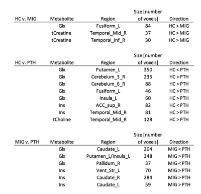 |
61 | A Novel Approach to Metabolic Imaging of Migraine and Post Traumatic Headache Using Full Brain 3D-MRSI
Simona Nikolova1, Todd Schwedt1, Yuxiang Zhou2, Brian Chong2, Gina Dumkrieger1, and Catherine Chong1
1Neurology, Mayo Clinic, Phoenix, AZ, United States, 2Radiology, Mayo Clinic, Phoenix, AZ, United States Keywords: Traumatic brain injury, Spectroscopy, migraine, 3D-MRSI, PTH Magnetic resonance spectroscopy provides a means to quantify brain metabolites. Metabolites of interest are N-acetyl aspartate (NAA, neuronal marker), Choline (Cho, marker of cell membrane turnover), Myoinositol (Ins, astrocyte marker), Glutamate+Glutamine (Glx, neurotransmitters). Studies demonstrate metabolic changes associated with migraine such as increased Glx levels (signifying excitotoxicity), decreased NAA (suggestive of neuronal damage), as well as abnormal GABA in pain processing areas. The main goal of this work was to use using a novel, whole-brain three-dimensional magnetic resonance spectroscopy imaging (MRSI) technique to collect preliminary data on metabolic differences between migraine, post traumatic headache (PTH), and healthy controls. |
|
1704.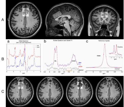 |
62 | Assessment of Glx and GABA levels in primary dysmenorrhea patients in menstruation and periovulatory phases
Xue Chen1, Zhou Huang1, Peng Wu2, and Yonggang Li1,3,4
1Department of Radiology, The First Affiliated Hospital of Soochow University, Suzhou, China, 2Philips Healthcare, Shanghai, China, 3Institute of Medical Imaging, Soochow University, Suzhou, China, 4National Clinical Research Center for Hematologic Diseases, The First Affiliated Hospital of Soochow University, Suzhou, China Keywords: Nerves, Brain The pathogenesis of primary dysmenorrhea (PDM) and the central nervous system (CNS) mechanisms leading to poorer mode and pain sensitization remain totally unclear, which was explored in our study using MEGA-PRESS. PDM patients showed significantly higher Glx level and mildly higher GABA+ level (not significantly) in ACC in menstruation phase. In menstruation phase, the SDS/PCS scores of the PDM patients had a positive correlation with ACC GABA+ levels. The imbalances in ACC GABA+/Glx levels in PDM patients in menstruation phase may be the mechanism mediating depressive symptoms and pain catastrophe.
|
|
1705.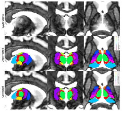 |
63 | Multi-parametric characterization of thalamic nuclei based on manual delineation using the Synthetic MPRAGE images
Alexa Gail Colinco1, Rosy Linda Njonkou Tchoquessi1, Steven Roys1, Li Jiang1, Prashant Raghavan1, Jerry Prince2, Rao P. Gullapalli1, and Jiachen Zhuo1
1Department of Diagnostic Radiology and Nuclear Medicine, University of Maryland School of Medicine, Baltimore, MD, United States, 2Department of Electrical and Computer Engineering, Johns Hopkins University, Baltimore, MD, United States Keywords: Traumatic brain injury, Tissue Characterization The accurate delineation of thalamic nuclei is important in understanding the underlying pathophysiology associated with neurological disorders and in targeting specific thalamic nuclei in image guided interventions. However, sub-parcellation of the thalamic nuclei is challenging given the lack of contrast within the thalamus provided by conventional T1 or T2 images. In this study, we use synthesized MPRAGE images with different inversion time (TI) (SynTI) to accurately parcellate thalamic nuclei, as outlined by the Morel atlas. Additionally, we examined the distinct magnetic susceptibility and diffusion properties of individual nuclei, which can be used to further improve thalamic nuclei segmentation. |
|
1706.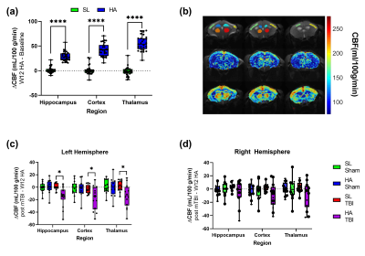 |
64 | A longitudinal neuroimaging study in mice on the effects of chronic high altitude exposure and repetitive traumatic brain injury
Alexandru Korotcov1,2, Caroline A Browne2,3, Asamoah Bosomtwi1,4, Shalini Jaiswal1,2, Nathan Cramer2,5,6, Xiufen Xu2,6, Kathleen Whiting2,7, Cheryl D Stimpson2,8, Bernard J Dardzinski1,9, Daniel Perl8, Dara L Dickstein2,8, and Zygmunt Galdzicki6
1Department of Radiology and Radiological Sciences, Uniformed Services University, Bethesda, MD, United States, 2The Henry M. Jackson Foundation for the Advancement of Military Medicine, Bethesda, MD, United States, 3Department of Pharmacology and Molecular Therapeutics, Uniformed Service University, Bethesda, MD, United States, 4Georgia Cancer Center, Augusta University, Augusta, GA, United States, 5Department of Anatomy and Neurobiology, University of Maryland School of Medicine, Baltimore, MD, United States, 6Department of Anatomy, Physiology and Genetics, Uniformed Service University, Bethesda, MD, United States, 7Graduate Program in Neuroscience, Uniformed Service University, Bethesda, MD, United States, 8Department of Pathology, Uniformed Service University, Bethesda, MD, United States, 9National Institute of Arthritis and Musculoskeletal and Skin Diseases, NIH, Bethesda, MD, United States Keywords: Traumatic brain injury, Preclinical, hypobaria, hypoxia, DTI, PET/CT, cerebral blood flow Chronic exposure to high altitude (HA) can lead to maladaptive physiological and pathological changes, resulting in an increased risk for neurological impairment. Whether the occurrence of mild traumatic brain injury (mTBI) at HA exacerbates neurobehavioral deficits remains uncertain. A series of experiments was conducted to test the impact of mTBI in the context of HA using established models of repetitive close head injury and simulated HA (5000m). A longitudinal study design was applied to identify the effects of chronic HA exposure (12 weeks) and mTBI in mice on neurobehavioral and neuroimaging biomarkers using PET and MRI. |
|
1707. |
65 | Feasibility of diffusion tensor imaging of the brachial plexus using iZOOM
Takayuki Sada1, Hajime Yokota2, Ryuna Kurosawa1, Takafumi Yoda1, Keisuke Nitta1, Koji Matsumoto1, Takashi Namiki3, Masami Yoneyama3, Adam Wu4, Yoshitada Masuda5, and Takashi Uno2
1Department of Radiology, Chiba University Hospital, Chiba, Japan, 2Diagnostic Radiology and Radiation Oncology, Graduate School of Medicine, Chiba University, Chiba, Japan, 3Philips Japan, Tokyo, Japan, 4Philips Healthcare (Shanghai) Ltd., Shanghai, China, 5Chiba University Hospital, Chiba, Japan Keywords: Nerves, Diffusion Tensor Imaging The brachial plexus is difficult to obtain diffusion tensor image (DTI) with good image quality due to the inhomogeneity of the magnetic field and motion artifacts. Zoom imaging based on 2D RF (iZoom) could both reduce the distortion dramatically and has better fat suppression effects. In this study, we compared DTI using conventional Zoom with that using iZoom for the brachial plexus and diffusion tractography with iZoom created higher mean length and fiber count than the conventional Zoom sequence, suggesting its feasibility as a new method for diffusion tensor imaging for the brachial plexus. |
|
1708.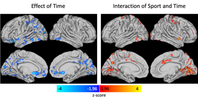 |
66 | The effects of repetitive head impact exposure on cerebrovascular reactivity in middle and high-school athletes
Alexander D. Cohen1, Benjamin L. Brett2, Milan D. Patel1, Kelly D. Ristow1, Michael A. McCrea2, and Yang Wang1
1Radiology, Medical College of Wisconsin, Milwaukee, WI, United States, 2Neurosurgery, Medical College of Wisconsin, Milwaukee, WI, United States Keywords: Traumatic brain injury, fMRI, Cerebrovascular reactivity, Breath hold Cerebrovascular reactivity (CVR) can provide a marker of vascular injury. The effects of repetitive head impact exposure (RHIE) during contact sports remains to be fully elucidated. We investigated the effect of sport, time, and their interaction on CVR in contact sport (CS) and non-contact sport (NCS) middle school and high school athletes. There was a significant effect of time and a significant interaction effect between contact group and time on CVR with CVR decreasing pre to post season in the NCS group but remaining unchanged in the CS group. |
|
1709.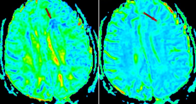 |
67 | Diagnostic value of combined DTI and DKI imaging for diffuse axonal injury of brain
Ke-ning XU1 and Wenjia Wang2
1Zhangjiakou First Hospital, Zhangjiakou Hebei, China, 2GE Healthcare, Beijing, China Keywords: Traumatic brain injury, Brain The values of fractional anisotropy (FA) in diffusion tensor imaging (DTI) and the mean kurtosis (MK) and mean diffusivity(MD) values in diffusion kurtosis imaging (DKI) were used to analyze diffuse axonal injury of brain Injury (DAI). We explored the diagnostic value of these three parameters in DAI and found that MK was superior to FA and MD. DTI and DKI could quantitatively analyze the severity of DAI patients and reduce the misdiagnosis rate of DAI.
|
|
1710.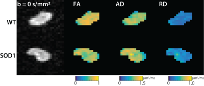 |
68 | Validation of Diffusion MRI Biomarkers of Peripheral Nerve Degeneration in ALS
Margaret M. McCann1,2, Ethan Mathew1,3, Alberto Fuentes1, Samuel G. Ferrante1, Charles Quarles4, Richard D. Dortch1, and Gregory Turner1
1Translational Neuroscience, Barrow Neurological Institute, Phoenix, AZ, United States, 2Biomedical Sciences, Creighton University, Omaha, NE, United States, 3School of Biological and Health System Engineering, Arizona State University, Phoenix, AZ, United States, 4Cancer Systems Imaging, University of Texas MD Anderson Cancer Center, Houston, TX, United States Keywords: Nerves, Diffusion Tensor Imaging ALS is a fatal, neurodegenerative disease that affects motor neurons. Assessment of disease progression is difficult and relies on tests that lack specificity/responsiveness. While imaging biomarkers may meet this need, previous work has focused on the motor neurons or skeletal muscles, with nerves that form the link between these tissues being largely understudied. Previous studies suggest DTI in human nerves may be sensitive to nerve degeneration in ALS and here we sought to validate these measures in a rat model. Preliminary findings showed a progressive decrease in FA, which correlated to the onset of motor neuron loss and muscle wasting. |
|
1711.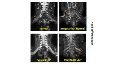 |
69 | Differentiation of brachial plexus caliber irregularities with simultaneous neurography and T2 mapping from MIXTURE
Hajime Yokota1, Takayuki Sada2, Ryuna Kurosawa2, Takafumi Yoda2, Koji Matsumoto2, Takashi Namiki3, Masami Yoneyama3, and Takashi Uno1
1Department of Diagnostic Radiology and Radiation Oncology, Chiba Univerisity, Chiba, Japan, 2Department of Radiology, Chiba Univerisity Hospital, Chiba, Japan, 3Philips Japan, Tokyo, Japan Keywords: Nerves, Neurography Multifocal chronic inflammatory demyelinating polyneuropathy (CIDP) is characterized by asymmetrical and irregular nerve thickening. However, the nerves are sometimes irregular in caliber even without obvious neuropathy. MIXTURE can simultaneously acquire neurography and T2 mapping. The diameter and T2 values from MIXTURE were significantly wider and higher in multifocal CIDP than in cases with normal but irregular caliber (P = 0.029 and 0.004). The AUCs of ROC analyses were 0.889 and 0.972 in diameter and T2 values for differentiating the two groups. Diameter and T2 values acquired with MIXTURE help differentiate whether caliber irregularities in the brachial plexus are pathological. |
|
1712.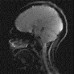 |
70 | Assessment of dynamic shear strain in the brain caused by mechanical transients using multi-scale MR elastography
Yuan Le1, Jun Chen1, Yi Sui1, Phillip J. Rossman1, Armando Manduca1, Kevin J. Glaser1, John Huston III1, Richard L. Ehman1, and Ziying Yin1
1Radiology, Mayo Clinic, Rochester, MN, United States Keywords: Traumatic brain injury, Traumatic brain injury, Motion encoding gradient, transient motion To study how the human brain moves and deforms under a sub-injury level transient impact, we tested the feasibility of measuring in-vivo brain transient motion resulting from a small amplitude impact using the multi-scale MR Elastography technique. Transient motion was encoded by wavelet-based motion encoding gradients at two wavelet scales plus a scaling function, and displacement was calculated using the inverse Haar transform. Brain motion over a wide frequency range was detected and was found to be consistent with previous results from a much higher impact. |
|
1713.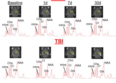 |
71 | Characterization of a controlled cortical impact brain injury and metabolic changes using magnetic resonance spectroscopy
Asamoah Bosomtwi1, Roxan Ara2, Fengchong Kong2, Yujiao Lu3, and Krishnan M DHANDAPANI3
1Georgia medical College, Augusta, GA, United States, 2Georgia Cancer Center, Augusta University, Augusta, GA, United States, 3Neurosurgery, Augusta University, Augusta, GA, United States Keywords: Traumatic brain injury, Spectroscopy Traumatic brain injury (TBI) diagnosis and treatment is a top priority for military medicine. Several preclinical models of TBI have been developed to help elucidate the etiology of brain injury but these models typically depend on behavioral outcomes to indicate injury severity. In this project we used magnetic resonance spectroscopy (MRS) to investigate a noninvasive, longitudinal technique to detect changes in brain metabolites after TBI
|
|
1714.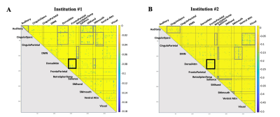 |
72 | Altered resting state functional connectivity in high-contact sports
Mahta Karimpoor1, Marios Georgiadis1, Brian Mills1, Hossein Moein Taghavi1, Narvin Phouksouvath1, Maged Goubran2, Nicole Mouchawar1, Sohrab Sami1, Max Wintermark1, Charles Liu3, John Van Horn4, Gerald Grant5, David Camarillo6, and Michael Zeineh1
1Radiology, Stanford University, Stanford, CA, United States, 2Medical Biophysics, University of Toronto, Toronto, ON, Canada, 3Neurological Surgery, University of Southern California, Los Angeles, CA, United States, 4Psychology, University of Virginia, Charlottesville, VA, United States, 5Neurosurgery, Stanford University, Stanford, CA, United States, 6Bioengineering, Stanford University, Stanford, CA, United States Keywords: Traumatic brain injury, Brain Connectivity, high-contact sports, resting state functional connectivity Repetitive head impact exposure during contact sports may increase risk of cognitive decline and neurodegenerative disease. We compared resting state functional connectivity (rsFC) networks between a group of high-contact collegiate PAC-12 athletes and a low-contact group from two institutions (groups comprised athletes of multiple sports and both sexes). A community chi-squared analysis evaluated differences in connectivity within/between 12 brain networks comparing high- vs. low-contact sports. Across both institutions, rsFC in the high-contact cohort was significantly increased (hyperconnectivity) between dorsal attention and default mode network (DMN), and significantly decreased (hypoconnectivity) between dorsal attention and frontoparietal networks, compared to the low-contact cohort. |
|
1715.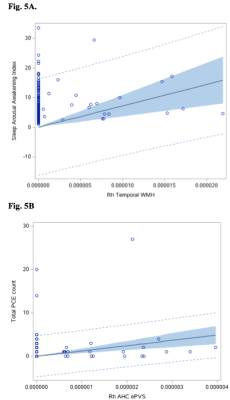 |
73 | Association of brain macrostructural changes and objective sleep measures in military service members after a remote mild traumatic brain injury
Ping-Hong Yeh1, J. Kent Werner2,3, Rujirutana Srikanchana1, Kimbra Kenney1,2, Chihwa Song1, Treven Pickett1,2, Grant Bonavia1,2, and John Ollinger1,2
1National Intrepid Center of Excellence,Walter Reed National Military Medical Center, Bethesda, MD, United States, 2Uniformed Services University of the Health Sciences, Bethesda, MD, United States, 3Walter Reed National Military Medical Center, Bethesda, MD, United States Keywords: Traumatic brain injury, Traumatic brain injury In this study, we quantitated the size of white matter hyperintense (WMH), enlarged perivascular space (ePVS) and diffusion tensor imaging analysis along the perivascular space (DTI_ALPS) index in service members after a remote mild traumatic brain injury (mTBI), and associated them with objective sleep measures. There was no significant difference of the regional size of WMH, ePVS or hemispheric DTI_ALPS index between controls and mTBI. In mTBI participants, lifetime TBI/PCE events was associated amygdala-hippocampal complex ePVS load; and sleep arousal / awakening index was associated with temporal WMH load, periodic limb movement during sleep with basal ganglia WMH load. |
|
1716.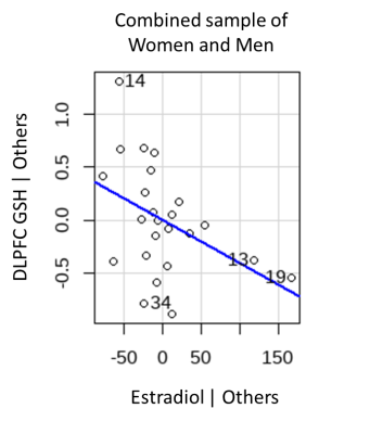 |
74 | Relationships of oxidative stress markers in the brain with circulating sex hormones
Jessica Busler1, Sarah Rose Slate2, Monica Foneska2, Huijun Liao2, Stanley Lyndon1, Jacob Taylor1, Alexander Lin1, and Pamela Mahon1
1Brigham and Women's Hospital/Harvard Medical School, Boston, MA, United States, 2Brigham and Women's Hospital, Boston, MA, United States Keywords: Psychiatric Disorders, Spectroscopy, Oxidative Stress Oxidative stress is implicated in multiple psychiatric and neurodegenerative conditions, differs by biological sex and covaries with circulating estrogens. However, limited knowledge exists on the association of brain markers of oxidative stress via glutathione (GSH) with circulating sex hormones. Therefore, we conducted brain magnetic resonance spectroscopy, assayed blood serum for circulating sex hormones, and measured brain GSH-sex hormones associations. We found that dorsolateral prefrontal cortex GSH relates to estradiol in women and men and anterior cingulate cortex GSH relates to estradiol, estrone, and total testosterone in women. This study highlights the importance of considering sex in GSH MRS studies. |
|
1717.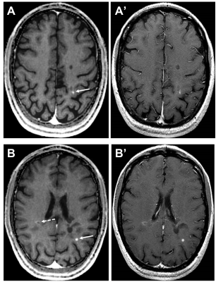 |
75 | Pathological contrast enhancement in different brain diseases in synthetic T1-weigthed images derived from 3D quantitative transient imaging
Graziella Donatelli1,2, Gianmichele Migaleddu1, Matteo Cencini3, Paolo Cecchi1,2, Luca Peretti3,4, Claudio D'Amelio5, Guido Buonincontri3, Michela Tosetti2,3, Mirco Cosottini5, and Mauro Costagli3,6
1Neuroradiology Unit, Azienda Ospedaliero-Universitaria Pisana, Pisa, Italy, 2IMAGO 7 Research Foundation, Pisa, Italy, 3Laboratory of Medical Physics and Magnetic Resonance, IRCCS Stella Maris, Pisa, Italy, 4University of Pisa, Pisa, Italy, 5Neuroradiology Unit, University of Pisa, Pisa, Italy, 6DINOGMI, University of Genoa, Genoa, Italy Keywords: Head & Neck/ENT, Contrast Agent Contrast enhancement, a marker of blood-brain barrier breakdown and active inflammation, provides crucial information in brain disease. Quantitative Transient Imaging (QTI) enables robust quantitative T1, T2 and PD mapping. 30 patients with brain tumors, multiple sclerosis and limbic encephalitis underwent a 3T-MRI brain exam which included conventional T1-weighted and QTI sequences acquired before and after contrast media administration. Synthetic T1-weighted images were obtained from the QTI maps. At radiological inspection, all pathological contrast enhancements in conventional images were visible in the synthetic T1-weighted images obtained from postcontrast QTI and showed the same patterns of contrast enhancement. |
|
| 1718. | 76 | Micro Fiber Disruption and Thalamus GABA Level Reduction in Lifelong Premature Ejaculation
Jiaming Lu1, Xin Zhang1, and Bing Zhang1
1The Affiliated Drum Tower Hospital, Medical School of Nanjing University, Nanjing, China Keywords: Head & Neck/ENT, Metabolism In this study, we using non invasion GABA-edited MEGA-PRESS MRS and NODDI model to measure the GABA content in bilateral thalamus and the water diffusion pattern in premature ejaculation patient. The PE patients have more dispersed neurites in the right putamen, more hindered water, and less space within axons in the orbitofrontal cortex. The bilateral thalamus GABA content in PE group is significantly lower than NC group. These findings may help us have a better neuroimage upstanding of PE from micro-structure to metabolism level. |
|
1719.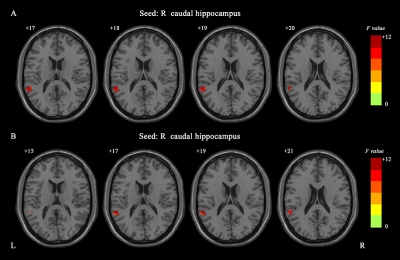 |
77 | Interaction Between ACTH and Hippocampal Dynamic Functional Connectivity onPersonality Traits in Bipolar Disorder with Suicidal Attempt
Pan Chen1, Ying Wang1, Shuming Zhong2, Guanmao Chen1, and Long Qian3
1Medical Imaging Center, First Affiliated Hospital of Jinan University, Guangzhou, China, 2MR Research, GE Healthcare, Guangzhou, China, 3MR Research, GE Healthcare, Beijing, China Keywords: Neuroinflammation, Brain Connectivity The goal of this study was to investigate the underlying mechanisms of suicidal behavior by detecting the dynamic functional connectivity (dFC) variability of hippocampus and HPA axis activity, as well as their relationship with personality traits in BD with suicidal attempt (SA). We assessed the activity of the HPA axis by measuring morning plasma adrenocorticotropic hormone (ACTH) and cortisol (CORT) levels. All participants underwent personality assessment using Minnesota Multiphasic Personality Inventory-2. BD with SA exhibited increased dFC variability of the hippocampal-temporal cortex and less HPA axis hyperactivity, which may both together lead to the possibility of changes in personality traits. |
|
1720.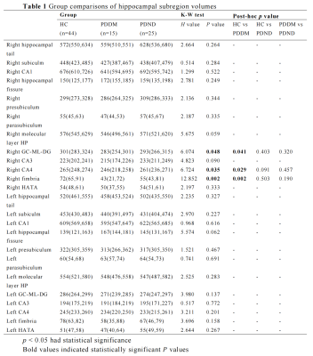 |
78 | Atrophy patterns of hippocampal subregions based on automated volumetry in Parkinson's disease with type 2 diabetes mellitus patients
Mingrui Qu1, Bingbing Gao1, Yuhan Jiang1, Yuan Li1, Lizhi Xie2, and Yanwei Miao1
1The First Affiliated Hospital of Dalian Medical University, Dalian, China, 2GE Healthcare, Shanghai, China Keywords: Nerves, Diabetes The objective of this study was to evaluate the atrophy pattern of the hippocampus subregions in Parkinson's disease with type 2 diabetes mellitus patients (PDDM) patients based on the 3DTI sequence and FreeSurfer software. The FreeSurfer provides extensive and automated neuroimaging analysis and has been successfully used to precisely segment the hippocampus. In this study, automated volumetry of hippocampal subfields in PDDM patients was performed, compared with Parkinson's disease without diabetes mellitus (PDND) patients and healthy controls . The results showed that PDDM patients have hippocampal subregions atrophy, especially in the right fimbria, right GC-ML-DG and right CA4. |
|
 Back to Meeting Home
 Back to the Program-at-a-Glance
The International Society for Magnetic Resonance in Medicine is accredited by the Accreditation Council for Continuing Medical Education to provide continuing medical education for physicians.
 Back to Meeting Home
 Back to the Program-at-a-Glance
 View Presentation Video