Digital Poster
Neurodegeneration II
ISMRM & ISMRT Annual Meeting & Exhibition • 03-08 June 2023 • Toronto, ON, Canada

| Computer # | |||
|---|---|---|---|
1721.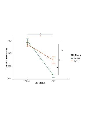 |
81 | Interaction of Alzheimer’s Disease Status and History of Traumatic Brain Injury on Measures of Cortical Thickness
Gina D'Souza1,2, Nathan W. Churchill2,3,4, Dylan X. Guan5, Marc A. Khoury1,2, Corinne E. Fischer1,2,6, and Tom A. Schweizer1,2,3,7,8
1Institute of Medical Science, University of Toronto, Toronto, ON, Canada, 2Keenan Centre for Biomedical Research, Li Ka Shing Knowledge Institute, St Michael's Hospital, Toronto, ON, Canada, 3Neuroscience Research Program, St Michael's Hospital, Toronto, ON, Canada, 4Physics Department, Toronto Metropolitan University, Toronto, ON, Canada, 5Cumming School of Medicine, University of Calgary, Calgary, AB, Canada, 6Department of Psychiatry, Faculty of Medicine, University of Toronto, Toronto, ON, Canada, 7Division of Neurosurgery, Department of Surgery, Faculty of Medicine, University of Toronto, Toronto, ON, Canada, 8The Institute of Biomaterials & Biomedical Engineering, University of Toronto, Toronto, ON, Canada Keywords: Alzheimer's Disease, Traumatic brain injury Traumatic Brain Injury (TBI) is associated with an accelerated course of dementia, although biological relationships are still incompletely understood. We characterized the differences in cortical thickness, for Alzheimer’s Disease (AD) patients with and without a history of TBI. Among individuals diagnosed with AD, a history of TBI was associated with a smaller decrease in cortical thickness in frontal-temporal regions, relative to their non-AD counterparts, with analyses controlling for the effects of age, sex, and education. TBI may lower the susceptibility threshold for cognitive decline related to AD by decreasing an individuals’ ability to cope with aging and/or AD pathology. |
|
1722.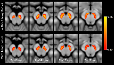 |
82 | Nigral volume as a marker for Lewy body pathology in Alzheimer’s disease
Jason Langley1 and Xiaoping Hu1,2
1Center for Advanced Neuroimaging, University of California Riverside, Riverside, CA, United States, 2Department of Bioengineering, University of California Riverside, Riverside, CA, United States Keywords: Alzheimer's Disease, Dementia, lewy body Up to 60% of Alzheimer's disease cases have Lewy body pathology. In Alzheimer's disease, Lewy body pathology is associated with more rapid cognitive decline, parkinsonism, and younger symptom onset. In this abstract, we examine nigral volume in Alzheimer's disease patients and patients with mild cognitive impairment with Lewy body pathology. We find that cognitively impaired patients with Lewy bodies have greater neuronal loss in substantia nigra pars compacta as well as reduced nigral volume. |
|
1723.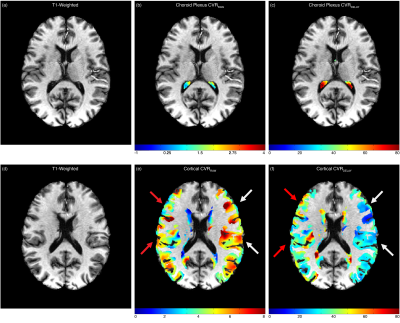 |
83 | Vascular reactivity of the choroid plexus in non-atherosclerotic vasculopathy: choroid plexus activity as a possible marker of ischemic stress
Caleb J. Han1, Spencer Waddle1, Maria Garza1, L. Taylor Davis2, Jarrod Eisma1, Rohan Chitale3, Matthew Fusco3, Colin D. McKnight2, Sky Jones1, Lori C. Jordan1,4, and Manus J. Donahue1,2,5
1Neurology, Vanderbilt University Medical Center, Nashville, TN, United States, 2Radiology, Vanderbilt University Medical Center, Nashville, TN, United States, 3Neurosurgery, Vanderbilt University Medical Center, Nashville, TN, United States, 4Pediatrics, Vanderbilt University Medical Center, Nashville, TN, United States, 5Psychiatry, Vanderbilt University Medical Center, Nashville, TN, United States Keywords: Neurofluids, Stroke, Moyamoya, glymphatic, choroid plexus, cerebrovascular reactivity, CSF This work applies hypercapnic reactivity and deep-learning techniques to evaluate choroid plexus (ChP) vascular compliance dependence on large arterial patency in intracranial vasculopathy. ChP reactivity was found to be preserved regardless of macrovascular vasculopathy, despite dependencies of resting ChP perfusion on cortical ischemia. Findings support the possibility that changes in resting ChP function in other studies in the presence of arterial vasculopathy and cerebral ischemia may be a response to circulating biochemical markers of ischemic stress, prompting the ChP to attenuate CSF production levels through feedback, rather than vascular mechanisms, to support glial health in ischemia. |
|
1724.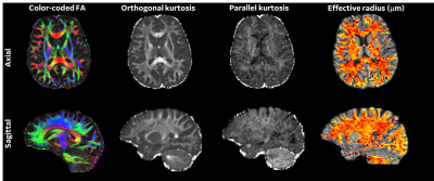 |
84 | White Matter Alterations Evident with DTI, DKI and Axonal Radius Measurements in Clinical Presentation of Chronic Symptomatic mTBI
Nastaren Abad1, Chitresh Bhushan1, Luca Marinelli1, Afis Ajala1, H. Doug Morris2, Ante Zhu1, Eric Fiveland1, Seung-Kyun Lee1, J Kevin DeMarco2,3, Robert Shih2,3, Maureen Hood2,3, Gail Kohls3, Kimbra Kenney2, Vincent Ho2, and Thomas K.F Foo1,2
1GE Research, Niskayuna, NY, United States, 2Uniformed Services University of the Health Sciences, Bethesda, MD, United States, 3Walter Reed National Military Medical Center, Bethesda, MD, United States Keywords: Traumatic brain injury, Diffusion/other diffusion imaging techniques Diffusion MRI based microstructural evaluation of DTI, DKI, and intra-axonal radii, enabled by the ultra-high performance MAGNUS gradients, was leveraged in this study to assess differences between healthy controls and chronic mild traumatic brain injury presentations. Parcel-wise group and brain WM asymmetry analysis highlighted specific sub-region involvement differing from healthy controls in evaluated metrics. Subject specific analysis highlighted specific anatomical regions that could be more susceptible in TBI with the effect size potentially masked with central tendency analysis. |
|
1725.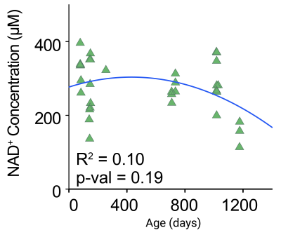 |
85 | Downfield 1H MRS can quantify age and hypoxia induced changes in brain NAD+
Emine Can1, Baby Martin-McNulty1, Ganesh Kolumam1, and Johannes Riegler1
1Calico Life Sciences LLC, South San Francisco, CA, United States Keywords: Neurodegeneration, Aging, Lifespan, Stroke, metabolism, Spectroscopy, Brain NAD+ is required for vital cellular processes such as redox regulation, DNA damage repair and cell signaling. There has been a growing interest in modifying NAD+ concentrations in the hope that it might increase longevity. However, there is no clear consensus on changes in NAD+ concentration due to healthy aging. Commonly used in vitro assays for NAD quantification are not suitable for longitudinal studies. We therefore implemented an in vivo downfield 1H MRS method to characterize NAD+ concentrations during aging in mice and showed that 1H MRS can detect hypoxia induced changes in brain NAD+ concentrations. |
|
1726.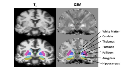 |
86 | Elevated Magnetic Susceptibility Suggests Distinct Neurodegenerative Pathology in World Trade Center First Responders
Thomas Hagan1, Jia Ying1, Chuan Huang1,2, Minos Kritikos3, Sean Clouston3, and Benjamin Luft4
1Department of Biomedical Engineering, Stony Brook University, Stony Brook, NY, United States, 2Department of Radiology, Stony Brook Medicine, Stony Brook, NY, United States, 3Program in Public Health and Department of Family, Population, and Preventative Medicine, Stony Brook Medicine, Stony Brook, NY, United States, 4Department of Medicine, Stony Brook Medicine, Stony Brook, NY, United States Keywords: Neurodegeneration, Degenerative Mounting evidence has shown that first responders to the 9/11 terrorist attack on the World Trade Center(WTC) are developing early-onset dementia but the underlying pathology driving this remains to be understood. Our team assessed the relationship between cognitive impairment and susceptibility differences in the brains of responders for the first time using quantitative susceptibility mapping(QSM). While research on Alzheimer’s disease has reported changes in QSM across several regions, we found elevated susceptibility located in the amygdala of responders with CI. Our findings bolster evidence suggesting that WTC responders are experiencing unique pathology which could point to a novel neurodegenerative disease. |
|
1727.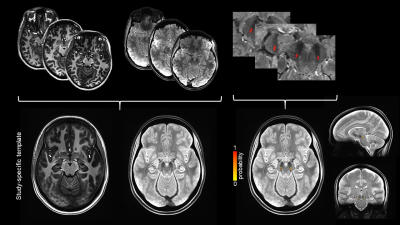 |
87 | High resolution probabilistic in-vivo atlas of Nigrosome 1
Marta Lancione1, Graziella Donatelli2,3, Matteo Cencini1, Paolo Bosco1, Mauro Costagli1,4, Mirco Cosottini5, Laura Biagi1, and Michela Tosetti1
1IRCCS Stella Maris, Pisa, Italy, 2IMAGO7 Foundation, Pisa, Italy, 3Azienda Ospedaliero-Universitaria Pisana, Pisa, Italy, 4University of Genoa, Genoa, Italy, 5University of Pisa, Pisa, Italy Keywords: Neurodegeneration, Neurodegeneration, Atlas, Nigrosome Iron accumulation in Nigrosome 1 (N1) may represent an important biomarker in neurodegenerative disorders but its small dimension and the vanishing swallow tail sign in patients make the segmentation of this region challenging. We propose a probabilistic atlas of N1 created on a multimodal (T1-w and T2*-w) template using ROIs that were manually drawn on 0.6mm-isotropic T2*-weighted images of twenty healthy subjects. Its usability and accuracy were tested on four additional subjects by comparing the mean N1 susceptibility and T2* obtained using the atlas to those obtained from manual ROIs and by measuring their geometrical proximity. |
|
1728.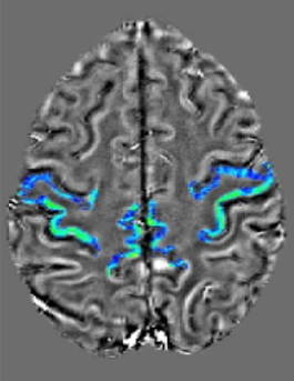 |
88 | IRON OVERLOAD IN AMYOTROPHIC LATERAL SCLEROSIS: DIAGNOSTIC ACCURACY OF VISUAL AND AUTOMATIC ASSESSMENTS
Valeria Elisa Contarino1, Francesco Maria Lo Russo1, Giorgio Conte1, Claudia Morelli2, Francesca Trogu2, Silvia Casale1, Sara Sbaraini3, Luca Caschera1, Valentina Genovese1, Chunlei Liu4, Claudia Maria Cinnante2, Vincenzo Silani2, and Fabio Maria Triulzi1
1Fondazione IRCCS Ca' Granda Ospedale Maggiore Policlinico Milano, Milano, Italy, 2Auxologico, Milano, Italy, 3ASST Santi Paolo e Carlo, San Carlo Borromeo Hospital, milano, Italy, 4UC Berkeley, Milano, Italy Keywords: Neurodegeneration, Brain, motor cortex; quantitative susceptibility mapping; amyotrophic lateral sclerosis vVisual SWI-based and automatic QSM-based assessments show high diagnostic accuracy in UMN- predominant ALS |
|
1729.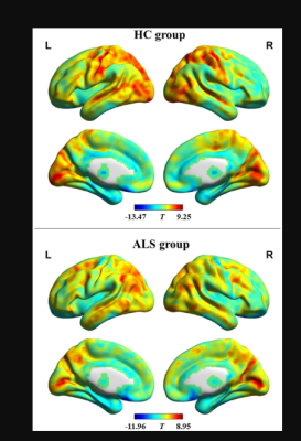 |
89 | Altered Dynamic in Functional Connectivity Density in Amyotrophic Lateral Sclerosis: A Resting-State FMRI Study
Jia-Hui Lin1, Jia-Hui Lin1, Qiu-Yi Dong1, Yun-Bin Cao1, and Hua-Jun Chen1
1Fujian Medical University Union Hospital, Fuzhou, China Keywords: Neurodegeneration, fMRI (resting state) This is the first study on alterations in the patterns of dynamic functional connection density (dFCD) involving ALS. We obtained resting-state fMRI data from ALS and healthy controls (HCs). We calculated the functional connectivity (FC) and the functional connection density (FCD) value. dFCD was assessed by sliding-window correlation method. The standard deviation of dFCD can measure dFCD variability. The dFCD variability was reduced in some brain region in HC group, whereas increase was observed in some brain region of ALS patients. dFCD variability can distinguish two groups. ALS patients exhibit aberrant dynamic property in brain functional architecture. |
|
1730.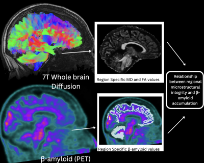 |
90 | Identifying multimodal imaging biomarkers: a framework exploring association of β-Amyloid Accumulation and Microstructural integrity at 7T
Mackenzie Langan1,2, Em Rose Triolo3, Oleksandr Khegai1,2, Carolina Ferreira-Atuesta4, Johnathan Sutkowski4, Trey Hedden4, Mehmet Kurt3, and Priti Balchandani2,5,6
1Icahn School of Medicine at Mount Sinai, New York, NY, United States, 2Biomedical Engineering and Imaging Institute, Icahn School of Medicine at Mount Sinai, New York, NY, United States, 3Department of Mechanical Engineering, University of Washington, Seattle, WA, United States, 4Department of Neurology, Icahn School of Medicine at Mount Sinai, New York, NY, United States, 5Diagnostic, Molecular and Interventional Radiology, Icahn School of Medicine at Mount Sinai, New York, NY, United States, 6Nash Family Department of Neuroscience, Icahn School of Medicine at Mount Sinai, New York, NY, United States Keywords: Neurodegeneration, Diffusion Tensor Imaging, High-Field MRI Here we outline a preliminary method using a novel method which leverages UHF neuroimaging to measure detectable correlations in two measures: microstructural (mean diffusivity and fractional anisotropy) acquired at 7T and amyloid beta (β-amyloid) accumulation, acquired using 3T PET-MR.The combination of these methods aids in the ability to map tissue structure and achieve unprecedented visualization of the consequences of β-amyloid accumulation as it relates to neurodegenerative disorders. We show the feasibility of leveraging high resolution diffusion to advance out understand of the relationship between fractional anisotropy and mean diffusivity within brain regions that may be affected by β-amyloid deposition. |
|
1731.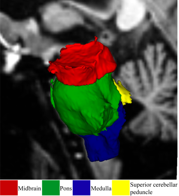 |
91 | Assessment of Brainstem volume in Myalgic Encephalomyelitis/Chronic fatigue syndrome and long-COVID patients
KIRAN THAPALIYA1, Sonya Marshall-Gradisnik1, Markus Barth2, Natalie Eaton-Fitch1, and Leighton Barnden1
1Griffith University, Gold coast, Australia, 2The University of Queensland, Brisbane, Australia Keywords: Neurodegeneration, Brain COVID -19 caused by the novel Severe Acute Respiratory Syndrome Coronavirus 2 (SARS-CoV-2) has infected more than 600 million and caused the deaths of over six million people worldwide. The majority of the infected patients do not recover fully from the COVID-19 infections and develop post-COVID conditions also known as long-COVID has similar symptoms compared to Myalgic Encephalomyelitis/Chronic fatigue syndrome. In this study, we evaluated the volumetric changes in the brainstem regions in ME/CFS, long-COVID patients compared to healthy controls. Our study showed that brainstem volumes higher in ME/CFS and long-COVID patients compared to healthy controls. |
|
1732.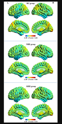 |
92 | Changes in the dynamic spontaneous neural activity of brain activity in minimal hepatic encephalopathy
Jia-Hui Lin1, Yun-Bin Cao1, Qiu-Yi Dong1, and Hua-Jun Chen1
1Fujian Medical University Union Hospital, Fuzhou, China Keywords: Neurodegeneration, fMRI (resting state) Amplitude of low-frequency fluctuation (ALFF) can identify abnormal regional neural activity in minimal hepatic encephalopathy (MHE). This work sought to evaluate the temporal variability of ALFF to reveal MHE-related alterations in the dynamics of spontaneous neural activity. Healthy controls and patients with cirrhosis [including MHE and without MHE (NHE)] were enrolled in this investigation. Utilizing a sliding-window approach to calculate the dynamic ALFF (dALFF) variability. The dALFF variability in some brain region progressively decreased from NHE to MHE group and it can distinguish NHE and MHE patients. Our findings highlight aberrant dynamic brain function in MHE. |
|
1733.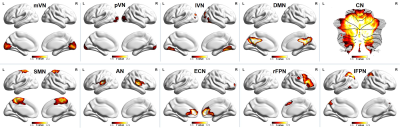 |
93 | Modifications of Large-Scale Resting-State Functional Connectivity in Spinocerebellar Ataxia Type 3
Bing LIU1,2, Linwei Zhang3, Aocai Yang1, Jixin Luan1, Kuan Lv1, and Guolin Ma1
1Department of Radiology, China Japan Friendship Hospital, Beijing, China, 2Graduate School of Peking Union Medical College, Beijing, China, 3Department of Neurology, China Japan Friendship Hospital, Beijing, China Keywords: Neurodegeneration, Brain Connectivity, functional MRI, independent component analysis, spinocerebellar ataxia type 3, large-scale brain networks, functional connectivity Large-scale resting-state functional network connectivity changes in spinocerebellar ataxia type 3 patients were found in both inter- and intranetwork functional connectivity. Especially, the increased intranetwork functional connectivity within the lateral visual network may potentially be a compensatory mechanism of visual-related symptoms in SCA3. |
|
1734.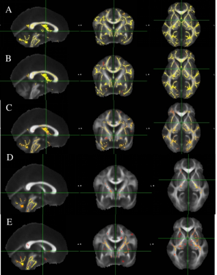 |
94 | Mapping of neurodegenerative changes with Diffusion Tensor Imaging of Spinocerebellar ataxia type 1, 2 and 12 patients
Pankaj Pankaj1, S Senthil Kumaran1, Achal Kumar Srivastava2, Ramesh Kumar Agrawal3, Ajay Garg4, Ashima Nehra5, and Roopa Rajan2
1Nuclear Magnetic Resonance, All India Institute of Medical Sciences, New Delhi, New Delhi, India, 2Neurology, All India Institute of Medical Sciences, New Delhi, New Delhi, India, 3School of Computer & Systems Sciences, Jawaharlal Nehru University, New Delhi- 110 067, New Delhi, India, 4Neuro-radiology, All India Institute of Medical Sciences, New Delhi, New Delhi, India, 5Neuropsychology, All India Institute of Medical Sciences, New Delhi, New Delhi, India Keywords: Neurodegeneration, Diffusion Tensor Imaging The study assessed changes in brain tissue microstructures in SCA type 1, 2 and 12 patients using diffusion tensor imaging. Our findings exhibited widespread reduced fractional anisotropy (FA), increased radial (RD) and axial diffusivity (AD) in SCA with respect to that in healthy subjects. FA was decreased in SCA1 and SCA2 in the superior and inferior longitudinal fasciculus, anterior thalamic radiation, inferior fronto-occipital fasiculus, forceps minor, corticospinal tract, cingulum, uncinate fasciculus as compared to controls. Abnormal white matter structure may be linked to cognitive and behavioural impairment in SCA 1 and SCA 2 patients. |
|
1735.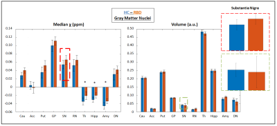 |
95 | Quantitative Susceptibility Mapping of cortical and subcortical brain regions in patients with REM sleep Behavior Disorder
Cristiana Fiscone1, Fiorina Bartiromo2, Luca Baldelli1, Luisa Sambati3, Serena D'Aniello4, Stefania Evangelisti1, Micaela Mitolo2,5, Claudia Testa2,6, Pietro Cortelli1,3, Raffaele Lodi1,2, Federica Provini1,3, and Caterina Tonon1,2
1Department of Biomedical and Neuromotor Sciences, University of Bologna, Bologna, Italy, 2Functional and Molecular Neuroimaging Unit, IRCCS Istituto delle Scienze Neurologiche di Bologna, Bologna, Italy, 3Clinica Neurologica Rete Metropolitana, IRCCS Istituto delle Scienze Neurologiche di Bologna, Bologna, Italy, 4Department of Advanced Biomedical Sciences, University of Naples Federico II, Napoli, Italy, 5Department of Experimental, Diagnostic and Specialty Medicine, University of Bologna, Bologna, Italy, 6Department of Physics and Astronomy, University of Bologna, Bologna, Italy Keywords: Neurodegeneration, Brain, RBD REM sleep Behavior Disoder (RBD) is a parasomnia, possibly converting in a neurodegenerative α-synucleinopathy. We explored to role of iron accumulation in this condition exploiting Quantitative Susceptibility Mapping (QSM), a quantitative imaging technique returning susceptibility values voxel-per-voxel. RBD patients were compared to healthy controls; cortical and sub-cortical areas were segmented with automatic and semi-automatic methods and susceptibility distributions were compared. Significant increase of iron deposition resulted in multiple cortical areas, in the brainstem and in the gray matter nuclei of the limbic system, suggesting that QSM may help in identifying biomarkers that predict the conversion from RBD to neurodegeneration. |
|
1736.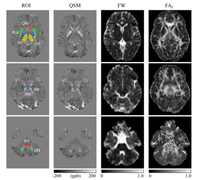 |
96 | Free Water Imaging in deep gray matter nucleus of Wilson’s Disease
Xiao-Zhong Jing1, Gai-Ying Li2, Yu-Peng Wu3, Xiang-Zhen Yuan4, Jia-Lin Chen2, Reyisha Taximaimaiti1, Jian-Qi Li2, and Xiao-Ping Wang5
1Department of Neurology, Tongren Hospital, Shanghai Jiao Tong University School of Medicine, Shanghai, China, 2Shanghai Key Laboratory of Magnetic Resonance, East China Normal University, Shanghai, China, 3East China Normal University, Shanghai, China, 4Department of Neurology, Weifang People's Hospital, Weifang, China, 5Department of Neurology, Jiading Branch of Shanghai General Hospital, Shanghai Jiao Tong University School of Medicine, Shanghai, China Keywords: Neurodegeneration, Diffusion/other diffusion imaging techniques, Wilson’s disease; free water imaging; quantitative susceptibility mapping. This is the first study to use a bi-tensor free water imaging to evaluate microstructural changes in deep gray matter (DGM) nuclei of Wilson’s disease (WD). Despite the shortcomings our study manifested that free water imaging detects microstructural alterations in both normal and abnormal appearing DGM nuclei of WD patients. Correlations between free water imaging indices and neurological impairment in WD patients were also noticed. Therefore, as a promising tool, free water imaging deserves further investigation in longitudinal studies to evaluate its role in monitoring disease onset, progression, and treatment efficacy in WD patients. |
|
1737.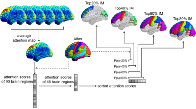 |
97 | Predicting brain age using partition modeling strategy and Atlas-based attentional enhancement in Chinese population
Yang Yang1, Bingsheng Huang2, Yingqian Chen3, Yingtong Wu2, Chuxuan Lin2, Zhiyun Yang3, and Xia Liu4
1Department of Radiology, Suining Central Hospital, Suining, China, 2Medical AI Lab, School of Biomedical Engineering, Health Science Center, Shenzhen University, Shenzhen, China, 3Department of Radiology, the First Affiliated Hospital, Sun Yat-sen University, Guangzhou, China, 4Department of Radiology, Shenzhen Kangning Hospital, Shenzhen, China Keywords: Neurodegeneration, Neurodegeneration, Brain age, Partition modeling This study aimed to develop and construct a MRI-based full-age-range brain age prediction model that can be applied in the Chinese health care system. We proposed a brain age prediction method based on transfer learning and partition modeling , which was using Atlas attention enhancement. The performance of models with different image masks were compared and the model constructed based on top60% image mask achieved the best prediction performance. The brain age prediction method proposed in this study can provide objective brain age for assessing brain health status in Chinese population. |
|
1738.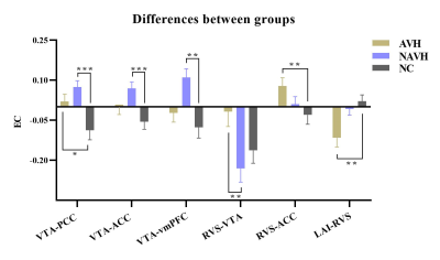 |
98 | Abnormal effective connectivity of reward network in first-episode schizophrenia with auditory verbal hallucinations
Jingli Chen1,2, Yarui Wei1,2, Kangkang Xue1,2, and Jingliang Cheng1,2
1Department of Magnetic Resonance Imaging, The First Affiliated Hospital of Zhengzhou University, Zhengzhou, China, 2Laboratory for Functional Magnetic Resonance Imaging and Molecular Imaging of Henan Province, Magnetic Resonance Imaging, Zhengzhou, China Keywords: Neurodegeneration, fMRI (resting state), Auditory verbal hallucinations /effective connectivity/ reward network/ dopamine Our study employs the dynamic causal modeling (DCM) approach to perform effective connectivity (EC) analysis of the reward network to investigate the mechanisms underlying schizophrenia patients with auditory verbal hallucinations (AVHs). This study enrolled 86 first-episode drug-naïve schizophrenia patients with AVHs (AVH), 93 patients without AVHs (NAVH), and 88 normal controls (NC), undergoing resting-state functional magnetic resonance. Our findings suggest that there are some common and different EC abnormalities in the reward network of AVH and NAVH. Particularly, the abnormalities of mesolimbic and mesocortex pathways in AVH may provide guidance for understanding the neurobiological mechanisms of AVHs and treatment. |
|
1739.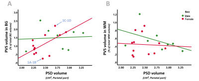 |
99 | Structural waste clearance markers in the elderly: sex differences in the relation between perivascular and parasagittal dural space volume
Merel M. van der Thiel1,2,3, Kilian Hett4, Nathalie A. Roos1,5, Colin D. Mcknight6, Melanie K. Leguizamon4, Sofia Venturini1,7, Inez H.G.B. Ramakers3,8, Walter H. Backes1,2,9, and Jacobus F.A. Jansen1,2,10
1Department of Radiology & Nuclear Medicine, Maastricht University Medical Center, Maastricht, Netherlands, 2School for Mental Health and Neuroscience, Maastricht University, Maastricht, Netherlands, 3Department of Psychiatry & Neuropsychology, Maastricht University, Maastricht, Netherlands, 4Department of Neurology, Vanderbilt University Medical Center, Nashville, TN, United States, 5Department of Biomedical Engineering, Eindhoven University of Technology, Eindhoven, Netherlands, 6Department of Radiology and Radiological sciences, Vanderbilt University Medical Center, Nashville, TN, United States, 7Faculty of Psychology and Neuroscience, Maastricht University, Maastricht, Netherlands, 8School for Mental Health & Neuroscience, Maastricht University, Maastricht, Netherlands, 9Cardiovascular Research Institute Maastricht, Maastricht University, Maastricht, Netherlands, 10Department of Electrical Engineering, Eindhoven University of Technology, Eindhoven, Netherlands Keywords: Neurodegeneration, Neurofluids, Sex differences, Perivascular spaces, Waste clearance Cerebral waste clearance reduces with healthy aging and occurs in various neurodegenerative diseases. Both perivascular spaces (PVS) and the parasagittal dural (PSD) space play important roles in waste clearance, where the former transports waste products through the parenchyma, the latter is associated with cerebrospinal fluid efflux from the cranial compartment. The current 7T MRI study investigated sex differences in the relation between PVS and PSD volume in an elderly sample. By identifying a relationship between PVS and PSD solely in females, this study illustrates the possibility of a different impairment mechanism of the clearance system between elderly men and women. |
|
 Back to Meeting Home
 Back to the Program-at-a-Glance
The International Society for Magnetic Resonance in Medicine is accredited by the Accreditation Council for Continuing Medical Education to provide continuing medical education for physicians.
 Back to Meeting Home
 Back to the Program-at-a-Glance
 View Presentation Video