Digital Poster
Brain Connectivity in Disease I
ISMRM & ISMRT Annual Meeting & Exhibition • 03-08 June 2023 • Toronto, ON, Canada

| Computer # | |||
|---|---|---|---|
3331.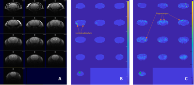 |
101 |
Altered brain function and structure in a transgenic mouse which
overexpress MTH1 hydrolase following an oxidative stimulus by
rs-fMRI and DTI
Taljinder Singh1,2,
Francesco de Pasquale3,
Gabriele De Luca4,
Paola Fortini5,
Valeria Simonelli5,
and Rossella Canese1
1MRI Unit, Core Facilities, Istituto Superiore di Sanità, Rome, Italy, 2Department of Basic and Applied Sciences for Engineering, Sapienza University of Rome, Rome, Italy, 3University of Teramo, Teramo, Italy, 4Oncology and Molecular Medicine Dept., Istituto Superiore di Sanità, Rome, Italy, 5Environmental and Health Dept., Istituto Superiore di Sanità, Rome, Italy Keywords: Brain Connectivity, Animals, DTI, oxidant agent Oxidative stress is involved in the pathogenesis of cancer, neurodegeneration and aging. hMTH1 is a hydrolase able to protect cells by oxidative damage. Overexpression of hMTH1 in transgenic mice confers significant protection against oxidative damage. Our study showed alterations in the brain networks of hMTH1-Tg mice with respect to their controls. The chronical exposure to an oxidant agent (Paraquat, most widely used herbicide) causes differences in the brain function (measured by rs-fMRI) and structure (measured by DTI) of transgenic mice. |
|
3332.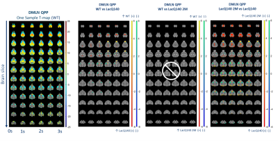 |
102 |
Impact of early suppression of mutant Huntingtin in LacQ140
mouse model of Huntington’s disease on resting-state dynamics
Tamara Vasilkovska1,2,
Mohit H. Adhikari1,2,
Joëlle van Rijswijk1,2,
Eline Van Doninck1,2,
Johan Van Audekerke1,2,
Dorian Pustina3,
Roger Cachope3,
Haiying Tang3,
Deanna M. Marchionini3,
Ignacio Munoz-Sanjuan3,
Annemie Van der Linden1,2,
and Marleen Verhoye1,2
1Bio-Imaging Lab, Deparment of Biomedical Sciences, University of Antwerp, Antwerp, Belgium, 2μNEURO Research Centre of Excellence, University of Antwerp, Antwerp, Belgium, 3CHDI Management/CHDI Foundation, Princeton, NJ, United States Keywords: Brain Connectivity, Preclinical, Huntington's Disease, dynamic resting-state fMRI Dynamic analyses of resting-state (RS) fMRI reveal transient constituents of RS networks such as the quasi-periodic patterns (QPPs), and co-activation patterns (CAPs) that were shown to be sensitive markers of neurodegenerative diseases in rodent models and humans. We investigated the effect of early suppression of mutant huntingtin (mHtt) expression in the LacQ140 mouse model of Huntington’s disease (HD) on QPP and CAP alterations at the manifest state. In both QPPs and CAPs, the observed genotypic changes in local activity were reduced in the mHtt suppressed group. Additionally, a cross-validated, three-class classification using CAP activations successfully predicted the mHtt suppressed group |
|
3333.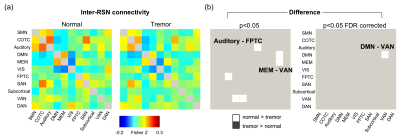 |
103 |
Altered Resting-State Network Connectivity in Essential Tremor
Sheng-Min Huang1,
Yu-Chin Huang2,
Nan-Hao Chen3,
Chun-Ying Shen4,
Ting-Kai Leung4,5,
and Li-Wei Kuo1,6
1Institute of Biomedical Engineering and Nanomedicine, National Health Research Institutes, Miaoli, Taiwan, 2Department of Neurology, Taoyuan General Hospital, Ministry of Health and Welfare, Taoyuan, Taiwan, 3Department of Biomedical Engineering and Environmental Sciences, National Tsing Hua University, Hsinchu, Taiwan, 4Department of Radiology, Taoyuan General Hospital, Ministry of Health and Welfare, Taoyuan, Taiwan, 5Graduate Institute of Biomedical Materials and Tissue Engineering, College of Biomedical Engineering, Taipei Medical University, Taipei, Taiwan, 6Institute of Medical Device and Imaging, National Taiwan University College of Medicine, Taipei, Taiwan Keywords: Brain Connectivity, fMRI (resting state), essential tremor We aim to characterize the functional connectivity in essential tremor to portray the resting-state network organization and its correlation to tremor features. Reduced functional connectivity between default mode network and ventral attention network was found in essential tremor subjects. The inter-network connectivity and intra-network connectivity were also shown to be associated with tremor features. Our result suggest that analysis of resting-state network is a potential approach in detecting the cerebral alterations in ET. |
|
3334.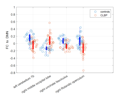 |
104 |
Functional Connectivity between Right Rolandic Operculum and
Default Mode Network Is Associated with Chronic Low Back
Pain-Related Disability
Chang-Min Chen1,
Shwu-Fen Wang2,
Dar-Ming Lai3,
and Wen-Chau Wu1
1Institute of Medical Device and Imaging, National Taiwan University, Taipei, Taiwan, 2Department of Physical Therapy, National Taiwan University, Taipei, Taiwan, 3Department of Surgery, National Taiwan University, Taipei, Taiwan Keywords: Brain Connectivity, fMRI (resting state) This study aimed to explore the relationship between emotion and pain-related disability in patients with chronic low back pain (CLBP) by using resting-state functional connectivity (FC). Our results showed that as compared with the control group (n = 26), the patient group (n = 26) had decreased FC with the default mode network in multiple brain regions. Specifically, the right Rolandic operculum was identified, where FC was inversely correlated with the Oswestry disability index (r = -0.616, p < 0.001), suggesting the interplay between emotion processing and pain perception. |
|
3335.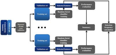 |
105 |
Brain connectivity markers for the diagnosis of non-suicidal
self-injury in adolescents: a DTI and resting-state fMRI study
Ping Jiang1,
Zhiang Niu1,
Hai Lin2,
Yongming Dai2,
and Jiajun Xu1
1West China Hospital, Sichuan University, Chengdu, China, 2Central Research Institute, United Imaging Healthcare, Shanghai, China Keywords: Brain Connectivity, Psychiatric Disorders, Non-suicidal self-injury Non-suicidal self-injury (NSSI), defined as the deliberate damage or destruction of body tissue without the intent to die, is a common behavior amongst adolescents. We conducted connectome-based analysis to investigate neural correlates of self-injury and identified several potential brain connectivity markers of NSSI diagnosis. On basis of these markers, the random forest model achieved an accuracy of 78.2% to discriminate between NSSI and non-NSSI patients. Our study indicated that structural and functional connectivity could characterize the behavior of self-injury and help early NSSI diagnosis in adolescents. |
|
3336.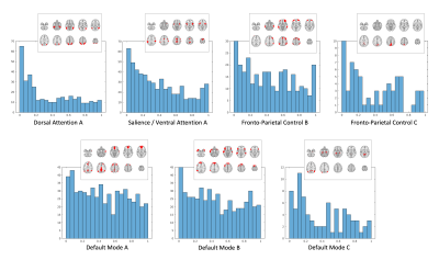 |
106 |
Cognitive Brain Networks in Adult Fetal Alcohol Syndrome:
Analyses of Functional Connectivity with a Higher Criticism
Approach
Benedikt Sundermann1,2,3,
Reinhold Feldmann4,
Christian Mathys1,3,
Johanna Rau5,
Stefan Garde2,6,
Anna Braje2,
Josef Weglage4,
and Bettina Pfleiderer2
1Institute of Radiology and Neuroradiology, Evangelisches Krankenhaus Oldenburg, Medical Campus University of Oldenburg, Oldenburg, Germany, 2Clinic of Radiology, Medical Faculty, University of Münster, Münster, Germany, 3Research Center Neurosensory Science, University of Oldenburg, Oldenburg, Germany, 4Department of General Pediatrics, University Hospital Münster, Münster, Germany, 5Department of Neurology with Institute of Translational Neurology, University Hospital Münster, Münster, Germany, 6Bergman Clinics Augenklinik Universitätsallee Bremen, Bremen, Germany Keywords: Brain Connectivity, fMRI (resting state) Functional connectivity (FC) between and within a majority of cognition-related brain networks is altered in young adults with Fetal Alcohol Syndrome. Findings in a network-level analysis of resting-state fMRI data with a Higher Criticism approach were most obvious within a dorsal attention subnetwork and to a lesser extent in a salience / ventral attention subnetwork. When analyzing these effects further by looking at single connections, no individual FC alteration was statistically significant when adjusting for multiple comparisons. The finding of a wide distribution of FC alterations across networks might help resolve partially contradictory results in previous studies. |
|
3337.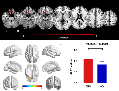 |
107 |
Intrinsic brain abnormalities in chronic rhinosinusitis and the
effect of mood and cognitive function: a resting-state
functional MRI study
SIMIN LIN1,
MIAOMIAO NIE2,
BINGSHAN WANG3,
and YI HAN4
1Radiology, Xiamen Cardiovascular Hospital of Xiamen University, Xiamen, China, 2Radiology, Zhongshan Hospital of Xiamen University, Xiamen, China, 3Zhongshan Hospital of Xiamen University, Xiamen, China, 4Fujian Provincial Key Laboratory of Ophthalmology and Visual Science, Eye Institute of Xiamen University, Xiamen, China Keywords: Brain Connectivity, fMRI (resting state), Amplitude of low-frequency fluctuation Chronic rhinitis (CRS) is one of the most prevalent chronic diseases that increases the risk of anxiety, depression and cognitive disorders. Here we collected rs-fMRI data to assess global brain activity and functional connectivity (FC) in CRS patients. Compared with controls, CRS patients demonstrated increased ALFF in the left orbital superior frontal cortex and decreased FC in the right precuneus cortex, with the former positively correlated with the severity of inflammation and the scores of anxiety and depression. These rs-fMRI studies provided several important hints into the potential neural mechanism related to mood and cognitive dysfunctions in CRS patients. |
|
3338.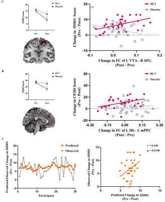 |
108 |
Bright Light Therapy Alters Brain Functional Connectivity in
Subthreshold Depression: A Randomized Clinical Trial
Zibin Yang1,
Guanmao Chen1,
Long Qian2,
and Ying Wang1
1Medical Imaging Center, First Affiliated Hospital of Jinan, Guangzhou, China, 2GE Healthcare, Beijing, China, Beijing, China Keywords: Brain Connectivity, fMRI (resting state), Subthreshold depression The underlying mechanisms of bright light therapy in prevention of individuals with subthreshold symptoms are unclear. This study aimed to assess the midbrain monoamine-producing nuclei treatment–related functional connectivity changes and their correlation to depressive symptom improvements in subthreshold depression. A total of 74 young adults with subthreshold depression were randomly assigned to receive 8-week BLT (N = 38) or placebo (N = 36). The dorsal raphe nucleus, ventral tegmental area, and habenula seed-based whole-brain FC were analyzed. In addition, a multivariate regression model examined whether baseline brain FC was associated with changes in scores on HDRS during BLT treatment. |
|
3339.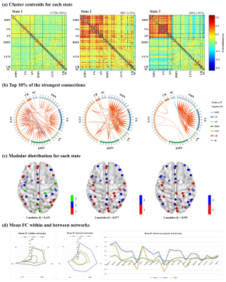 |
109 |
Altered intrinsic brain functional network dynamics in
drug-naïve, first-episode adolescents with major depressive
disorder
Baolin Wu1,
Zhiyun Jia1,2,
and Qiyong Gong1
1Huaxi MR Research Center (HMRRC), Department of Radiology, West China Hospital of Sichuan University, Chengdu, China, 2Department of Nuclear Medicine, West China Hospital of Sichuan University, Chengdu, China Keywords: Psychiatric Disorders, fMRI (resting state) This study aimed to explore the dynamic FC in adolescent MDD patients, with a focus on the temporal properties of functional connectivity states as well as the variability of network topological organization. We found that adolescent MDD patients spent more time in the weakly-connected and relatively highly-modularized State 1, spent less time in the strongly-connected and low-modularized State 2, and had higher variability in the network efficiency than healthy controls. These findings suggest impaired local segregation and global integration of functional networks, as well as segregation-integration imbalance in adolescent MDD patients. |
|
3340.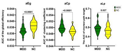 |
110 |
Altered network pattern of cortical microarchitecture in major
depressive disorder
Ziyun Xu1,
Gangqiang Hou1,
Yingli Zhang1,
Bo Peng1,
Long Qian2,
and wentao Lai1
1Shenzhen Kangning Hospital, Shenzhen, China, 2MR Research, GE Healthcare, Beijing, China Keywords: Brain Connectivity, Brain Connectivity Although major depressive disorder (MDD) has been deeply studied in decades, there are still no reliable biological markers. Here, we combine the T1-wighted, diffusion tensor imaging (DTI) and inhomogeneous magnetization transfer imaging (IhMT) to detect cortical morphometric changes in individuals with MDD. Morphometric similarity network (MSN) was established for each subject. Network properties and rich-club organizations were assessed and analyzed between groups. MDD showed significant alteration in global and nodal properties, as well as reorganization of rich clubs. Consequently, topological structure of the morphometric similarity network is disrupted, which may be a potential biomarker for major depressive disorder. |
|
3341.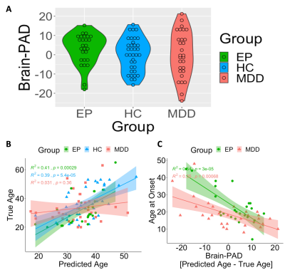 |
111 |
Older Brain Age Prediction Using Functional Brain Network
Efficiency Linked to Early Disorder Onset among Depression and
Epilepsy Patients
Yael Jacob1,
Gaurav Verma1,
Laurel S Morris1,
Lara Marcuse1,
Madeline Fields1,
James W Murrough W Murrough1,
and Priti Balchandani1
1Icahn School of Medicine at Mount Sinai, New York, NY, United States Keywords: Brain Connectivity, Aging Brain network organization, and specifically its efficiency, is known to be associated with age, implying older brain exhibit reduced network efficiency. Using resting-state functional magnetic resonance imaging, we investigate if estimated brain age according to functional network efficiency is deviant from chronological age in major depressive disorder (MDD) and Epilepsy patients (EP). Global network efficiency significantly predicted chronological age among HC. Importantly, we found the differences between predicted and true brain age among MDD and EP patients were significantly associated with age at onset, indicating that the prediction of an older age brain is associated with early age disorder onset. |
|
3342.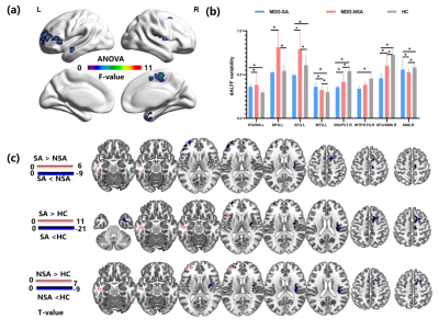 |
112 |
Altered static and dynamic spontaneous neural activity in
adolescent with first episode major depressive disorder and
previous suicide attempts
Xiaofang Cheng1,2,
Jianshan Chen1,
Xiaofei Zhang1,
Ting Wang3,
Jiaqi Sun1,
Yanling Zhou1,
Ruilan Yang1,
Yeyu Xiao3,
Amei Chen3,
Ziyi Song1,
Pinrui Chen1,
Chanjuan Yang1,
Qiuxia Wu1,
Taifeng Lin1,
Yingmei Chen1,
Yongzhou Xu4,
Liping Cao1,
and Xinhua Wei2
1The Affiliated Brain Hospital of Guangzhou Medical University, Guangzhou, China, 2South China University of Technology, Guangzhou, China, 3Guangzhou Integrated Traditional Chinese and Western Medicine, Guangzhou, China, 4Philips Healthcare, Guangzhou, China Keywords: Brain Connectivity, Adolescents, Major depressive disorder; suicide; frontal cortex This study combined dynamic and static amplitude of low-frequency fluctuations (ALFF) approaches to investigate the differences in local spontaneous brain activities between depressed adolescents with and without suicide attempts (SA). Our findings suggest that alterations in brain activities in regions involved in emotional processing, decision-making, and response inhibition are associated with an increased risk of suicidal behaviors in depressed adolescents. |
|
3343.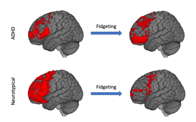 |
113 |
Functional magnetic resonance imaging reveals fidgeting in ADHD
improves prefrontal cortex activation during executive
functioning
Xirui Zhao1,
Paul Condron2,
Haribalan Kumar1,2,3,
Eryn Kwon1,2,4,5,
Gina Waters 4,6,
Christopher Erb6,
Makarena Dudley4,6,
Davidson Taylor2,7,
Gil Newburn2,
Jerome Maller3,
Vickie Shim1,2,
Alan Wang1,4,5,
Samantha Holdsworth2,4,5,
Karen Waldie4,6,
and Justin Fernandez1,2,8
1Auckland Bioengineering Institute, University of Auckland, Auckland, New Zealand, 2Mātai Medical Research Institute, Tairāwhiti/Gisborne, New Zealand, 3General Electric Healthcare AUS/NZ, Melbourne, Australia, 4Centre for Brain Research, University of Auckland, Auckland, New Zealand, 5Department of Anatomy and Medical Imaging, Faculty of Medical and Health Sciences, University of Auckland, Auckland, New Zealand, 6Department of Psychology, Faculty of Science, University of Auckland, Auckland, New Zealand, 7Ngai Tāmanuhiri, Rongowhakaata, Ngāti Porou, Tūranganui-a-Kiwa, Tairāwhiti, New Zealand, 8Department of Engineering Science, Faculty of Engineering, University of Auckland, Auckland, New Zealand Keywords: Brain Connectivity, fMRI (task based), ADHD, fidgeting MRI People with Attention Deficit Hyperactivity Disorder (ADHD) are often observed to fidget or display repetitive fine motor skills. Though it has been proposed that fidgeting may assist with concentration and sustained attention, there is little objective evidence to support this idea. This is the first study to evaluate the impact of fidgeting, using functional magnetic resonance imaging (fMRI), with neurotypical and ADHD adult volunteers. Comparative analyses revealed significant increases in medial prefrontal cortex brain activation while fidgeting during executive functioning in the ADHD participant. This distinction may provide additional information in future MRI diagnosis of ADHD. |
|
| 3344. | WITHDRAWN | ||
3345.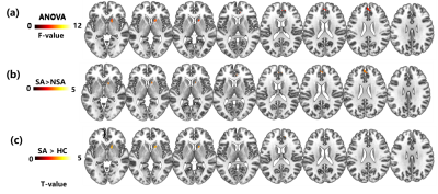 |
114 |
Alterations of static and dynamic functional connectivity in
adolescents with first-episode major depression and suicidal
tendencies
Xiaofang Cheng1,2,
Jianshan Chen1,
Xiaofei Zhang1,
Ting Wang3,
Jiaqi Sun1,
Yanling Zhou1,
Ruilan Yang1,
Yeyu Xiao3,
Amei Chen3,
Ziyi Song1,
Pinrui Chen1,
Chanjuan Yang1,
Qiuxia Wu1,
Taifeng Lin1,
Yingmei Chen1,
Yongzhou Xu4,
Liping Cao1,
and Xinhua Wei2
1The Affiliated Brain Hospital of Guangzhou Medical University, Guangzhou, China, 2South China University of Technology, Guangzhou, China, 3Guangzhou Integrated Traditional Chinese and Western Medicine, Guangzhou, China, 4Philips Healthcare, Guangzhou, China Keywords: Brain Connectivity, Adolescents, major depressive disorder; suicide; FCS This study used graph theory-based dynamic and static functional connectivity strength (FCS) approaches to investigate the differences in global brain connectivity patterns in depressed adolescents with and without suicide attempts. Our findings suggest that abnormal connectivity patterns in multiple brain regions involving cognition and emotion control, and self-referential processing are associated with the increasing risk of vulnerability to a suicide attempt in depressed adolescents. |
|
3346.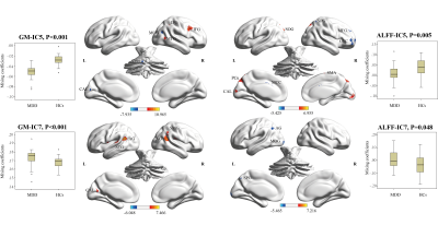 |
115 |
Multimodal structural and functional features underlying
cognitive function in first-episode major depressive disorder
Qian Zhang1,
Youjin Zhao1,
Chenyang Yao1,
Yaxuan Wang1,
Ziyuan Zhao1,
Aoxiang Zhang1,
and Qiyong Gong1
1Department of Radiology, West China Hospital of Sichuan University, Huaxi MR Research Center, Chengdu, China Keywords: Psychiatric Disorders, Brain, major depressive disorder Multimodal joint differences in the right dorsal lateral prefrontal cortex, bilateral superior and inferior parietal lobules, and bilateral calcarine cortex contributed to identification of patients with major depressive disorder (MDD), offering promise in the multimodal psychoradiological characterization for MDD diagnosis. In addition, widespread gray matter alterations across neocortex were associated with cognitive impairments, and partially mediated the age-related cognitive difficulties associated with MDD, deepening the understanding of the relationships between brain alterations and neurocognitive changes in MDD. |
|
3347.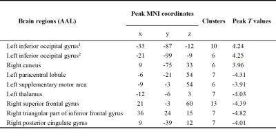 |
116 |
Brain activity alterations in premature ejaculation patients
before and after treatment with sertraline
Shaowei Liu1,
Jianhuai Chen2,
and Weiqiang Dou3
1Radiology, Jiangsu Province Hospital of Chinese Medicine, Affiliated Hospital of Nanjing University of Chinese Medicine, Nanjing, China, 2Andrology, Jiangsu Province Hospital of Chinese Medicine, Affiliated Hospital of Nanjing University of Chinese Medicine, Nanjing, China, 3GE Healthcare, MR Research China, Beijing, China Keywords: Brain Connectivity, Brain Connectivity This study aimed to explore the central neural mechanisms underlying sertraline in the treatment of premature ejaculation (PE). Resting-state functional magnetic resonance imaging (rs-fMRI) of PE patients before and after treatment with sertraline were acquired. The brain regions with altered regional homogeneity (ReHo) values were found. Increased activity in the occipital lobe and decreased activity in the prefrontal and parietal lobe and thalamus might be the mechanisms of sertraline in the treatment of PE. |
|
3348.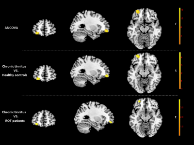 |
117 |
Altered Brain Activity and Functional Connectivity in transition
from recent-onset to chronic tinnitus, a rs-fMRI study
Shuting Han1,
Peng Wu2,
and Yonggang Li1
1Department of Radiology, First Affiliated Hospital of Soochow University, Suzhou, China, Suzhou city, China, 2Philips Healthcare, Shanghai, China, Shanghai, China Keywords: Brain Connectivity, fMRI (resting state), tinnitus As a common disorder, the development of tinnitus deserves the attention of neuroscientists. The present study combined fractional amplitude of low-frequency fluctuations and functional connectivity to explore brain functional abnormalities in transition from recent-onset to chronic tinnitus. Abnormal intraregional neural activity and functional connectivity were observed in the left middle frontal gyrus and left dorsolateral superior frontal gyrus during the development of chronic tinnitus, and these regions are major components of attention network and executive control network. These findings provide us with a better understanding of the aberrant brain changes and neuropathophysiological mechanisms of the progression of tinnitus. |
|
3349.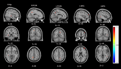 |
118 |
Abnormal brain function changes in chronic tinnitus with hearing
loss: A Resting-State Functional Magnetic Resonance Imaging
Study
Jiapei Xie1 and
Meiyun Wang1
1Henan Provincial People's Hospital, Zhengzhou, China Keywords: Brain Connectivity, Brain Connectivity To investigate the differences of spontaneous brain activity and brain functional connectivity between chronic tinnitus patients with hearing loss and healthy control group. Resting state functional magnetic resonance imaging (rs-fMRI) was performed to obtain the low-frequency fluctuation amplitude (ALFF), regional homogeneity (ReHo) and functional connectivity (FC). In this study, compared with healthy controls, chronic tinnitus patients with hearing loss showed aberrant brain activity increased in the temporal lobe, frontal lobe, hippocampus, precuneus, amygdala and cingulate cortex. It provided additional evidence to understand the neuropathophysiological mechanism of chronic tinnitus with hearing loss. |
|
3350.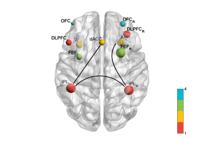 |
119 |
Effects on Granger causality Connectivity of Deaf Children
during Working Memory Task after Aerobic Exercise Intervention
Hang Qu1,
Wei-qiang Dou2,
and Wang Wei3
1RADIOLOGY, Affiliated Hospital of Yangzhou University, Yangzhou, China, 2GE Healthcare, MR Research China, Beijing, China, 3Radiology, Affiliated Hospital of Yangzhou University, Yangzhou, China Keywords: Brain Connectivity, fMRI (task based) The cross-modal plasticity was truly involved in theWorking memory (WM) that related to decision-making and memory processing after exercise intervention. However, we know little about the functional connectivity changes after the exercise intervention. The purpose of this study was to examine the cross modal plasticity and Granger causality connectivity among the internetwork in deaf children when performing a WM task before and after exercise intervention. Our results show that aerobic exercise intervention could improve working memory performance in reaction time and mean accuracy rate accompanied with increased internetwork casual flow connectivity. |
|
 Back to Meeting Home
 Back to the Program-at-a-Glance
The International Society for Magnetic Resonance in Medicine is accredited by the Accreditation Council for Continuing Medical Education to provide continuing medical education for physicians.
 Back to Meeting Home
 Back to the Program-at-a-Glance
 View Presentation Video