Digital Poster
Neurodegeneration III
ISMRM & ISMRT Annual Meeting & Exhibition • 03-08 June 2023 • Toronto, ON, Canada

| Computer # | |||
|---|---|---|---|
1918.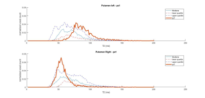 |
101 | T2 mapping with GRAPPATINI for the characterization of adult-onset and juvenile-onset Huntington Disease
Maria Eugenia Caligiuri1, Maria Celeste Bonacci1, Tobias Kober2, Domenico Zacà3, Ferdinando Squitieri4,5, Aldo Quattrone1, and Umberto Sabatini1
1Neuroscience Research Center, Università degli Studi Magna Graecia di Catanzaro, Catanzaro, Italy, 2Advanced Clinical Imaging Technology, Siemens Healthcare AG, Lausanne, Switzerland, 3Siemens Healthcare, Milan, Italy, 4Italian League for Research on Huntington Disease, Rome, Italy, 5IRCSS Casa Sollievo della Sofferenza/CSS-Mendel, Rome, Italy Keywords: Neurodegeneration, Rare disease, Huntington Disease T2 mapping is a quantitative MRI technique that may provide further insight regarding the changes that differentially underlie Huntington Disease in two of its clinical forms, adult-onset and juvenile-onset. Here we present pilot evidence that the study of T2 properties of the brain might be of great benefit to the research focused on this rare disease. |
|
1919.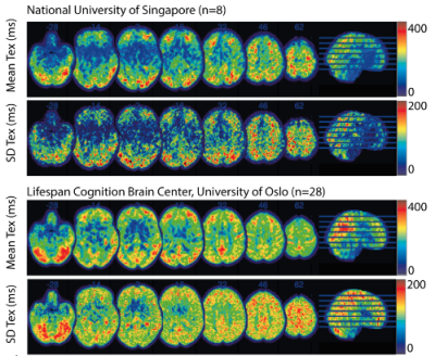 |
102 | Blood-brain barrier permeability changes over the lifespan
Beatriz Padrela1, Markus H Sneve2, Sanne Zelhorst1, Mervin Tee3, Amnah Mahroo4, Joost Kuijer1, Kristine Walhovd2, Simon Konstandin4, Klaus Eickel4,5, Frederik Barkhof1,6, Saima Hilal3, Matthias Günther4, Henk J.M.M. Mutsaerts1, and Jan Petr7
1Radiology and Nuclear Medicine, Amsterdam UMC, Amsterdam, Netherlands, 2Department of Psychology, Center for Lifespan Changes in Brain and Cognition, Oslo, Norway, 3National University of Singapore and National University Health System, Saw Swee Hock School of Public Health, Singapore, Singapore, 4Fraunhofer-Insitute for Digital Medicine MEVIS, Bremen, Germany, 5mediri GmbH, Heidelberg, Germany, 6University College London, Queen Square Institute of Neurology and Centre for Medical Image Computing (CMIC), London, United Kingdom, 7Helmholtz-Zentrum Dresden-Rossendorf, Dresden, Germany Keywords: Neurodegeneration, Arterial spin labelling, Blood-brain barrier Blood-brain-barrier (BBB) dysfunction is a hallmark of aging-related disorders, including cerebral small vessel disease and Alzheimer’s disease. An emerging biomarker of BBB dysfunction is time of exchange (Tex) of water across the BBB as measured by multi-echo arterial spin labeling MRI. We evaluated Tex across the age spectrum in 40 adults from two cohorts of healthy controls, and demonstrated that Tex is higher in gray than in white matter, higher in females than in males, and that Tex decreases with age. These findings suggest that BBB permeability changes over the lifespan can be investigated using arterial spin labeling approaches. |
|
| 1920. | 103 | Choroid plexus volume can predict dementia conversion in non-dementia older subjects
Won Jin Moon1, Younghee Yim2, Byeong Kyu Park1, Jinho Yang1, Yeonsil Moon3, Seol-Heui Han3, Hee-Jin Kim4, and Jongho Lee5
1Radiology, Konkuk University Medical Center, Seoul, Korea, Republic of, 2Radiology, Chung-Ang University hospital, Seoul, Korea, Republic of, 3Neurology, Konkuk University Medical Center, Seoul, Korea, Republic of, 4Neurology, Hanyang University Hospital, Seoul, Korea, Republic of, 5Electrical and Computer Engineering, Seoul National University, Seoul, Korea, Republic of Keywords: Neurodegeneration, Alzheimer's Disease Prediction or prevention of progression to dementia for non-demented patients is critical for management and prevention strategy of dementia. Recent evidences suggest that larger choroid plexus (CP) volume was associated with the severity of cognitive impairment in Alzheimer disease (AD) spectrum. We evaluated clinical data and the volume, permeability and susceptibility of choroid plexus of 62 consecutive non-demented prospective cohorts with follow-up up to 2 years. Dementia converter group showed larger CP volume than that of non-converters. Thus, CP volume could be utilized as a potential imaging marker for patients who are likely to progress to dementia. |
|
1921.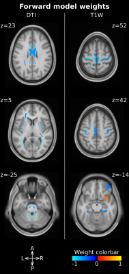 |
104 | Identification of brain alterations specifically associated with Duchenne Muscular Dystrophy using multi-modal MRI and multivariate analysis
Denis Peruzzo1, Tommaso Ciceri1, Sara Mascheretti2, Valentina Lampis3, Filippo Arrigoni4, Nivedita Agarwal1, Alice Giubergia1, Filippo Maria Villa3, Alessandro Crippa3, Maria Nobile3, Elisa Mani3, Annamaria Russo5, and Maria Grazia D'Angelo5
1Neuroimaging Unit, Scientific Institute IRCCS Eugenio Medea, Bosisio Parini, Italy, 2Department of Brain and Behavioral Sciences, University of Pavia, Pavia, Italy, 3Child Psychopathology Unit, Scientific Institute IRCCS Eugenio Medea, Bosisio Parini, Italy, 4Paediatric Radiology and Neuroradiology Department, V. Buzzi Children’s Hospital, Milano, Italy, 5NeuroMuscular Unit, Scientific Institute IRCCS Eugenio Medea, Bosisio Parini, Italy Keywords: Neurodegeneration, Genetic Diseases Duchenne Muscular Dystrophy (DMD) is a genetic disease caused by an abnormal dystrophin expression and it is often associated with other cognitive and behavioral impairments, which are an important confounding factor while investigating the effects of dystrophin abnormal expression in the central nervous system. We applied a Machine Learning based analysis to T1-weighted and Diffusion Tensor Imaging data of 36 subjects accounting also for their demographic, cognitive and behavioral profiles. DMD is specifically associated with a reduced microstructural integrity of long fiber bundles and a reduced cortical thickness of the motor cortex, cingulate cortex, hippocampus and insula. |
|
1922.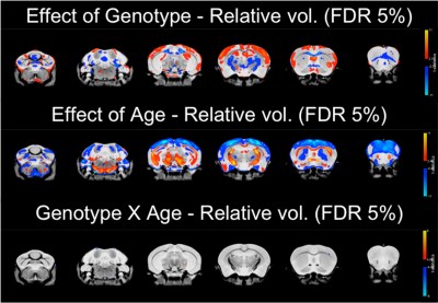 |
105 | Altered brain structure in a mouse model of ALS with mutation in Tardbp is observed from early adulthood
Aurea Martins Bach1, Shoshana Spring2, Zeinab Ali3, Brian J. Nieman2, John Sled2, Remya R. Nair3,4, Elizabeth Fisher5, Silvia Corrochano6, Thomas Cunningham3,7, Jason Lerch1, and Karla Miller1
1Wellcome Centre for Integrative Neuroimaging, FMRIB, Nuffield Department of Clinical Neurosciences, University of Oxford, Oxford, United Kingdom, 2Mouse Imaging Centre, The Hospital for Sick Children, Toronto, ON, Canada, 3Mammalian Genetics Unit, MRC Harwell Institute, Oxford, United Kingdom, 4Department of Physiology, Anatomy and Genetics, University of Oxford, Oxford, United Kingdom, 5Department of Neuromuscular Diseases, UCL Queen Square Institute of Neurology, University College London, London, United Kingdom, 6Neurological Disorders Group, Hospital Clínico San Carlos, IdISSC, Madrid, Spain, 7MRC Prion Unit and Institute of Prion diseases, University College London, London, United Kingdom Keywords: Neurodegeneration, Preclinical Amyotrophic lateral sclerosis (ALS) is a progressive neurodegenerative disease characterized by aggregates of TDP-43 in the brain. The TDP-M323K mouse model of ALS has a mutation in Tardbp and presents progressive motor, neurological and behavioural phenotypes, in addition to widespread changes in brain volume at 12 months of age. Here, we assessed if these volumetric changes are progressive or if they are already present before other symptoms start to present. Post-mortem structural MRI in 3- and 12 months-old TDP-M323K mice revealed that brain volume is already altered in young adults despite the absence of major clinical and pathological phenotypes. |
|
1923.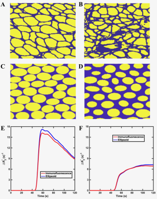 |
106 | Investigation of the biophysical basis of relaxivity contrast imaging as a biomarker for myofiber microstructural changes in an ALS model
Natenael B Semmineh1, Ethan Mathew2, Bruce Damon3, and C Chad Quarles1
1Cancer Systems Imaging, The university of Texas MD Anderson Cancer Center, Houston, TX, United States, 2Barrow Neuroimaging Innovation Center, Barrow Neurological Institute, Phoenix, TX, United States, 3Carle Clinical Imaging Research Program Stephens Family Clinical Research Institute Carle Health, Urbana, IL, United States Keywords: Neurodegeneration, Microstructure, amyotrophic lateral sclerosis - ALS Amyotrophic lateral sclerosis (ALS) is a fatal upper and lower motor neuron degradation disease that leads to progressive myofiber abnormalities (e.g., decreased size and distribution). The relaxivity contrast imaging (RCI) parameter, TRATE (tissue transverse relaxivity at tracer equilibrium), has been shown to decrease during ALS induced myofiber degradation. The goal of this study is to expand a validated computational method to investigate the biophysical basis of TRATE in muscles of ALS patients and to optimize RCI acquisition parameters. |
|
1924.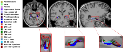 |
107 | Hippocampal subfields atrophy based on automated volumetry analysis in end-stage-renal disease patients
Yuhan Jiang1, Yuan Li1, Qingwei Song1, and Yanwei Miao1
1the First Affiliated Hospital of Dalian Medical University, Dalian, China Keywords: Neurodegeneration, Neurodegeneration Cognitive impairment is very common in patients with end-stage renal disease (ESRD). Hippocampal atrophy has been proven to be an effective marker for distinguishing the normal population from the cognitively impaired patients. However, there are almost no studies on changes in hippocampal structure, especially the volume of hippocampal subfields, in ESRD patients. In this study, automated volumetry of hippocampal subfields in ESRD patients was performed and compared with healthy controls, and found that ESRD patients have hippocampal subfield atrophy, especially in bilateral fimbria and right hippocampal tail. |
|
| 1925. | 108 | 3 T MR Neuroimagingand CT Guided Injection for Atypical Facial Pain: A multimodality analysis of Pain Response
YANG MaoJiang1, Liu JianHao2, ANUP Bhetuwal1, QIONG Xian3, ZHANG HanWen1, YANG HanFeng1, Chen Meining4, and XU XiaoXue1
1Affiliated Hospital of North Sichuan Medical College, Nanchong 637000, Nanchong, China, 2The Fifth People's Hospital of Chengdu, Chengdu, China, 3Second Affiliated Hospital of North Sichuan Medical College,Nanchong 637100, Nanchong, China, 4MR Scientific Marketing, Siemens Healthcare, Shanghai, China, Shanghai, China Keywords: Neurodegeneration, Neurodegeneration At present, the pathogenesis of atypical facial pain is unknown. We conducted pain related analysis on the included patients through magnetic resonance imaging and trigeminal neurolysis, and the results suggest that trigeminal neuropathy may be a pathogenesis of atypical facial pain. |
|
1926.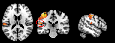 |
109 | Olfactory Oddball Detection Task Activates the Left Temporoparietal Junction
Prasanna Karunanayaka1, Biyar Ahmed1, Rommy Elyan1, and Qing Yang1
1Pennsylvania State University College of Medicine, Hershey, PA, United States Keywords: Neurodegeneration, Neuro, Olfactory fMRI Both the medial and inferior temporal lobes have been previously implicated in odor-identification. However, the precise neural substrate remains unclear. We used a novel oddball detection olfactory fMRI task to probe the neural basis of odor identification. fMRI activation was detected in the left temporoparietal junction along with known olfactory brain structures. Given the presence of odor identification deficits in early Alzheimer’s disease (AD), our paradigm has the potential to establish relationships between olfactory deficits, neurodegeneration, and memory impairment. |
|
1927.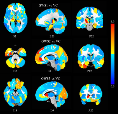 |
110 | Connectomics Biomarkers of Gulf War Illness
Guangming Yang1, Bruce Crosson1, Robert Haley2, Kaundinya Gopinath1, and Ying Guo1
1Emory University, Atlanta, GA, United States, 2UT Southwestern Medical Center, Dallas, TX, United States Keywords: Neurodegeneration, fMRI (resting state) Around 200,000 veterans of the 1991 Gulf War (GW) suffer from GW illness (GWI). GWI is a poorly understood chronic medical condition, characterized by multiple symptoms. One factor that hampers mechanistic investigations into GWI is that there is considerable heterogeneity in symptoms across the GW veteran population. Only one case definitions of GWI addresses this heterogeneity. The Haley GWI case definition addresses this by further breaking down GWI into three main syndrome variants (Syn1, Syn2, and Syn3) based on factor analysis of symptoms presented by GWI veterans. In this study, we extracted rsfMRI connectomics biomarkers for different syndromes of GWI. |
|
1928.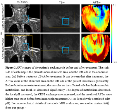 |
111 | Multiparametric MRI assessment of botulinum-toxin treatment for a patient with cervical dystonia
Xubin Chai1,2, Le He1, Changhao Zhu3, Wanting Hu3, Rong Xue2, Xiaolei Song1, and Li Wang4
1Center for Biomedical Imaging Research, Department of Biomedical Engineering, School of Medicine, Tsinghua University, Beijing, Beijing, China, 2State Key Laboratory of Brain and Cognitive Science, Institute of Biophysics, Chinese Academy of Sciences, Beijing, China, 3China School of Information Sciences and Technology, Northwest University, Xi'an, China, 4Department of Neurology, The First Hospital of Tsinghua University, Beijing, China Keywords: Neurodegeneration, Molecular Imaging Cervical dystonia (CD) has been regarded as the most common form of focal dystonia, the affected neck muscles always in sustained situation lead to an abnormal rotation of the head[1]. The mechanisms of CD have not yet been thoroughly illustrated. Botulinum toxin (BT)is considered as the recommended first-line therapeutic method for the focal dystonia[2].Traditional MRI Scan has been used to evaluate any pathophysiological changes of brain anatomy or the affected neck muscles[3].However, seldom MRI have provided the cervical muscles metabolic messages[4].The CEST MRI could provide the metabolic aspects of the cervical muscles before and after the treatment of the BT |
|
1929.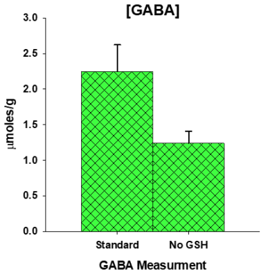 |
112 | Glutathione+γ-Aminobutyric Acid Detection Improves Inter-Subject Concentration Variability at 3T
Jack Knight-Scott1, Marie Caillaud2, Isabelle Gallagher2, Yanrong Li2, and Andreana P. Haley2
1Radiology, Children's Healthcare of Atlanta, Atlanta, GA, United States, 2Psychology, The University of Texas at Austin, Austin, TX, United States Keywords: Neurodegeneration, Spectroscopy Quantification of the combination glutathione (GSH) + γ-aminobutyric acid (GABA) at 3T in medium TE but high signal-noise brain spectra yields a stable concentration with a low coefficient of variation (CV) of only 6%, smaller than individual CV values for GSH or GABA. While an indication of the interdependency of GSH and GABA quantification, [GSH+GABA] also suggests a pathway for the study of perturbations in GABA and GSH at 3T while maintaining the high information content typical of un-edited spectra. |
|
1930.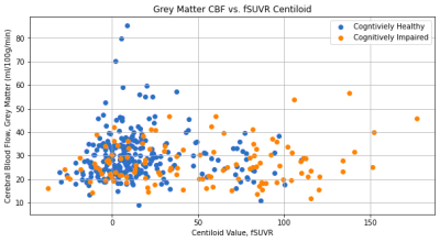 |
113 | Comparing brain amyloid load using PET to grey matter perfusion using ASL on the OASIS-3 dataset
Sierra Sparks1, Daniel P. Bulte1, and Joana Pinto1
1Institute of Biomedical Engineering, Department of Engineering Science, University of Oxford, Oxford, United Kingdom Keywords: Neurodegeneration, Alzheimer's Disease, Tracers Alzheimer’s disease (AD) is the leading cause of dementia, and it is associated with cerebral amyloid beta accumulation and changes in cerebral perfusion. Amyloid PET imaging can be used to measure the amyloid beta load, while ASL-MRI is preferred for cerebral perfusion measurements, which may occur before amyloid beta accumulation. This study compares the amyloid beta load with the CBF in grey matter, in both cognitively healthy and cognitively impaired subjects from the OASIS-3 dataset. A logistic regression model using this data was able to classify the cognitive status of subjects with a 78.48% test accuracy. |
|
1931. |
114 | Age-related cell size and cellularity variation in deep gray matter in vivo
Yufan Chen1, Yishi Wang2, Changyuan Xu3, Diwei Shi4, and Guangbin Wang1
1Shandong Provincial Hospital, Shandong University, Jinan, China, 2Philips Healthcare, Beijing, China, 3Shandong Provincial Hospital Affiliated to Shandong First Medical University, Jinan, China, 4Center for Nano & Micro Mechanics, Department of Engineering Mechanics, Tsinghua University, Beijing, China Keywords: Neurodegeneration, Degenerative, aging The aging progress of neuron is characterized by degenerative changes of structure and function. Few studies have focus on the structure in cellular level. In this study, we quantified cell size and cellularity in globus pallidus, putamen and substantia nigra pars compacta using time dependent diffusion imaging. Subgroup analysis by age showed the increased intracellular volume fraction of putamen in older people while diameter does not change over age. These findings might demonstrate the potential of MRI cell size imaging to assess the degeneration of neuron on cellular level. |
|
1932.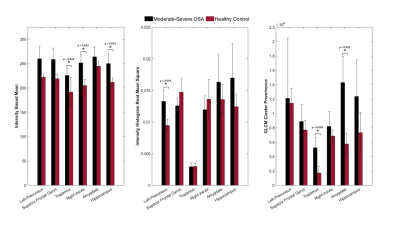 |
115 | 3D MRI Texture Analysis of the Brain in Obese Subjects with OSA: Analysis of Brain Injury and Relationship to Cognitive Impairments
Daniel Sare1, Amartei Brocke1, and Andrea Kassner1,2
1Translational Medicine, The Hospital for Sick Children, Toronto, ON, Canada, 2Department of Medical Imaging, The University of Toronto, Toronto, ON, Canada Keywords: Neurodegeneration, Brain Up to 60% of obese youths with obstructive sleep apnea (OSA) are afflicted with episodes of nocturnal hypoxia, a known risk factor for structural cerebral alterations and neurocognitive problems leading to cognitive impairment. By applying grey-level co-occurrence-based texture analysis in children with and without OSA, we were able to show that the presence of OSA is associated with microscopic changes in normal appearing white matter in regions impacted by cognitive impairment. The findings support the lower tissue homogeneity, and decrease in cortical density and thickness seen in moderate-severe OSA groups. |
|
1933.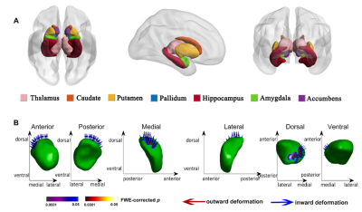 |
116 | Alterations of cortical and subcortical structures in mild cognitive impairment
Junxia Wang1 and Bing Zhang1
1Nanjing Drum Tower Hospital, Nanjing, China Keywords: Neurodegeneration, Alzheimer's Disease In this study, we investigated alterations of cortical morphology, subcortical nuclei volume and morphology in MCI using multiple morphological analysis methods. Moreover, we explored these imaging features relationship with cognitive performances and their efficiency in classification of MCI using support vector machine (SVM). We speculate that combining volumetric and morphological analysis methods will outperform than single analysis method in MCI identification. |
|
1934.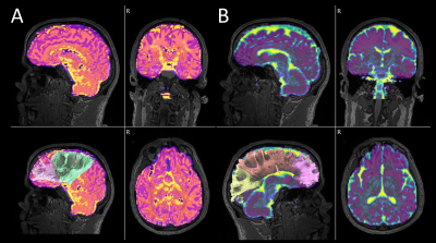 |
117 | Combined Microstructural Assessment of Progressive Apraxia of Speech by Diffusion Tensor Imaging-Based Tractography and Multi-Shell NODDI
Rodolfo G. Gatto1, Joseph R. Duffy1, Rene L. Utianski1, Heather M. Clark1, Hugo Botha1, Mary M. Machulda2, Val J. Lowe3, Keith A. Josephs1, and Jennifer L. Whitwell3
1Division of Neurology, Mayo Clinic at Rochester, Rochester, MN, United States, 2Division of Psychiatry and Psychology, Mayo Clinic at Rochester, Rochester, MN, United States, 3Division of Radiology, Mayo Clinic at Rochester, Rochester, MN, United States Keywords: Neurodegeneration, Rare disease, Progressive Apraxia of Speech Progressive apraxia of speech (PAOS) is a tauopathy characterized by difficulties with motor speech programming and planning. Grey and white matter (GM & WM) brain regions were interrogated by diffusion tensor imaging (DTI) and neurite orientation dispersion and density imaging (NODDI). Twenty-three patients with PAOS and 22 matched controls underwent diffusion MRI. Global WM differences in PAOS were better attained by intracellular volume fraction (ICVF), whereas GM global differences were better attained by mean diffusivity (MD) and isotropic volume fraction (isoVF). DTI and NODDI represent unique aspects of brain tissue microstructure and can be used as PAOS biomarkers. |
|
1935.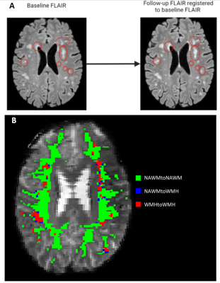 |
118 | Cerebrovascular reactivity is associated with longitudinal white matter hyperintensity progression in small vessel disease on 7T MRI
Stanley D.T. Pham1, Danique E. Versluis1, Hilde van den Brink2, Nikki Dieleman2, Jaco J.M. Zwanenburg1, Geert Jan Biessels2, and Jeroen C.W. Siero1
1Radiology, UMC Utrecht, Utrecht, Netherlands, 2Neurology, UMC Utrecht, Utrecht, Netherlands Keywords: Neurodegeneration, White Matter, small vessel disease, white matter hyperintensities We studied the association between cerebrovascular reactivity measured with blood oxygenation level-dependent (BOLD-CVR) to hypercapnia and progression of white matter hyperintensities (WMH) on 7T MRI in cerebral small vessel disease (SVD). BOLD-CVR was assessed in voxels of four different categories which represent the fate of a voxel after two-year follow-up. The BOLD-CVR in normal appearing white matter (NAWM) that converted into WMH was significantly lower than the BOLD-CVR of voxels that remained WMH (0.39 [0.16-0.63] vs 0.98 [0.75-1.21]) (p<0.0001). The association between BOLD-CVR and WMH progression signifies the potential of BOLD-CVR to be a predictor of disease progression in SVD. |
|
1936.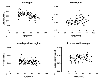 |
119 | Regional age-related changes of neuromelanin and iron in the substantia nigra based on neuromelanin accumulation and iron deposition
Yufan Chen1, Tao Gong2, Cong Sun3, Fei Gao2, Tong Chen1, Weibo Chen4, and Guangbin Wang2
1Shandong Provincial Hospital, Shandong University, Jinan, China, 2Shandong Provincial Hospital Affiliated to Shandong First Medical University, Jinan, China, 3Department of Radiology, Beijing Hospital, National Center of Gerontology, Institute of Geriatric Medicine, Chinese Academy of Medical Sciences, Beijing, China, 4Philips Healthcare, Shanghai, China Keywords: Neurodegeneration, Aging The study investigated age-related neuromelanin signal variation and iron content changes in the subregions of substantia nigra simultaneously by magnetization transfer contrast neuromelanin-sensitive multi-echo fast field echo sequence in a normal population. We manually delineated neuromelanin, iron deposition regions and the overlap regions. The correlation analysis revealed contrast ratio increased over age, while volume showed an age-related decline in the overlap region which were similar to those in the neuromelanin region. No significant correlations were found between susceptibility and age in any subregion. This study may provide more insight for the future degenerative elucidations of substantia nigra. |
|
1937.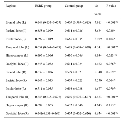 |
120 | Intravoxel incoherent motion diffusion weighted imaging to assess brain microstructure and perfusion in patients with end-stage renal disease
Xiangxiang Wu1, Zijian Jiang1, Jiahui Zheng1, Zhuqing Jiao2, Tongqiang Liu1, Weiqiang Dou3, and Haifeng Shi1
1Changzhou Second People’s Hospital, Changzhou, China, 2Changzhou University, Changzhou, China, 3GE Healthcare, MR Research China, Beijing, China Keywords: Neurodegeneration, Diffusion/other diffusion imaging techniques This study aimed to investigate the clinical value of intravoxel incoherent motion (IVIM) diffusion-weighted imaging in evaluating the brain microstructural and perfusion changes in end-stage renal disease (ESRD) patients. 40 ESRD patients and 30 healthy subjects were recruited in this study and underwent IVIM MRI. The microstructure and perfusion of the brain showed significantly differences in the left frontal lobe, bilateral temporal lobe, left hippocampus, right occipital lobe, represented by IVIM derived parameters of slow apparent diffusion coefficient (ADCslow) and fast apparent diffusion coefficient (ADCfast). Therefore, we concluded that the brain microstructure and perfusion were impaired in ESRD patients. |
|
 Back to Meeting Home
 Back to the Program-at-a-Glance
The International Society for Magnetic Resonance in Medicine is accredited by the Accreditation Council for Continuing Medical Education to provide continuing medical education for physicians.
 Back to Meeting Home
 Back to the Program-at-a-Glance
 View Presentation Video