Digital Poster
Blood Vessels I
ISMRM & ISMRT Annual Meeting & Exhibition • 03-08 June 2023 • Toronto, ON, Canada

| Computer # | |||
|---|---|---|---|
2294.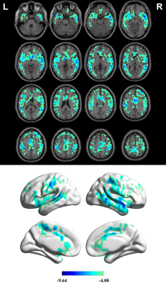 |
121 | Dobutamine-induced Stress Altered Cerebral Blood Flow in Healthy Adults: a 3D Pseudocontinuous Arterial Spin Labeling Study
Pengling Ren1, Tingting zhang1, Haijun Niu2, Yawen Liu2, Linkun Cai2, erwei zhao3, Yishi Wang4, Yi Zhu4, Dong Liu1, Min Li1, Wenjuan Liu1, Penggang Qiao1, Wei Zheng3, Zhenghan Yang1, and Zhenchang Wang1
1Beijing Friendship Hospital, Capital Medical University, Beijing, China, 2School of Biological Science and Medical Engineering, Beihang University, Beijing, China, 3National Space Science Center, Chinese Academy of Sciences, Beijing, China, 4Philips Healthcare, Beijing, China Keywords: Blood vessels, Arterial spin labelling, Dobutamine Stress It is not fully known whether dobutamine commonly clinically used in echocardiography and short-term treatment of congestive heart failure has effect on brain microcirculatory behavior. In this study, we sought to investigate the effect of dobutamine on cerebral haemodynamics in healthy volunteers. The results showed that dobutamine-induced stress significantly decreased the CBF in the anterior circulation, mainly in frontal lobe. Furthermore, volunteers with high BMI, large BA diameter and low SBP during dobutamine stress test are more likely to have stress-induced decrease of CBF. |
|
2295.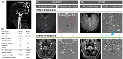 |
122 | Real-time phase contrast sequences versus conventional phase contrast sequences in cerebral blood flow quantification: an in vivo study
Pan LIU1,2, Heimiri Monnier1, Kimi Piedad Owashi1, Serge Metanbou3, Cyrille Capel4, and Olivier Balédent1,2
1CHIMERE UR 7516, Jules Verne University of Picardy, Amiens, France, 2Medical Image Processing Department, Amiens Picardy University Hospital, Amiens, France, 3Radiology Department, Amiens Picardy University Hospital, Amiens, France, 4Neurosurgery Department, Amiens Picardy University Hospital, Amiens, France Keywords: Blood vessels, Velocity & Flow, real time phase contrast, phase contrast, cerebral blood flow Real-time phase contrast sequences (RT-PC) appear to have great potential in clinical applications. However, it is important to validate RT-PC in the quantification of cerebral blood flow prior to its use in clinical applications. In this study, we analyzed RT-PC accuracy by comparing the segmentation area, flow rate and pulsatility index of cerebral vessels obtained from RT-PC and conventional phase contrast sequences. RT-PC with 2×2 mm2 spatial resolution and 75ms/image temporal resolution can accurately quantify cerebral blood flow rate with an error of less than 3%. Higher temporal resolution in RT-PC could improve accuracy in cerebral arterial flow quantification. |
|
2296.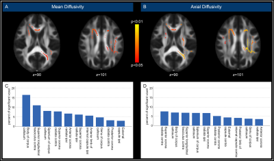 |
123 | Effects of high-grade asymptomatic carotid artery stenosis on grey and white matter structure
Lena Schmitzer1, Stephan Kaczmarz1,2, Jens Göttler1, Michael Kallmayer3, Hans-Henning Eckstein3, Dennis Hedderich1, Jan Kufer1, Claus Zimmer1, Christine Preibisch1,4, and Nico Sollmann1,5
1Department of Neuroradiology, School of Medicine, Technical University of Munich, Munich, Germany, 2Philips GmbH Market DACH, Hamburg, Germany, 3Department for Vascular and Endovascular Surgery, School of Medicine, Technical University of Munich, Munich, Germany, 4Department of Neurology, School of Medicine, Technical University of Munich, Munich, Germany, 5Department of Diagnostic and Interventional Radiology, University Hospital Ulm, Ulm, Germany Keywords: Blood vessels, Diffusion/other diffusion imaging techniques Internal carotid artery stenosis (ICAS) is a known risk factor for stroke, additionally affecting brain structure and function. We investigated ICAS effects on white and grey matter structure using diffusion tensor imaging (DTI) and cortical thickness assessment, revealing alterations in various indices focused on the corpus callosum without signs of atrophy in grey matter. In addition, small vessel disease (SVD) burden, assessed by peak width of skeletonized mean diffusivity (PSMD), was increased in ICAS patients. Taken together, the structural alterations in white matter might precede those in grey matter, supporting the relevance of assessment of microstructure to detect early changes. |
|
2297.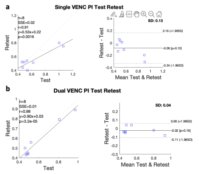 |
124 | Comparison of Arterial Pulsatility of Cerebral Perforating Arteries between Dual-VENC and Single-VENC Phase-Contrast MRI at 7T
Jianing Tang1,2, Elizabeth Joe3, Helena Chui3, and Lirong Yan2,3
1Biomedical Engineering, Northwestern University, Evanston, IL, United States, 2Radiology, Northwestern University, Evanston, IL, United States, 3Department of Neurology, University of Southern California, Los Angeles, CA, United States Keywords: Blood vessels, High-Field MRI Dysfunction of cerebral perforating arteries is a major pathology of small vessel disease. Previous study has demonstrated the pulsatility of cerebral perforating arteries can be measured by 7T high-resolution PC-MRI. In this study, we systematically compared the performance of PC-MRI with single and dual-VENC on pulsatility index (PI) measurements of cerebral perforating arteries. Our results showed that dual-VENC provides more reliable PI measurements than single VENC. PI measurements were insensitive to the temporal resolution of PC-MRI acquisition but showed greater variations with single VENC. The pilot study suggests that PI of cerebral perforating arteries was significantly higher in aged participants. |
|
2298. |
125 | Construction of a contrast enhancement map of intracranial vessel wall MRI with 3D volume rendering
Yin Guo1, Xin Wang2, Gador Canton2, Duygu Baylam Geleri 2, Kaiyu Zhang2, Niranjan Balu2, Thomas S. Hatsukami2, Mahmud Mossa-Basha2,3, and Chun Yuan2,4
1Bioengineering, University of Washington, Seattle, WA, United States, 2University of Washington, Seattle, WA, United States, 3University of North Carolina, Chapel Hill, NC, United States, 4University of Utah, Salt Lake City, UT, United States Keywords: Vessel Wall, Atherosclerosis Contrast enhancement (CE), by the administration of gadolinium, has been shown as an important intracranial vessel wall MRI biomarker. In this study we have developed a CE map with 3D volume rendering, extended from a previously developed intracranial atherosclerosis analysis pipeline MOCHA. 3D CE map can help clinicians easily identify plaque contrast enhancement. It demonstrates CE’s spatial location relative to the vessel and provides reliable quantification of its intensity. It is a promising tool for future longitudinal studies to focus on the association between 3D CE map features and disease development. |
|
2299. |
126 | The relationship between unruptured intracranial aneurysm morphology and hemodynamics - 7T 4D flow MRI
Rick J van Tuijl1, Kimberley M Timmins1, Birgitta K Velthuis1, Pim van Ooij2, Jaco J M Zwanenburg1, Ynte M Ruigrok3, and Irene C van der Schaaf1
1Radiology, UMC Utrecht, Utrecht, Netherlands, 2Pediatric Cardiology, UMC Utrecht, Utrecht, Netherlands, 3Neurology, UMC Utrecht, Utrecht, Netherlands Keywords: Blood vessels, Velocity & Flow, High-Field MRI, Aneurysm We investigated relationships between unruptured intracranial aneurysm (UIA) morphology and hemodynamics parameters measured with 7T 4D flow imaging in 35 patients with an UIA. Blood flow measured in the UIA was positively correlated with UIA size, and negatively correlated with elongation and shape index of UIA (a decrease in shape index is characteristic for unstable UIA (either growing or rupture)). Therefore, blood flow is an important parameter for risk evaluation of UIA. The described relations help toobtain better insight in risk parameters of UIA development, growth, and possible rupture. |
|
2300. |
127 | Alterations of cerebrovascular reactivity and morphologies in de novo Parkinson's disease
Hongwei Li1, Xiali Shao2, Bingyi Wang1, Jian Wang2,3, Jia Jia4, Zhensen Chen1,5, He Wang1,5,6, and Lirong Jin4
1Institute of Science and Technology for Brain-Inspired Intelligence, Fudan University, Shanghai, China, 2Department of Radiology, Zhongshan Hospital, Fudan University, Shanghai, China, 3Department of Radiology, Zhongshan Hospital, Fudan University (Xiamen Branch), Xiamen, China, 4Department of Neurology, Zhongshan Hospital, Fudan Universit, Shanghai, China, 5Key Laboratory of Computational Neuroscience and Brain-Inspired Intelligence (Fudan University), Ministry of Education, Shanghai, China, 6Human Phenome Institute, Fudan University, Shanghai, China Keywords: Blood vessels, Brain Increasing evidence showed that vascular changes might be involved in Parkinson's disease. However, the altered markers of neurovascular unit and cerebral vessels that serve as blood flow adaptors, remain unclear in early stage of de novo PD. In this study, we aimed to investigate regional cerebral blood flow (CBF), cerebrovascular reactivity (CVR) and artery morphological changes in de novo PD patients including cognitive impairment (PD-MCI) and normal cognition (PD-NC). Our data revealed that in the patient group, CVR was significantly reduced in left superior occipital gyrus, whose feeding arteries were dilated, but there was no significant change in CBF. |
|
2301. |
128 | Altered amide proton transfer weighted signal in different Suzuki grading and preinfarction period stages in patients with moyamoya disease
Chao Xia1,2, Jiaxin Zeng1,2, Ziyu Li1,2, Xia Wei1,2, Xing Li1,2, Yuan Sun1,2, Na Hu1,2, Yi Liu3, Kai Ai4, and Su Lui1,2
1Functional and Molecular Imaging Key Laboratory of Sichuan Province, Department of Radiology, West China Hospital, Chengdu, China, 2Research Unit of Psychoradiology, Chinese Academy of Medical Sciences, Chengdu, China, 3Department of Neurosurgery, West China Hospital, Sichuan University, Chengdu, China, 4Philips Healthcare, Xi'an, China Keywords: Blood vessels, Vessels, moyamoya disease, amide proton transfer To explore the alteration of amide proton transfer weighted (APTw) signal in different Suzuki stages and preinfarction period stages in patients with moyamoya disease (MMD), we enrolled 24 patients who underwent computed tomography perfusion (CTP), digital subtraction angiography (DSA), and APTw imaging. Although our results demonstrated that APTw values in the cerebral hemispheres of MMD patients with Suzuki stages V-VI and preinfarction period stage III are significantly higher than those in other stages, suggesting that the microenvironment of cerebral hemispheres in MMD patients with different Suzuki stages and preinfarction period stages suffers different severity of acidosis penumbra. |
|
2302.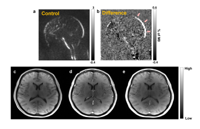 |
129 | Correlation of volume transfer constant measured from WEPCAST and DCE-MRI: A pilot study
Dinil Sasi Sankaralayam1, Wen Shi1, Zixuan Lin2, Anna DuVal3, Kaisha Hazel1, Katie Ecoff3, Dengrong Jiang2, Shiv Saidha3, and Hanzhang Lu1
1Department of Radiology, Johns Hopkins University, Baltimore, MD, United States, 2Johns Hopkins University, Baltimore, MD, United States, 3Department of Neurology, Johns Hopkins University, Baltimore, MD, United States Keywords: Blood vessels, Contrast Agent There is an unmet need of a non-invasive technique to compute the water exchange between vascular and extra-vascular space in the brain since blood brain barrier (BBB) disruption is common in many pathologies and the state of art exogenous contrast agent based method cann’t be applied to all clinical population. So, the current study is aimed at computing a volume transfer constant (Ktrans) from water-extraction-with-phase-contrast-arterial-spin-labelling (WEPCAST) method and to compare it with the global Ktrans from DCE-MRI data. Results of the study finds a good correlation between Ktrans computed from WEPCAST and DCE-MRI data. |
|
2303. |
130 | Magnetic resonance imaging of ultrasound-induced strain fields for discrimination of solid and liquid phases
Ryan Willoughby1, Henrik Odéen2, Jesse Jones3, and Mark Bolding4
1Department of Radiology, University of Alabama at Birmingham, Birmingham, AL, United States, 2Department of Radiology and Imaging Sciences, University of Utah, Salt Lake City, UT, United States, 3Department of Neurosurgery, University of Alabama at Birmingham, Birmingham, AL, United States, 4Radiology, University of Alabama at Birmingham, Birmingham, AL, United States Keywords: Blood vessels, Focused Ultrasound Focused ultrasound (FUS) has gained clinical utility as a non-invasive treatment option, but applications to diseases affecting blood vessels in the brain are just now being investigated. MR acoustic radiation force imaging (MR-ARFI) of FUS-induced displacement and strain fields were evaluated along with diffusion imaging as a method of differentiating solid and liquid material inside a tissue-mimicking gelatin phantom. Apparent diffusion coefficient (ADC) performed best as a solid-liquid classifier, and axial displacement performed worst. Normal and shear strain components performed better than displacement measurements alone. |
|
2304.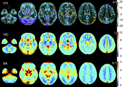 |
131 | Vascular architecture mapping reveals distinct microvascular profiles in healthy grey and white matter
Anja Hohmann1, Ke Zhang2, Christoph Mooshage3, Johann Jende3, Lukas Rotkopf4, Heinz-Peter Schlemmer4, Martin Bendszus3, Wolfgang Wick1,4, and Felix T. Kurz3,4
1Department of Neurology, Heidelberg University Hospital, Heidelberg, Germany, 2Department of Radiology, Heidelberg University Hospital, Heidelberg, Germany, 3Department of Neuroradiology, Heidelberg University Hospital, Heidelberg, Germany, 4German Cancer Research Center, Heidelberg, Germany Keywords: Blood vessels, Quantitative Imaging Vessel architecture mapping (VAM) is a quantitative MR imaging technique based on dynamic changes in gradient-echo (R2*) and spin-echo (R2) relaxation rates during contrast agent administration, that can characterize cerebral blood vessel microstructure and their changes in vivo. We aimed to establish VAM parameter reference ranges in healthy brain tissue by scanning n=135 patients with whole-brain high resolution VAM at 3.0 Tesla MRI. Subsequent analysis revealed distinct microvascular profiles for cortical gray matter, supratentorial white matter and examined subcortical nuclei. These reference values may serve as a starting point for future VAM studies on cerebrovascular pathologies. |
|
2305.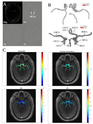 |
132 | Voxel-wise Parametric Mapping of Cerebrovascular Hemodynamics using 4D Flow MRI: A Scan-Rescan Reproducibility Study at 3T
Jackson E Moore1, Deima Koko1, Adam Richter1, Ann Ragin1, Fan Caprio2, Sameer A Ansari1, Susanne Schnell1,3, Michael Markl1, and Kelly Jarvis1
1Radiology, Feinberg School of Medicine, Northwestern University, Chicago, IL, United States, 2Neurology, Feinberg School of Medicine, Northwestern University, Chicago, IL, United States, 3Department of Medical Physics, University of Greifswald, Greifswald, Germany Keywords: Blood vessels, Velocity & Flow Cerebrovascular dual-venc 4D flow MRI enables measurement of global and local intracranial 3D hemodynamics with full volumetric coverage of the circle of Willis. Hemodynamic analysis was completed using a semi-automated voxel-wise parametric mapping tool, enabling assessment of velocity-based parameters, including mean velocity, peak velocity, kinetic energy and time-to-peak velocity. The workflow reproducibility was evaluated with scan-rescan analysis for 10 subjects who underwent two MRI scans within 30 days. Global circle of Willis and hemispheric analysis regions showed good to excellent reproducibility for all parameters except for time-to-peak velocity. Regional analysis showed better reproducibility for parameters derived from systolic cardiac timepoints. |
|
2306.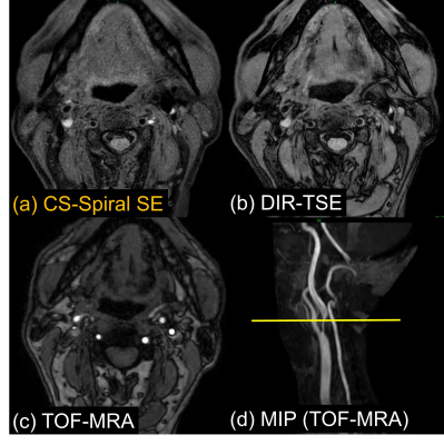 |
133 | Improved visualization of T1-weighted carotid artery plaque imaging using time-efficient spiral spin-echo DIXON with Compressed SENSE
Masahiro Enzaki1, Minako Azuma2, Yoshihito Kadota2, Tenma Takahashi1, Masami Yoneyama3, Hiroshi Hamano3, and Masanori Komi1
1Division of Radiology, Miyazaki University Hospital, Miyazaki, Japan, 2Department of Radiology, Faculty of Medicine, University of Miyazaki, Miyazaki, Japan, 3Philips Japan, Tokyo, Japan Keywords: Blood vessels, Vessels In MR carotid plaque imaging, black-blood imaging can diagnose plaque components and predict plaque hardness with a high degree of accuracy. In this study, we proposed T1-weighted black-blood carotid plaque images using non-ECG gated time-efficient Spiral spin-echo (SE) with Compressed SENSE (CS-Spiral SE) and compared with conventional ECG-gated DIR-TSE. We have demonstrated that the non-gated CS-Spiral SE could provide improved visualization in carotid plaque imaging with superior contrast to DIR-TSE with sufficient fat and blood signal suppression within a shorter scan time. It has a great potential to help more accurate assessment and characterization of the carotid atherosclerotic plaque. |
|
2307.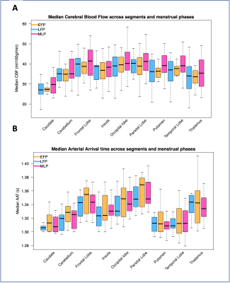 |
134 | Hormone-driven CBF and AAT changes across the menstrual cycle
Melissa Emily Wright1, Andrew Crofts1, Saajan Davies1, Michael Germuska1, Jessica J Steventon2, and Kevin Murphy1
1Cardiff University Brain Research Imaging Centre (CUBRIC), School of Physics and Astronomy, Cardiff University, Cardiff, United Kingdom, 2Cardiff University Brain Research Imaging Centre (CUBRIC), School of Medicine, Cardiff University, Cardiff, United Kingdom Keywords: Blood vessels, Arterial spin labelling The current study investigated the influence of progesterone and oestrogen on cerebral blood flow (CBF) and arterial arrival time (AAT) across a menstrual cycle and multiple cortical and subcortical regions. 21 healthy female participants were scanned in a Siemens MAGNETOM Prisma 3T scanner and their bloods were taken to measure circulating hormones. Linear models found that while there was no interactive effect with region for either hormone, circulating oestrogen explained a significant portion of CBF value, while progesterone explained a significant portion of AAT value. Recommendations for future research are discussed. |
|
2308.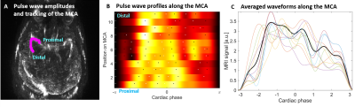 |
135 | MRI derived pulse wave indexes of the middle cerebral arteries
Henning U. Voss1,2, Liangdong Zhou2, and Yi Li2
1Cornell University, Ithaca, NY, United States, 2Weill Cornell Medicine, New York, NY, United States Keywords: Blood vessels, Vessels, Pulsatility, Pulse waves The pulse pressure wave has been used as a marker for cardiovascular health but cannot be measured easily within the brain. We have developed an MRI protocol to image, track, and process intracranial pulse waves of the middle cerebral artery. The protocol allows for the estimation of pulsatility along the MCA and extends the estimation of standard pulse waveform indexes that are normally being used in the periphery to the brain. The reliability and reproducibility of this protocol is being tested on normal subjects and subjects with mild cognitive impairment.
|
|
2309.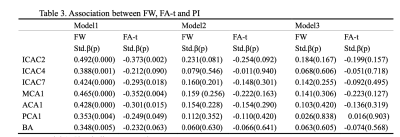 |
136 | Intracranial arterial pulsatility assessed by 4D-flow imaging is associated with white matter degeneration
Linyun Xie1, Yao Zhang2, Hui Hong1, Shan Xu1, Lei Cui2, Luwei Hong2, Jixuan Li2, and Peiyu Huang1
1Department of Radiology, The Second Affiliated Hospital, Zhejiang University School of Medicine, Hangzhou, China, 2The Second Affiliated Hospital, Zhejiang University School of Medicine, Hangzhou, China Keywords: Blood vessels, Velocity & Flow, Pulsatility We investigate the association between the intracranial pulsatility assessed by 4D-flow imaging and white matter degeneration in a community cohort. White matter free water and tissue fractional anisotropy were used to reflect white matter degeneration. We found that the intracranial arterial pulsatility in different vessel segments was positively associated with increased free water, and negatively associated with tissue fractional anisotropy. After adjusting for age and vascular risk factors, the associations disappeared. Further investigations in larger samples are warranted. |
|
2310.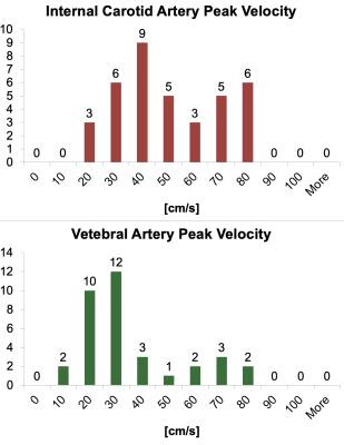 |
137 | Assessing Ranges of Cerebral Blood Flow Velocity and Vessel Area with Phase Contrast in Children and Adults
Eamon Doyle1,2, Hannah Wiseman3, Isabel Torres3, Payal Shah4, John Wood2,5, and Matthew Borzage2,6
1Radiology, Children's Hospital of Los Angeles, Los Angeles, CA, United States, 2Keck School of Medicine, University of Southern California, Los Angeles, CA, United States, 3Children's Hospital of Los Angeles, Los Angeles, CA, United States, 4Children's Hospital of Los Angeles, Los, CA, United States, 5Division of Cardiology, Children's Hospital of Los Angeles, Los Angeles, CA, United States, 6Pediatrics, Children's Hospital of Los Angeles, Los Angeles, CA, United States Keywords: Blood vessels, Blood, flow Measuring brain blood flow is possible with phase contrast (PC) imaging. We assessed blood flow through and area of internal carotid and vertebral arteries in pediatric and adult populations. Sequences parameters are suggested based upon the findings to facilitate PC acquisitions at other institutions. |
|
2311.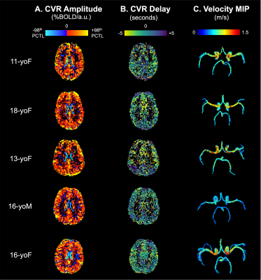 |
138 | Regional coupling of macrovascular flow velocity and cerebrovascular reactivity amplitude and delay in healthy adolescents
Kristina M Zvolanek1,2, Jackson E Moore2,3, Kelly Jarvis3, Adam Richter3, Sarah J Moum3,4, and Molly G Bright1,2
1Physical Therapy and Human Movement Sciences, Feinberg School of Medicine, Northwestern University, Chicago, IL, United States, 2Biomedical Engineering, McCormick School of Engineering and Applied Sciences, Northwestern University, Evanston, IL, United States, 3Radiology, Feinberg School of Medicine, Northwestern University, Chicago, IL, United States, 4Ann & Robert H. Lurie Children’s Hospital of Chicago, Chicago, IL, United States Keywords: Blood vessels, Velocity & Flow, 4D flow In a preliminary sample of healthy adolescents, we evaluated the relationship between hemodynamic function in large cerebral arteries and smaller cortical vessels. Specifically, we investigated correlations between 4D flow MRI-derived systolic blood velocity in the anterior, middle, and posterior cerebral arteries and BOLD cerebrovascular reactivity (CVR) in associated vascular territories. Both the amplitude and delay of CVR were negatively correlated with systolic velocity. These results provide new insights into the mechanism of CVR and measures of macrovascular flow, but more work is needed to better understand these relationships. Our work establishes important normative data for future comparisons in cerebrovascular pathology. |
|
2312.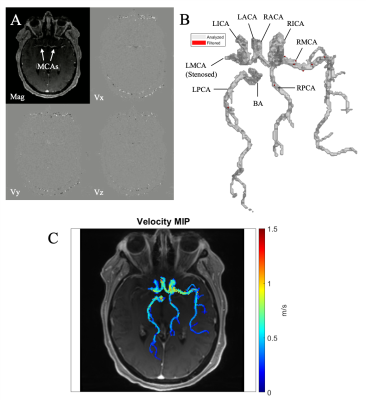 |
139 | Cerebrovascular Peak-Velocity Mapping in Intracranial Atherosclerotic Disease (ICAD) using 4D Flow MRI
Jackson E Moore1, Deima Koko1, Kelly Jarvis1, Abhinav Patel1, Adam Richter1, Ramez Abdalla1, Maria Aristova1, Ann Ragin1, Fan Caprio2, Sameer A Ansari1, Susanne Schnell1,3, and Michael Markl4
1Radiology, Feinberg School of Medicine, Northwestern University, Chicago, IL, United States, 2Neurology, Feinberg School of Medicine, Northwestern University, Chicago, IL, United States, 3Department of Medical Physics, University of Greifswald, Greifswald, Germany, 4Radiology, Northwestern University, Chicago, IL, United States Keywords: Blood vessels, Atherosclerosis Intracranial atherosclerotic disease (ICAD) accounts for 10-50% of acute ischemic strokes and is a leading cause of global morbidity and mortality. We used a semi-automated voxel-wise velocity-based mapping approach to visualize and quantify global and regional peak velocities (PVs) in the Circle of Willis (CoW) in 31 ICAD patients with moderate to severe MCA or ICA stenosis and 23 healthy controls. Our findings indicate 4D flow MRI-based cerebrovascular PV mapping can detect asymmetric PV distribution in the CoW on hemispheric and vessel levels. Future work will include investigation of PV asymmetry measures in the assessment of ICAD progression and outcomes. |
|
2313.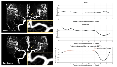 |
140 | Development of quantitative vessel analysis platform based on MR angiography to assess reversible cerebral vasoconstriction syndrome
Pei-Hsuan Kuo1, Shuu-Jiun Wang2,3,4, Jiing-Feng Lirng2,5, Shih-Pin Chen2,3,4, Chia-Hung Wu2,5, Yu Kuo2,5, and Chia-Feng Lu1
1Department of Biomedical Imaging and Radiological Sciences, National Yang Ming Chiao Tung University, Taipei, Taiwan, 2School of Medicine, National Yang Ming Chiao Tung University, Taipei, Taiwan, 3Department of Neurology, Neurological Institute, Taipei Veterans General Hospital, Taipei, Taiwan, 4Brain Research Center, National Yang Ming Chiao Tung University, Taipei, Taiwan, 5Department of Radiology, Taipei Veterans General Hospital, Taipei, Taiwan Keywords: Blood vessels, Visualization Reversible cerebral vasoconstriction syndrome (RCVS) shows reversible diffuse constriction of the cerebral arteries. Previous studies proposed manual measurement of vasoconstriction. However, the stenosis estimated by comparing the stenosed with proximally normal diameter might result in an underestimate of vasoconstriction for the long-segmental stenosis. Accordingly, we developed a quantitative vessel analysis platform to measure the vasoconstriction rate by comparing vessel diameters between acute and remission MR angiography. Our platform provided objective measures of vasoconstriction and further confirmed that acute RCVS presented not only local vasoconstriction but a large portion of short- and long-segmental vasoconstriction in all artery segments. |
|
 Back to Meeting Home
 Back to the Program-at-a-Glance
The International Society for Magnetic Resonance in Medicine is accredited by the Accreditation Council for Continuing Medical Education to provide continuing medical education for physicians.
 Back to Meeting Home
 Back to the Program-at-a-Glance
 View Presentation Video