Digital Poster
Blood Vessels II
ISMRM & ISMRT Annual Meeting & Exhibition • 03-08 June 2023 • Toronto, ON, Canada

| Computer # | |||
|---|---|---|---|
2471.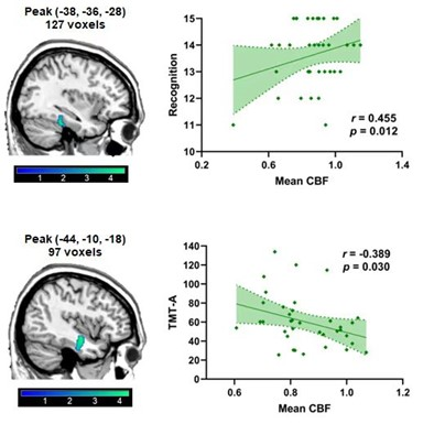 |
121 | Correlation of Cerebral Blood Flow with Cognitive Impairment in Hemodialysis Patients Based on 3D pCASL
Man Wang1, Yuhan Jiang1, Liangjie Lin2, Jiajun Cao1, and Yanwei Miao1
1Department of Radiology, the First Affiliated Hospital of Dalian Medical University, Dalian, China, 2Clinical and Technical Support, Philips Healthcare, Beijing, China Keywords: Blood vessels, Kidney, Cerebral Blood Flow、 Cognitive Impairment、 Hemodialysis Patients、 3D pCASL、 End-stage renal disease This work aimed to use 3D pseudo-continuous arterial spin labeling (3D pCASL) imaging technology to compare the difference of cerebral blood flow between hemodialysis patients (HDs) and healthy controls (HCs), and to analyze the relationship between cerebral blood flow changes and cognitive impairment. Results showed that decreased cerebral blood flow in the left Fusiform (Fusiform-L) and the left Temporal Inferior of HDs compared with HCs. Mean CBF in Fusiform-L was positively correlated with recognition, and mean CBF in Fusiform-L was negatively correlated with psychomotor speed and visual attention in HDs. |
|
2472.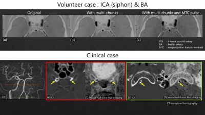 |
122 | Examination of the optimal imaging parameters for intracranial vessel wall bone-like imaging using FRACTURE
Keita Fukushima1, Miho Gomyo2, Kazuhiro Tsuchiya2,3, Shun Saito1, Tatsuya Yoshioka1, Takahiro Arai1, Takayuki Yonaha1, Ayaka Negishi1, Kosuke Sakaguchi1, Yuma Kumagai1, Makoto Obara4, Masatoshi Honda4, Takashi Namiki4, Yoshiyuki Nishimura4, Akihito Nakanishi1, and Kenichi Yokoyama2
1Section of Radiology, Kyorin University Hospital, Tokyo, Japan, 2Department of Radiology, Faculty of Medicine, Kyorin University, Tokyo, Japan, 3Department of Radiology, JR Tokyo General Hospital, Tokyo, Japan, 4Philips Japan, Ltd., Tokyo, Japan Keywords: Blood vessels, Bone, bone-like imaging Fast field echo resembling a CT using restricted echo-spacing (FRACTURE), which has been recently developed, has enabled visualization of calcification as well as cortical and spongy bones. However, it cannot be applied to intracranial vessel wall imaging due to insufficient suppression of cerebrospinal fluid (CSF) signal and intra-arterial flow artifact. In this study, by performing scans of an original phantom and healthy volunteers, we reveal that the application of multi-chunk and magnetization transfer contrast pulse to FRACTURE can suppress CSF signal and intra-arterial flow artifact and that FRACTURE can be efficiently applied to vessel wall bone-like imaging. |
|
2473.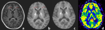 |
123 | Microstructural and microvascular alterations in the non-NPSLE patients: a DKI and 3D pCASL study
Xiaojuan Wang1, Lingling Huang1, Qi Lin1, Lian Yu1, Peng Wu2, and Xiance Zhao2
1Longyan First Hospital Affiliated to Fujian Medical University, Longyan, China, 2Philips Healthcare, Shanghai, China Keywords: Blood vessels, Brain, non-neuropsychiatric systemic lupus erythematosus DKI and 3D pCASL were employed to investigate whether white/gray matter microstructure and cerebral microcirculation were altered in patients suffering from non-neuropsychiatric systemic lupus erythematosus (SLE). MK, MKT and CBF values were obtained from 25 brain areas, including gray and white matter. We demonstrated decreased MK, MKT, and elevated CBF in some regions in SLE compared to controls. This study suggested that the brain lesions existed in SLE, DKI and pCASL might be useful for depicting SLE brain early damages, and MK seems to be more sensitive and helpful. |
|
2474.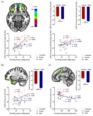 |
124 | Sex-specific grey matter atrophy and brain-behavior relationships in CADASIL
Xiuqin Jia1, Chen Ling2, Yingying Li1, Xuejia Jia1, Chen Zhang3, Danny JJ Wang4, Zihao Zhang5, Yun Yuan2, and Qi Yang1
1Beijing Chaoyang Hospital, Capital Medical University, Beijing, China, 2Peking University First Hospital, Beijing, China, 3MR Scientific Marketing, Siemens Healthineers, Beijing, China, 4USC Mark & Mary Stevens Neuroimaging and Informatics Institute, Keck School of Medicine, University of Southern California, Los Angeles, CA, United States, 5State Key Laboratory of Brain and Cognitive Science, Institute of Biophysics, Chinese Academy of Sciences, Beijing, China Keywords: Blood vessels, Brain, CADASIL The potential neurobiological substrates for sex differences in cerebral autosomal dominant arteriopathy with subcortical infarcts and leukoencephalopathy (CADASIL) are largely unexplored. In this study, we explored the sex-specific neuroanatomical mechanisms of CADASIL. Greater grey matter atrophy was found in the frontotemporal cortex in male CADASIL. Working memory was associated with volumes in the bilateral orbitofrontal cortex and left entorhinal cortex among female CADASIL. The current findings indicate that sex affects the pathogenesis of CADASIL, ranging from differences in neuroanatomy to those in behavioral performance. |
|
2475. |
125 | Impact of resting physiology on the repeatability of phase contrast MRI measures of cerebral blood flow
Hannah R Johnson1,2, Rachael C Stickland2, Kristina M Zvolanek1,2, Yufen Chen3, and Molly G Bright1,2
1Biomedical Engineering, McCormick School of Engineering, Northwestern University, Evanston, IL, United States, Evanston, IL, United States, 2Physical Therapy and Human Movement Sciences, Feinberg School of Medicine, Northwestern University, Chicago, IL, United States, 3Radiology, Feinberg School of Medicine, Northwestern University, Chicago, IL, United States Keywords: Blood vessels, Velocity & Flow Interpretation of clinically significant changes in total cerebral blood flow (CBF) can be confounded by normal variability in flow measures. We assessed the effects of resting end-tidal CO2 (PETCO2) on inter-session variability in total CBF as measured with phase contrast MRI. Participants were scanned twice, 1-3 weeks apart, with PETCO2 clamped at resting physiology. Clamping PETCO2 at participants’ resting respiratory physiology does not notably improve inter-session variability in flow, which was found to be consistent with previously reported values. Furthermore, inter-session changes in resting PETCO2 did not help to explain noted variability in total CBF. |
|
2476.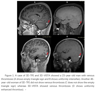 |
126 | The value of high resolution black blood 3D T1-VISTA-MSDE in the detection of cerebral venous sinus
Jianmin ZHENG1, Jianxiu Lian2, Hong Wang1, Leilei Li1, Yingjuan Chang1, Guorui Hou1, Gang Lin1, Yang Li1, Yang Li1, and Minwen Zheng1
1Xijing Hospital, Air Force Medical University, Xi 'an, China, 2Philips Healthcare, Beijing, China, Beijing, China Keywords: Blood vessels, Blood vessels, black blood, venous sinus thrombosis Cerebral venous sinus thrombosis (CVST) is one of ischemic cerebrovascular diseases with low morbidity and high mortality. Three-dimensional volumetric isotropic turbo spin echo acquisition(3D-VISTA)can be adopted, which has high resolution and most blood signals will be suppressed. In addition, a motion-sensitized and fault-driven equilibrium(MSDE) technology were applied for more thorough blood suppression. 3D-VISTA showed SNR and CNR were significantly higher than when compared with enhanced three-dimensional TFE sequence, which could more accurately identify CVST. 3D-VISTA sequence can be used to improve the recognition rate of craniocerebral veins, which could provide valuable evidence for accurate identification of clinical craniocerebral venous thrombosis. |
|
2477.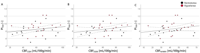 |
127 | Increased pulsatility is associated with higher blood flow in the cerebral microvasculature as assessed with 7T MRI
Elles P. Elschot1,2, Marieke van den Kerkhof1,2, Merel M. van der Thiel1,2,3, Robert J. van Oostenbrugge2,4,5, Abraham A. Kroon5,6, Walter H. Backes1,2,5, and Jacobus F. A. Jansen1,2,7
1Radiology & Nuclear Medicine, Maastricht University Medical Center +, Maastricht, Netherlands, 2School for Mental Health and Neuroscience, Maastricht University, Maastricht, Netherlands, 3Psychiatry & Neuropsychology, Maastricht University Medical Center +, Maastricht, Netherlands, 4Neurology, Maastricht University Medical Center +, Maastricht, Netherlands, 5School for Cardiovascular Diseases, Maastricht University, Maastricht, Netherlands, 6Internal Medicine, Maastricht University Medical Center +, Maastricht, Netherlands, 7Electrical Engineering, Eindhoven University of Technology, Eindhoven, Netherlands Keywords: Blood vessels, Hypertension We studied blood flow pulsatility and perfusion in the cerebral microvasculature to increase the understanding of the pathophysiological processes in hypertension. By exploiting the possibilities of ultra-high-field strength (7T), utilizing phase contrast MRI and spin-echo dynamic susceptibility MRI, we focused specifically on the microvasculature. We found that a higher blood flow pulsatility in the lenticulostriate arteries is correlated with higher cerebral blood flow. Furthermore, hypertension status seems to have an effect on varying pulsatility, which seems to be counteracted by medication usage. |
|
2478.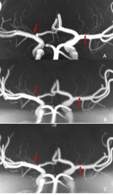 |
128 | Feasibility of pointwise encoding time reduction with radial acquisition (PETRA) MR angiography for assessment of intracranial arteries at 7T
Xiangchuang Kong1, Jun Ma2, Erin Westerhold1, Eric H. Middlebrooks1, Shengzhen Tao1, Chen Lin1, and Xiangzhi Zhou1
1Department of Radiology, Mayo Clinic, Jacksonville, FL, United States, 2Siemens Medical Solutions USA, Inc., Jacksonville, FL, United States Keywords: Blood vessels, High-Field MRI, PETRA-MRA, 7T PETRA-MRA is a non-contrast subtraction-based MRA technique for intracranial-vasculature assessment. Compared to TOF, PETRA-MRA is robust to turbulent-flow-related signal voids and metallic-susceptibility artifacts. 7T MRI should further improve the performance of PETRA-MRA due to higher SNR allowing improved spatial-resolution. Here we demonstrate the feasibility and performance of PETRA-MRA at 7T in evaluating intracranial-vasculature. 7T 3D-TOF and PETRA-MRA were performed on volunteers. The blood-to-background contrast ratio for PETRA-MRA was significantly higher than TOF. PETRA-MRA also has better cerebral-artery-visualization with more uniform artery blood signal and comparable sharpness. Overall, PETRA-MRA is a promising technique for evaluating cerebral-arteries at 7T. |
|
2479.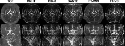 |
129 | Angiographic Comparison of Different Velocity-Selective Labeling Modules Directly on Cerebral Arteries
Dapeng Liu1,2, Dan Zhu1,2, and Qin Qin1,2
1Kennedy Krieger Institute, Baltimore, MD, United States, 2The Russell H. Morgan Department of Radiology and Radiological Science, Johns Hopkins School of Medicine, Baltimore, MD, United States Keywords: Blood vessels, Blood vessels, MR Angiography Velocity-selective labeling has been utilized in both arterial spin labeling (VSASL) and angiography (VSMRA). This study evaluated the labeling characteristics directly on various cerebral arteries using VSMRA with different VS labeling modules, including double refocused hyperbolic tangent (DRHT), eight-segment B1-insensitive rotation (BIR-8), delay alternating with nutation for tailored excitation (DANTE), Fourier transform based velocity-selective saturation (FT-VSS) and inversion (FT-VSI). Their effects on both the cerebral arterial vasculature and static brain tissue were visualized angiographically and compared quantitatively. And the implication for VSASL and VSMRA are discussed. |
|
2480.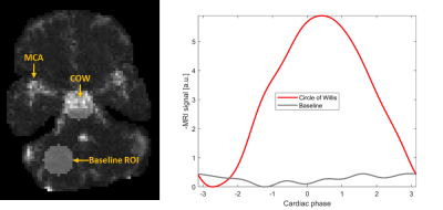 |
130 | Pulsatility in the circle of Willis increases with age
Henning U. Voss1,2 and Qolamreza R. Razlighi2
1Cornell University, Ithaca, NY, United States, 2Weill Cornell Medicine, New York, NY, United States Keywords: Blood vessels, Velocity & Flow, Pulsatility, Pulse waves As we get older, the elasticity of our blood vessels decreases significantly, which can lead to a diminished absorption of blood pressure pulsations, potentially causing damage including hemorrhagic stroke and microbleeds. We studied pulsatility in the circle of Willis, which has been hypothesized to be a pressure absorber, by the method of MRI hypersampling by analytic phase projection (APP). A significant correlation of pulsatility in the circle of Willis with age was found. This finding is interpreted as an ageing-related increase in pulsatile flow relative to the steady flow component of overall cerebral blood flow. |
|
2481.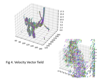 |
131 | 4DFlow Leap-Frog and FFE-DIXON analysis for aneurysm endovascular image-guided treatment
María Paula Del Popolo1,2,3, Rodrigo Nahuel Alcalá1,2,4, Chiara Lombardo1,4, Ezequiel Petra2, Federico Julián González1,5, Olivier Balédent6, Sebastián Moguilner7, Roberto Isoardi1,4,5, Valdir Fialkowski8, and Daniel Fino1,2,5
1Fundación Escuela de Medicina Nuclear, Mendoza, Argentina, 2Fundacion Argentina para el Desarollo en Salud, Mendoza, Argentina, 3Universidad de Mendoza, Mendoza, Argentina, 4FCEN, Universidad Nacional de Cuyo, Mendoza, Argentina, 5Instituto Balseiro, Universidad Nacional de Cuyo, Bariloche, Argentina, 6University Hospital Amiens, Amiens, France, 7Harvard Medical School, Boston, MA, United States, 8Philips, Sao Paulo, Brazil Keywords: Blood vessels, Velocity & Flow, Neuro 4Dflow has become a widely used sequence for dynamical characterization of the vascular system. This work shows that it is feasible to implement Leapfrog to acquire results that are colse to those obtained with Finite Volume Methods with excelent results in velocity, vortex and energy, but with certain limitations when estimating the WSS. In order to diminish the acquisition time, we will implement a postprocessing tool using only a 2D Phase Contrast acquisition and implement FSI (CFD), this results will be compared with those given by the LF method. |
|
2482.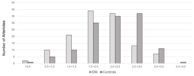 |
132 | Quantitative phase contrast MRI of penetrating arterioles in cerebral white matter in patients with diabetes at 7T
Xiaopeng Zong1, Jordan Jimenez2, Tengfei Li2, and William Powers3
1ShanghaiTech University, Shanghai, China, 2University of North Carolina at Chapel Hill, Chapel Hill, NC, United States, 3Duke University, Durham, NC, United States Keywords: Blood vessels, Aging, small vessel disease Brain lesions caused by cerebral small vessel disease (SVD) are commonly observed in the elderly. However, it remains challenging to noninvasively measure the early pathological changes of the underlying vessels. To this end, we evaluated the feasibility of detecting changes in white matter penetrating arterioles (PA) with 7T MRI in patients with diabetes, a known risk factor for SVD, but without severe SVD and in age and gender matched healthy controls (HC). We observed lower flow velocities in PAs in the patients, suggesting that early changes in PA that are discriminative of overt SVD risks can be detected at 7T. |
|
2483.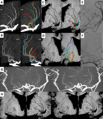 |
133 | Visualization of Lenticulostriate Arteries on High-Resolution Magnetic Resonance Imaging and Digital Subtraction Angiography
ZhongPing Chen1, Dan Tong1, Yang Sun1, Shiyu Guo2, Yueluan Jiang3, and Zechen Yu4
1The First Hospital of Jilin University Department of Radiology, Changchun, China, 2The First Hospital of Jilin University Department of Radiology, Chanchun, China, 3MR Scientific Marketing, Siemens Healthineers, Beijing, China, Beijing, China, 4Siemens Healthineers Digital Technology (Shanghai) Co., Ltd., Shanghai, China Keywords: Blood vessels, Vessels Morphological characteristics of lenticulostriate arteries (LSAs) have important clinical significance. TOF MRA and intracranial vessel wall imaging (VWI) are powerful noninvasive vascular imaging techniques routinely used in vascular visualization. However, due to the small caliber of LSAs, it is difficult for conventional 3T TOF MRA to achieve the visualization of LSAs. High-resolution magnetic resonance imaging based on compressed sensing (CS) technology is expected to solve this dilemma. DSA is the gold standard for vascular visualization. This study proposes an optimized high-resolution TOF MRA scanning scheme based on CS technology and shows its feasibility in LSA visualization by comparing with DSA. |
|
2484.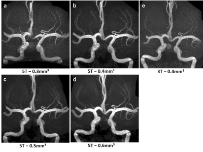 |
134 | Imaging of Lenticulostriate Arteries and Pontine Arteries using 5.0-Tesla Magnetic Resonance Angiography
Lei Zhang1, Fei Feng2, Long Yang1, Guanxun Cheng2, Na Zhang1, Xin Liu1, and Hairong Zheng1
1Shenzhen Institute of Advanced Technology, Chinese Academy of Sciences, Shenzhen, China, 2Department of Radiology, Beijing University Shenzhen Hospital, Shenzhen, China Keywords: Blood vessels, Blood vessels, ultra-high field, MRA Small arteries and arterioles, particularly the lenticulostriate arteries (LSAs) and pontine arteries (PAs) are known to be involved in small vessel disease, which contribute to progressive cognitive impairment in elderly persons. In this study, ultra-high field (5T) TOF-MRA was optimized and performed to visualize the LSAs and PAs. Our results show that 0.4mm3 is the optimal resolution for the visualization of LSAs and PAs. |
|
2485.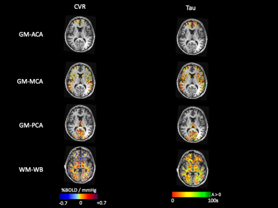 |
135 | Cerebrovascular reactivity differences in the anterior and posterior brain circulations in Healthy individuals
Harrison T. Levine1,2, Julien Poublanc2, Ece Su Sayin1,2, James Duffin1,3, Olivia Sobczyk2,3, Joseph A Fisher1,3, and David Mikulis2
1Department of Physiology, University of Toronto, Toronto, ON, Canada, 2Joint Department of Medical Imaging and the Functional Neuroimaging Lab, University Health Network, Toronto, ON, Canada, 3Department of Anaesthesiology and Pain Management, University Health Network, Toronto, ON, Canada Keywords: Blood vessels, fMRI (task based), Cerebrovascular Reactivity There is no consensus regarding the differences in Cerebrovascular Reactivity (CVR) magnitude differences between anterior and posterior circulations in healthy individuals. Forty-four healthy individuals underwent BOLD fMRI using a standardized blood vascular stress test achieved with sequential gas delivery. These results showed that cerebrovascular magnitude and speed of response tau) were stronger and faster in the posterior circulation compared to anterior circulation tissues. Therefore, the knowledge that there are significant regional differences in CVR is critical for investigating the regional impact of cerebrovascular pathologies. |
|
2486.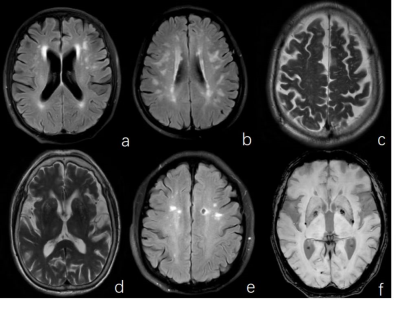 |
136 | Simple MRI score to evaluate the effect of cerebral small vascular disease on the mental state of patients with hemodialysis
Mengying Li1, Yuhan Jiang1, Dandan Zheng2, and Yanwei Miao1
1the First Affiliated Hospital of Dalian Medical University, Dalian, China, 2Philips Healthcare, Beijing, China Keywords: Blood vessels, Neuroscience, Psychiatric Disorders Hemodialysis(HD) can lead to hemodynamic changes and result in the occurrence of cerebral small vascular diseases(CSVD), which can cause changes in the mental state of patients. The objective of this study was to evaluate the effect of CSVD on the mental state of patients with hemodialysis by simple MRI score. Preliminary results show that HD is more likely to induce more serious CSVD and increase the prevalence of anxiety and depression, especially periventricular white matter hyperintensities (PVWMH) and cerebral microbleeds(CMB), which are closely related to the occurrence of anxiety and depression. |
|
2487.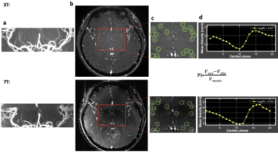 |
137 | Characterization of Cerebral Perforating Arteries using submillimeter-resolution PC-MRI with Dual-VENC at 3T: A Feasibility Study
Jianing Tang1,2, Helena Chui3, and Lirong Yan2,3
1Biomedical Engineering, Northwestern University, Evanston, IL, United States, 2Radiology, Northwestern University, Evanston, IL, United States, 3Department of Neurology, University of Southern California, Los Angeles, CA, United States Keywords: Blood vessels, Brain Direct characterization of cerebral perforating arteries, such as lenticulostriate arteries (LSAs), can provide valuable insight for understanding the pathology of small vessel diseases. Previous studies have demonstrated the feasibility of 7T PC-MRI for the characterization of LSA pulsatility. However, 7T MRI has not been in widespread use in both clinical and research studies, compared to conventional 3T. The current study aims to study the feasibility of using 3T dual-VENC PC-MRI to characterize LSA pulsatility. Our results show that 3T PC-MRI with dual-VENC offers reliable LSA pulsatility measurements that are comparable to 7T measurements. |
|
2488.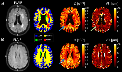 |
138 | Cerebral vessel size imaging reveals altered microvascular architecture in patients with vascular cognitive impairment
Paulien HM Voorter1, Maud van Dinther2, Gerhard S Drenthen1, Elles P Elschot1, Julie Staals2, Robert J van Oostenbrugge2, Walter H Backes1, and Jacobus FA Jansen1
1Department of Radiology and Nuclear Medicine, School for Mental Health and Neuroscience, Maastricht University Medical Center, Maastricht, Netherlands, 2Department of Neurology, CARIM School for Cardiovascular Diseases, Maastricht University Medical Center, Maastricht, Netherlands Keywords: Blood vessels, Blood vessels, Vessel size imaging With vessel size imaging, utilizing hybrid spin-echo gradient-echo perfusion scans, we studied microvascular architectural differences in patients with vascular cognitive impairment (VCI). We found lower vessel density and larger vessel radii in white matter hyperintensities compared to normal-appearing white matter. Moreover, we observed lower vessel density and larger vessel radii in deep gray matter of VCI patients compared to controls. Our findings suggest that the smallest capillaries are the first to collapse in VCI pathology and that the microvasculature not only alters in visibly injured tissue but also in regions that are less prone to hypoperfusion (gray matter). |
|
2489.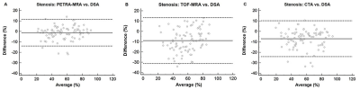 |
139 | Diagnostic Performances of PETRA-MRA, TOF-MRA, and CTA in Assessing Intracranial Arterial Stenosis with DSA as a Reference Standard
Junxia Niu1, Yuncai Ran2, Yan Zhang2, Yanglei Wu3, and Rui Chen2
1Department of Magnetic Resonance, The First Affiliated Hospital of Zhengzhou University, Zhengzhou, China, 2Department of Magnetic Resonance Imaging, The First Affiliated Hospital of Zhengzhou University, Zhengzhou, China, 3Siemens Healthineers Ltd., Beijing, China Keywords: Stroke, MR Value, Vessels This study compared the performance of three noninvasive techniques, namely, 3D pointwise encoding time reduction magnetic resonance angiography (PETRA-MRA), 3D time-of-flight (TOF) MRA and computed tomography angiography (CTA) in accurately measuring the degree of stenosis and lesion length in patients with intracranial arterial stenosis using DSA as the reference standard. This study demonstrated that the estimation of intracranial stenosis and lesion length by PETRA-MRA was more accurate and comparable with DSA than TOF-MRA and CTA. Therefore, PETRA-MRA is a promising noninvasive tool for the accurate assessment of ICAS. |
|
 Back to Meeting Home
 Back to the Program-at-a-Glance
The International Society for Magnetic Resonance in Medicine is accredited by the Accreditation Council for Continuing Medical Education to provide continuing medical education for physicians.
 Back to Meeting Home
 Back to the Program-at-a-Glance
 View Presentation Video