Digital Poster
Reconstruction: Body & Cardiovascular
ISMRM & ISMRT Annual Meeting & Exhibition • 03-08 June 2023 • Toronto, ON, Canada

Digital Poster
Reconstruction: Body & Cardiovascular
| Computer # | |||
|---|---|---|---|
3076.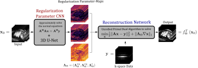 |
21 |
CNN-based Estimation of Spatio-Temporal Regularization
Parameter-Maps for TV-Reconstruction in Dynamic Cardiac MRI
Andreas Kofler1,
Clemens Sirotenko2,
Felix Frederik Zimmermann1,
David Schote1,
Christoph Kolbitsch1,3,
Fatima Antarou Ba4,
Fabian Altekrüger4,5,
Evangelos Papoutsellis6,7,
and Kostas Papafitsoros8
1Physikalisch-Technische Bundesanstalt (PTB), Braunschweig and Berlin, Germany, 2Weierstraß-Institut für Angewandte Analysis und Stochastik, Berlin, Germany, 3King’s College London, London, United Kingdom, 4Technische Universität Berlin, Berlin, Germany, 5Humboldt Universität zu Berlin, Berlin, Germany, 6Science and Technology Facilities Council (STFC), Oxford, United Kingdom, 7Finden Ltd, Oxford, United Kingdom, 8Queen Mary University of London, London, United Kingdom Keywords: Image Reconstruction, Machine Learning/Artificial Intelligence, Interpretable Machine Learning, Algorithmic Unrolling, Iterative Neural Networks We propose a method for estimating spatio-temporal regularization parameter-maps to be used for dynamic cardiac MR image reconstruction using total variation (TV)-minimization. Based on recent developments in algorithmic unrolling using Neural Networks (NNs), our approach uses two sub-networks. The first one predicts a spatio-temporal regularization parameter-map from an input image. Then, a second sub-network approximately solves a TV-reconstruction problem which is formulated with the estimated regularization parameter-map. We show that the proposed method can be used to further improve the TV-reconstructions compared to using only one single scalar regularization parameter or two regularization parameters for space and time. |
|
3077. |
22 |
Combining HOSVD-based joint reconstruction with shifted
acquisition sampling pattern enables liver DWI in a single
breath-hold
Guohao Jiang1,
Linzheng Hong1,
Zhaopeng Li2,
and Jun Xie2
1ShanghaiTech University, Shanghai, China, 2United Imaging Healthcare, Shanghai, China Keywords: Image Reconstruction, Diffusion/other diffusion imaging techniques Fast liver DWI scan has significant clinical relevance, however, motion artifacts, long scan time and low SNR remain as major technical challenges for liver DWI. In this study, we proposed a joint recon method based on global and local high order tensor SVD (HOSVD), combining with shifted acquisition sampling pattern across diffusion directions. The proposed method improves DWI image quality and enables fast liver DWI scan within a single breath-hold. |
|
3078. |
23 |
Accelerated 3D dynamic upper-airway MRI in naturally sleeping
obstructive sleep apnea patients.
Wahidul Alam1,
Junjie Liu2,
and Sajan Goud Lingala1,3
1Roy J. Carver Department of Biomedical Engineering, University of Iowa, iowa city, IA, United States, 2Department of Neurology, University of Iowa, iowa city, IA, United States, 3Department of Radiology, University of Iowa, Iowa city, IA, United States Keywords: Image Reconstruction, Head & Neck/ENT Obstructive sleep apnea (OSA) is characterized by breathing-related obstructions of the upper airway during sleep. In this work, we develop a motion resolved extra-dimension sparsity-based approach to resolve the kinematics of upper-airway in OSA during natural sleep. This approach is demonstrated on two adult OSA patients: one undergoing a Bi-directional positive airway pressure therapy during MRI scanning, and one without therapy |
|
3079. |
24 |
Water/Fat separation with spatio-temporal EPI-based acquisition
and reconstruction in body imaging
Xuetong Zhou1,2,
Philip K. Lee2,
and Brian A. Hargreaves1,2,3
1Department of Bioengineering, Stanford University, Stanford, CA, United States, 2Department of Radiology, Stanford University, Stanford, CA, United States, 3Department of Electrical Engineering, Stanford University, Stanford, CA, United States Keywords: Image Reconstruction, Image Reconstruction Water/fat separation is a reliable fat suppression technique. However, it is challenging in EPI-based imaging due to the large displacement along phase-encoding direction. We demonstrate the artifacts resulting from the poor-conditioning reconstruction of water/fat separation with EPI-based acquisition in body imaging. As a solution, unlike conventional multi-echo acquisition methods, our approach encodes the spectral components (water/fat) by acquiring and reconstructing the spatial and echo-time dimensions jointly. EPTI sampling trajectories and water/fat masking were used to improve the conditioning of the reconstruction. Phantom and in vivo (brain and breast) experiments were performed to show both results and limitations of this approach. |
|
3080.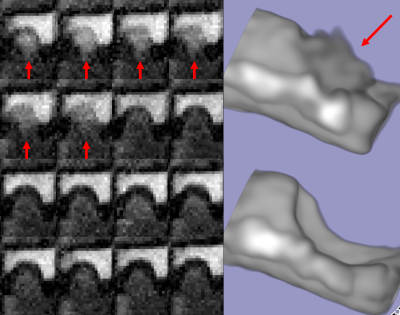 |
25 |
High resolution 4D-cine MRI of pulsatile aneurysmal motion with
radial acquisition at 7T
Thai Akasaka1,
Koji Fujimoto2,
Martijn Cloos3,
Tomohisa Okada1,
Shinichi Urayama1,
and Isa Tadashi1
1Human Brain Research Center, Kyoto University, Kyoto, Japan, 2Real World Data Research and Development, Kyoto University, Kyoto, Japan, 3University of Queensland, Brisbane, Australia Keywords: Image Reconstruction, Image Reconstruction The pulsation of intracranial aneurysms (IAs) is a novel risk factor of rupture. We aim to visualize the pulsation of IAs with 7T MRI using GRASP, a relatively novel MRI technique that provides high spatial and temporal resolution. Using thsis method, we were able to depict the pulsation of an aneurysm phantom as a 4D-cine movie. This technique may be useful in predicting the probability of IA rupture more accurately. |
|
3081.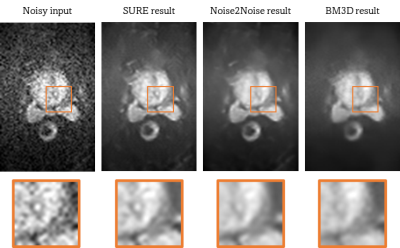 |
26 |
Unsupervised denoising of prostate DWI
Laura Pfaff1,2,
Fabian Wagner1,
Julian Hossbach1,2,
Elisabeth Preuhs1,
Fasil Gadjimuradov1,2,
Thomas Benkert2,
Dominik Nickel2,
Tobias Wuerfl2,
and Andreas Maier1
1Pattern Recognition Lab, Friedrich-Alexander-Universität Erlangen-Nürnberg, Erlangen, Germany, 2Magnetic Resonance, Siemens Healthcare GmbH, Erlangen, Germany Keywords: Image Reconstruction, Diffusion/other diffusion imaging techniques The diagnostic value of diffusion-weighted MR images is often degraded by their inherently low signal-to-noise ratio (SNR), especially for high b-values. In this context, the application of learning-based denoising methods is difficult since most methods require noise-free target images for training. We show how to denoise and evaluate diffusion-weighted MR images in a self-supervised manner by exploiting an adapted version of Stein’s unbiased risk estimator and specific properties of the data. Both quantitative and qualitative evaluations indicate increased performance over state-of-the-art unsupervised denoising methods. |
|
3082.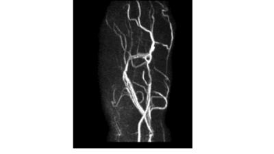 |
27 |
Non-contrast high-resolution 4D-peripheral MR angiography using
retrospective echo planar imaging with Compressed SENSE
Yasuhiro Goto1,
Michinobu Nagao2,
Masami Yoneyama3,
Johannes M Peeters4,
Yasutomo Katsumata3,
Isao Shiina1,
Kazuo Kodaira1,
Yutaka Hamatani1,
Takumi Ogawa1,
Mana Kato1,
and Shuji Sakai2
1Department of Radiological Services, Tokyo Women's Medical University, Tokyo, Japan, 2Department of Diagnostic imaging & Nuclear Medicine, Tokyo Women's Medical University, Tokyo, Japan, 3Philips Japan, Tokyo, Japan, 4Philips Healthcare, Best, Netherlands Keywords: Image Reconstruction, Blood vessels In this study demonstrated that the higher reduction factor (R=10.0) with a one-minute scan still provided sufficient image quality with significantly faster scan time compared with conventional REPI-SENSE. REPIX would be useful for further assessment of PAD pathology even with multiple VENC acquisitions still in a clinically feasible scan time.REPIX 4D-MRA well depicted peripheral arteries clearly in a significantly short time thanks to Compressed SENSE, compared with conventional SENSE. |
|
3083.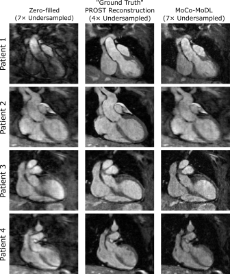 |
28 |
Accelerated whole-heart MRI for congenital heart disease
patients using a motion-corrected deep learning reconstruction
network
Andrew Phair1,
Anastasia Fotaki1,
Lina Felsner1,
Haikun Qi2,
René M. Botnar1,
and Claudia Prieto1
1School of Biomedical Engineering and Imaging Sciences, King's College London, London, United Kingdom, 2School of Biomedical Engineering, ShanghaiTech University, Shanghai, China Keywords: Image Reconstruction, Cardiovascular A deep learning reconstruction framework, trained in an end-to-end fashion and incorporating both a non-rigid respiratory motion estimation network and a motion-informed model-based reconstruction network, has been previously demonstrated to enable good quality images from seven-fold undersampled acquisitions for coronary magnetic resonance angiography applications. Herein, we apply the framework to whole-heart MRI scans of patients with congenital heart disease, enabling fast reconstruction of 7×-accelerated acquisitions and achieving image quality comparable to that of state-of-the-art patch-based low-rank iterative techniques. |
|
3084.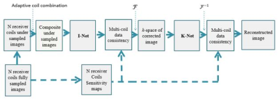 |
29 |
Dual Domain Deep Learning Framework for Cardiac MR Image
Reconstruction
Faisal Najeeb1,
Madiha Arshad 1,
Muhammad Shafique1,
and Hammad Omer1
1Electrical and Computer Engineering, COMSATS University Islamabad, Islamabad, Pakistan Keywords: Image Reconstruction, Artifacts, Deep Learning, Compressed Sensing , Parallel MRI Most deep learning methods apply U-Net either in image or k-space domain. Nevertheless, these methods have limitations: (1) Directly applying U-Net in k-space domain is not optimal for extracting features; (2) conventional image-domain oriented U-Net does not fully utilize the information of encoder part of the network for extracting features in the decoder part. In this paper, a dual-domain deep learning-based approach is presented, incorporating multi-coil data consistency layers for the reconstruction of cardiac MR images from 1-D Variable Density (VD) under-sampled data. Experiments show superior reconstruction results of the proposed method than conventional Compressed Sensing (CS) method. |
|
3085.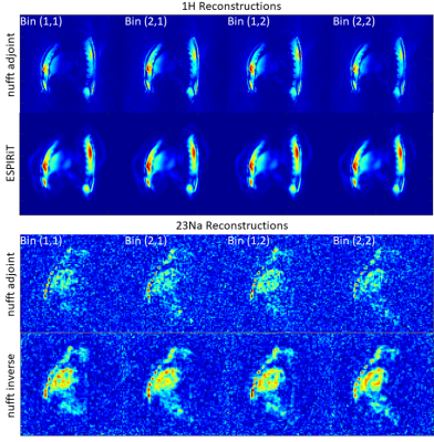 |
30 |
Towards motion-resolved interleaved radial 23Na/1H magnetic
resonance imaging of the human heart at 7 Tesla
Jörn Huber1,
Laurent Ruck2,3,
Matthias Günther1,4,5,
Armin Nagel2,6,
and Simon Konstandin1,4
1Fraunhofer Institute for Digital Medicine MEVIS, Bremen, Germany, 2Institute of Radiology, University Hospital Erlangen, Erlangen, Germany, 3Friedrich-Alexander-Universität Erlangen-Nürnberg (FAU), Erlangen, Germany, Erlangen, Germany, 4mediri GmbH, Heidelberg, Germany, 5Faculty 1 (Physics/Electrical Engineering), University of Bremen, Bremen, Germany, 6Division of Medical Physics in Radiology, German Cancer Research Center (DKFZ), Heidelberg, Germany, Erlangen, Germany Keywords: Image Reconstruction, Myocardium Motion correction in interleaved 23Na/1H MRI of the human heart is important to improve the diagnostic reliability of reconstructed images and derived quantitative parameters. Therefore, this work demonstrates and compares the application of different reconstruction techniques to undersampled and motion-gated 23Na/1H MRI data at 7 T. |
|
3086. |
31 |
FIRE and MATLAB for Free-Breathing Segmented LGE – A Novel
Sequence and Reconstruction Approach for Selecting and
Correcting Data by Respiration
Wolfgang G Rehwald1,2,
Kelvin Chow3,
Rafael Rojas4,
Nestor Mena4,
Diana Alexandrov4,
George Gamoneda4,
David Wendell4,
Ryan Seward4,
Jeana Dement4,
Vera Kimbrell4,
Han Kim4,
Indraneel Borgohain5,
Igor Klem4,
and Raymond Kim4
1Siemens Healthineers, Durham, NC, United States, 2Duke Cardiovascular MR Center, Duke University, Durham, NC, United States, 3Siemens Healthineers, Chicago, IL, United States, 4Duke Cardiovascular MR Center, Duke University Hospital, Durham, NC, United States, 5Siemens Healthineers, Princeton, NJ, United States Keywords: Image Reconstruction, Motion Correction, Reordering We introduce a free breathing segmented LGE technique combining a new acquisition, reordering and reconstruction scheme. The on-scanner reconstruction uses MATLAB and FIRE. The method produces image quality (IQ) and SNR otherwise only obtainable with breath held segmented interleaved LGE. In 27 patients, we show that IQ is superior to free breathing segmented interleaved LGE with multiple averages. SNR is higher compared to averaged motion corrected single shots when matching spatial and temporal resolution, the number of used measurements per image, and the readout type. Being a segmented technique, temporal and spatial resolution limitations of single shots do not apply.
|
|
3087.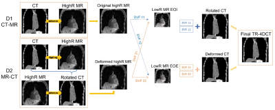 |
32 |
Super-resolution reconstruction of time-resolved
four-dimensional computed tomography (TR-4DCT) with multiple
breaths based on TR-4DMRI
Yilin Liu1,
Asala Ahamd1,
Xingyu Nie2,
and Guang (George) Li1
1Medical Physics, Memorial Sloan Kettering Cancer Center, New York, NY, United States, 2Radiology, University of Kentucky, Lexington, KY, United States Keywords: Image Reconstruction, Radiotherapy, time-resolved 4DMRI, time-resolved 4DCT, multi-breathing cycles This study has demonstrated that the feasibility to reconstruct multiple-breath TR-4DCT via the super-resolution reconstruction framework through either CT4D→(MRBH→MRFB) or (CT4D←MRBH)→ MRFB deformable image registration. Using TR-4DCT, potential dosimetry consequences in radiotherapy of lung, liver, and pancreatic patients due to patient breathing irregularities can be readily assessed. |
|
3088.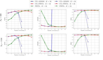 |
33 |
Convergent and Interpretable Dynamic Cardiac MR Image
Reconstruction with Neural Networks-based Convolutional
Dictionary Learning
Andreas Kofler1,
Christian Wald2,
Tobias Schaeffter1,3,4,
Markus Haltmeier5,
and Christoph Kolbitsch1,3
1Physikalisch-Technische Bundesanstalt (PTB), Braunschweig and Berlin, Germany, 2Charité - Universitätsmedizin Berlin, Berlin, Germany, 3King’s College London, London, United Kingdom, 4Technical University of Berlin, Berlin, Germany, 5University of Innsbruck, Innsbruck, Austria Keywords: Image Reconstruction, Machine Learning/Artificial Intelligence, Signal Processing, Sparsity-based Methods, Convolutional Dictionary Learning In this work we consider three different variants physics-informed Neural Networks (PINNs) which use the convolutional dictionary learning framework for image reconsruction in dynamic cardiac MRI. Although all three NNs share the same mechanism for regularization, the iterative schemes differ because they are derived from different problem formulations. We compare the methdos in terms of reconstruction performance as well as stability with respect to the number of iterations. All three methods yield similarly accurate reconstructions. However, by construction, only one of the three methods defines a convergent reconstruction algorithm and is therefore stable w.r.t. to the number of iterations. |
|
3089.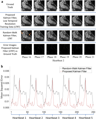 |
34 |
Two-Stage Kalman Filtering as a Framework for Accelerated
Cardiac MRI
Aaron Curtis1,2 and
Hai-Ling Margaret Cheng1,2,3
1Electrical and Computer Engineering, University of Toronto, Toronto, ON, Canada, 2Translational Biology & Engineering Program, Ted Rogers Centre for Heart Research, Toronto, ON, Canada, 3Institute of Biomedical Engineering, University of Toronto, Toronto, ON, Canada Keywords: Image Reconstruction, Heart Robust, real-time dynamic cardiac MRI (CMR) would provide information on the temporal signatures of disease that we currently cannot assess. We present a novel Kalman filtering framework that uses a priori statistics derived from a single cardiac cycle to adaptively predict temporal cardiac dynamics. Kalman filtering is ideal, as it ameliorates noise introduced from our maximum acceleration factor of 60, guarantees reconstruction fidelity, and enables flexible undersampling. Furthermore, reconstruction may be performed at an even higher temporal resolution than the training data. As such, our algorithm can be a foundation for true real-time dynamic CMR. |
|
3090.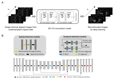 |
35 |
4D Cardiac MR Image Reconstruction by Deep Learning with Wavelet
Transform
Junhao Zhang1,2,
Yujiao Zhao1,2,
Jiahao Hu1,2,3,
Ye Ding1,2,
Christopher Men1,2,
Vick Lau1,2,
Alex T.L.Leong1,2,
and Ed X. Wu1,2
1Laboratory of Biomedical Imaging and Signal Processing, the University of Hong Kong, HongKong, China, 2Department of Electrical and Electronic Engineering, the University of Hong Kong, HongKong, China, 3Department of Electrical and Electronic Engineering, Southern University of Science and Technology, Shenzhen, China Keywords: Data Analysis, Cardiovascular, cardiac reconstruction,deep learning We present a CNN-based deep learning model to reconstruct the cardiac cine images from undersampled single-channel 4D MR data. The wavelet transform and spatial-temporal attention mechanisms are introduced in the model. The proposed model could reconstruct the cardiac images and recover the cardiac wall motion more robustly than the low-rank plus sparsity (i.e., L+S) reconstruction method. This approach presents one promising solution for accelerated cardiac dynamic imaging with one single channel through deep learning from the sparsity in spatial and temporal domains. |
|
3091.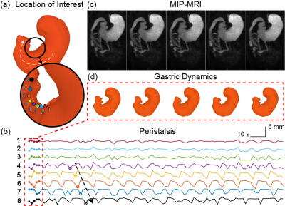 |
36 |
Tracking the moving stomach using MRI and neural ordinary
differential equations
Xiaokai Wang1,
Jiayue Cao2,
Kuan Han2,
Minkyu Choi2,
Yushi She2,
Ulrich Scheven2,
and Zhongming Liu2
1Biomedical Engineering, University of Michgan, Ann Arbor, MI, United States, 2University of Michigan, Ann Arbor, MI, United States Keywords: Data Analysis, Data Analysis, Neural Network We describe a method, namely neural ordinary differential equations, to track the movement of the stomach based on dynamic and contrast-enhanced gastrointestinal MRI. This model uses a neural network to learn the continuous biomechanical process that drives the shape change of the stomach wall over the course of digestion. This method allows us to represent gastric motor events on a generic surface template of the stomach and to further reveal the pattern of gastric motility with higher specificity and resolution than are previously attainable in vivo. This method should be also applicable to other organs, such as the heart. |
|
3092.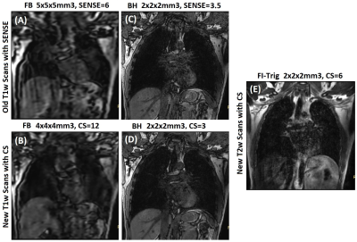 |
37 |
Improvement of the time-resolved 4DMRI image quality through
super-resolution reconstruction using compressed sensing
Can Wu1,
Guang (George) Li1,
and Yilin Liu1
1Medical Physics, Memorial Sloan Kettering Cancer Center, New York, NY, United States Keywords: Image Reconstruction, Radiotherapy, TR-4DMRI, multi-breathing cycles, breathing irregularities, image quality, compressed sensing Since the development of respiratory-correlated four-dimensional computed tomography (RC-4DCT) [1] and magnetic resonance imaging (RC-4DMRI) [2,3], patient-specific respiratory-induced tumor motion has been incorporated in radiotherapy for treating mobile tumors, such as lung, liver and pancreatic cancer. However, the RC-based snapshot 4D imaging only provides one breathing cycle, often contains severe binning motion artifacts, and may not represent tumor motion over 20-minute treatment, affecting treatment outcomes. Therefore, respiratory motion irregularities remain a challenge in radiotherapy. In this study, we report an improved time-resolved 4DMRI technique that captures multi-breath and can be used clinically and quantifies tumor motion irregularities. |
|
3093.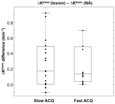 |
38 |
High temporal resolution DCE-MRI improves the performance of
model fitting parameters
Xin Li1,
Travis Rice-Stitt2,
Lina Gao3,
Marina Aguiñaga4,
Kevin R Turner5,
Bryan Foster6,
Fergus Coakley6,
Mark Garzotto4,7,
and Ryan Kopp4,7
1Advanced Imaging Research Center, Oregon Health & Science University, Portland, OR, United States, 2Pathology, Oregon Health & Science University, Portland, OR, United States, 3Knight Biostatistics Shared Resources, Oregon Health & Science University, Portland, OR, United States, 4Urology, Oregon Health & Science University, Portland, OR, United States, 5Providence Health and Service, Portland, OR, United States, 6Diagnostic Radiology, Oregon Health & Science University, Portland, OR, United States, 7Portland VA Medical Center, Portland, OR, United States Keywords: Data Analysis, DSC & DCE Perfusion Recent advance in data acquisition makes fast DCE-MRI data acquisition feasible. This work investigated the impact of DCE-MRI temporal resolution on the data’s pharmacokinetic modeling and the water exchange effect imparted into the model parameters. Using both the Tofts model and the water-exchange sensitized Shutter-Speed model, our results showed that DCE data with higher temporal resolution (shorter intersample interval) is beneficial in model parameter precision and in quantifying transcytolemmal water exchange effect. The later may offer additional lesion-detection specificity in clinical prostate MRI. |
|
3094.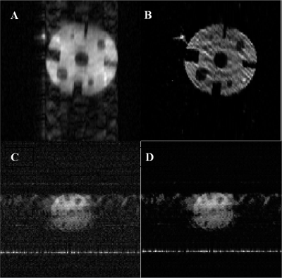 |
39 |
Implementation of Hyperpolarized 129Xe Accelerated MRI in
Phantom and Human Lungs: Preliminary Study and Troubleshooting
Samuel Perron1,
Matthew S. Fox1,2,
and Alexei V. Ouriadov1,2,3
1Physics and Astronomy, University of Western Ontario, London, ON, Canada, 2Lawson Health Research Institute, London, ON, Canada, 3School of Biomedical Engineering, University of Western Ontario, London, ON, Canada Keywords: Data Acquisition, Low-Field MRI Accelerated MRI could significantly improve image quality of lung imaging without increasing costs, especially for low field strengths. The signal decay of a series of undersampled xenon-129 images is fitted to the Stretched-Exponential-Model to yield higher SNR. The proposed method was implemented in undersampled phantom images at low field (0.074T) and human lung images at high field (3T) using the FGRE pulse sequence with hyperpolarized 129Xe; SNR was significantly improved within the same scan duration compared to a fully-sampled image. Potential issues with the compressed-sensing reconstruction are identified and possible solutions presented to promote robustness of method. |
|
3095.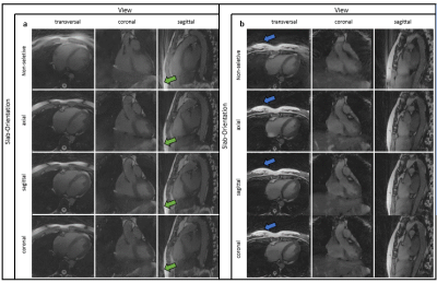 |
40 |
Slab-selection in free-running cardiac and respiratory
motion-resolved bSSFP 5D whole-heart MRI
Robin Ferincz1,
Ludovica Romanin1,
Jérôme Yerly2,
Davide Piccini1,3,4,
Matthias Stuber2,
and Christopher William Roy1
1Department of Radiology, Lausanne University Hospital (CHUV) and University of Lausanne (UNIL), Lausanne, Switzerland, 2Center for Biomedical Imaging (CIBM), Lausanne, Switzerland, 3Advanced Clinical Imaging Technology (ACIT), Siemens Healthineers International AG, Lausanne, Switzerland, 4LTS5, École Polytechnique Fédérale de Lausanne (EPFL), Lausanne, Switzerland Keywords: Data Acquisition, Artifacts Recent advances have enabled high resolution cardiac and respiratory motion-resolved 5D whole-heart MRI using non-contrast enhanced non-selective 3D radial bSSFP. However, the nature of bSSFP, the subject dependent anatomy, as well as the underlying sparse reconstruction can lead to banding and streaking artifacts, which degrade image quality and reduce diagnostic utility. In this work, the impact of slab-selective RF pulses in a programmable orientation that is independent of the k-space trajectory is assessed in a cohort of healthy volunteers. Preliminary results suggest that a subject-specific slab orientation can reduce artifacts and improve image quality. |
|
 Back to Meeting Home
 Back to the Program-at-a-Glance
The International Society for Magnetic Resonance in Medicine is accredited by the Accreditation Council for Continuing Medical Education to provide continuing medical education for physicians.
 Back to Meeting Home
 Back to the Program-at-a-Glance
 View Presentation Video