Digital Poster
Imaging Aging, Dementia & Alzheimer's Disease II
ISMRM & ISMRT Annual Meeting & Exhibition • 03-08 June 2023 • Toronto, ON, Canada

Digital Poster
Imaging Aging, Dementia & Alzheimer's Disease II
| Computer # | |||
|---|---|---|---|
3508.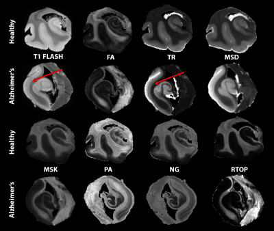 |
101 |
Differential Radiologic-Pathologic Correspondence of
Diffusivity, Restriction and Anisotropy with Plaque and Tangle
Cores
Courtney J Comrie1,
Laurel A Dieckhaus1,
Tom G Beach2,
Geidy E Serrano2,
and Elizabeth B Hutchinson1
1Biomedical Engineering, University of Arizona, Tucson, AZ, United States, 2Brain and Body Donation Program, Banner Sun Health Research Institute, Sun City, AZ, United States Keywords: Alzheimer's Disease, Alzheimer's Disease Alzheimer’s is an irreversible degenerative brain disease. Clinical MRI may visualize severe brain atrophy but fails to recognize earlier biomarkers associated with subtle microstructural changes. Microstructural MRI techniques such as DTI, MAP-MRI, and MSDKI may sensitively detect and distinguish tissue degeneration, tauopathies, and beta amyloid plaques in the hippocampus and entorhinal cortex. The capability of these techniques was investigated in post-mortem human temporal lobe specimens at high resolution and high image quality. Prominent findings seen were differences between DTI and MAP-MRI anisotropy metrics, and striking differences between the hippocampus and entorhinal cortex for restriction due to plaques. |
|
3509.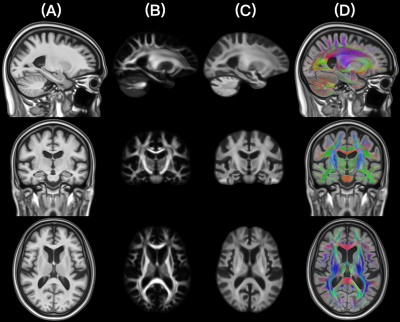 |
102 |
Development of an FOD template of the older adult brain for the
MIITRA atlas
Yingjuan Wu1,
Abdur Raquib Ridwan2,
Mohammad Rakeen Niaz1,
David A. Bennett2,
and Konstantinos Arfanakis1,2
1Biomedical Engineering, Illinois Institute of Technology, Chicago, IL, United States, 2Rush Alzheimer’s Disease Center, Rush University Medical Center, Chicago, IL, United States Keywords: Alzheimer's Disease, Aging, Atlas A high quality FOD template of the older adult brain can enhance the sensitivity and accuracy of white matter investigations in older adults. The present work compared several FOD template construction methods for the purpose of developing a high-resolution FOD template of the older adult brain in the space of the MIITRA atlas. The preferred method employed multichannel registration based on T1w, FA and FOD information, followed by additional FOD single modality registration. The resulting FOD template exhibited high image quality, was constructed based on precisely matched FOD data, and exhibited high spatial matching to other existing MIITRA templates. |
|
3510.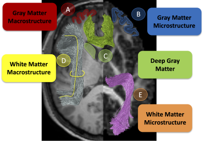 |
103 |
Detecting micro- and macro-structural deviations in individuals
with cognitive impairment
Kurt Schilling1,
Francois Rheault2,
Dmitri Shastin3,
Leon Y Cai4,
Andrea T Shafer5,
Susan M Resnick5,
Bennett A Landman4,
and Maxime Chamberland6
1VUMC, Nashville, TN, United States, 2Sherbrooke University, Sherbrooke, QC, Canada, 3Cardiff University Brain Research Imaging Centre (CUBRIC), Cardiff University, Cardiff, United Kingdom, 4Vanderbilt University, Nashville, TN, United States, 5National Institute on Aging, Baltimore, MD, United States, 6Donders Institute for Brain, Cognition and Behaviour, Radboud University, Nijmegen, Netherlands Keywords: Alzheimer's Disease, Alzheimer's Disease Medical imaging is a promising tool in detecting altered brain tissue states related to cognitive impairment and dementia, however, clinical heterogeneity challenges interpretation of these changes. Recent work in normative modeling has paved the way for not only group-based comparisons of control/cohorts, but detection of deviations in individual subjects. Here, we apply this framework to detect anomalies in individuals with cognitive impairment by assessing the classification power of microstructural and macrostructural features of different tissue types, and also attributing anomalies to specific features of brain tissue. |
|
3511.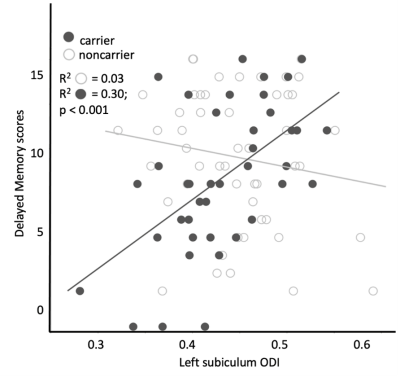 |
104 |
Relating left hippocampal microstructure and delayed memory in
cognitively intact adults at genetic-risk for developing
Alzheimer's disease
Jennapher Lingo VanGilder1,
Leslie C. Baxter2,
Andrew Hooyman1,
Leland S. Hu3,
Yuxiang Zhou3,
Richard J. Caselli4,
Kurt G. Schilling5,
and Scott C. Beeman1
1School of Biological and Health Systems Engineering, Arizona State University, Tempe, AZ, United States, 2Psychiatry and Psychology, Mayo Clinic, Scottsdale, AZ, United States, 3Radiology, Mayo Clinic, Scottsdale, AZ, United States, 4Neurology, Mayo Clinic, Scottsdale, AZ, United States, 5Radiology, Vanderbilt University, Nashville, TN, United States Keywords: Alzheimer's Disease, Alzheimer's Disease, ApoE4; genetic-risk We have recently shown that the functional connectivity of the left hippocampus is significantly related to memory trajectory in cognitively intact ApoE4 carriers, independent of hippocampal volume. The purpose of this preliminary study was to determine if dendritic orientation dispersion of the left hippocampus explains variance in delayed verbal memory in this cohort. Results indicated that dendritic complexity of the left hippocampus was related to delayed memory in ApoE4 carriers. These findings suggest that ApoE4 carriers may experience subtle microstructural declines in the left hippocampus at the cognitively-intact stage. |
|
3512.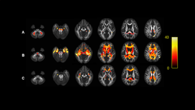 |
105 |
White matter fiber characteristics in Alzheimer's disease using
fixel-based analysis
Meng Li1,
Shanwen Liu2,
Yuqi Zhi1,
Rong Liu1,
Zhen Jiang1,
Xiaoyun Liang3,4,
Hua Hu2,
Yunzhu Wu5,
Yang Song5,
and Jiangtao Zhu1
1Department of Radiology, The Second Affiliated Hospital of Soochow University, Suzhou, China, 2Department of Neurology, The Second Affiliated Hospital of Soochow University, Suzhou, China, 3Institute of Artificial Intelligence and Clinical Innovation, Neusoft Medical Systems Co., Ltd., Shanghai, China, 4Florey Institute of Neuroscience and Mental Health, The University of Melbourne, Melbourne, Australia, 5MR Scientific Marketing, SIEMENS Healthineers Ltd., Shanghai, China Keywords: Alzheimer's Disease, Alzheimer's Disease We collected diffusion-weighted images of all subjects and divided them into four groups: HC, MCI, mild AD and moderate AD. The differences of FD, FC and FDC were analyzed by FBA method. The results showed that the three metrics in AD group were significantly lower than those in HC Group, but there was no significant difference in MCI Group. This study indicates that there is a microstructural or macrostructural degeneration in the white matter of AD patients. |
|
3513. |
106 |
The value of diffusion kurtosis imaging in evaluating the mild
cognitive impairment of occupational aluminum workers
Wenji Xu1,
Xiaochun Wang2,
Hui Zhang2,
and Yan Tan*2
1College of Medical Imaging, Shanxi Medical University, Taiyuan, China, 2Department of Radiology, First Hospital of Shanxi Medical University, Taiyuan, China Keywords: Alzheimer's Disease, Alzheimer's Disease, Alzheimer’s disease; aluminum exposure; diffusion kurtosis imaging In this work, we focused on the Al-exposed workers and confirm the findings that DKI can discriminate MCI from NC, furthermore we assess the severity of cognitive impairment in Al-exposed workers, and find the MK, Kr, MD and FA values are correlated with MoCA scores,which may provide quantitative imaging biomarkers for Al-exposed MCI workers. |
|
3514.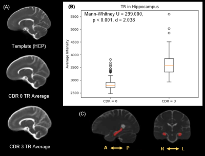 |
107 |
Processing Pipeline and Analytic Framework for Diffusion and
Morphometric Analyses of Alzheimer’s Disease Repository Data
Samantha N Schatz1,
Courtney J Comrie1,
Laurel A Dieckhaus1,
and Elizabeth B Hutchinson1
1University of Arizona, Tucson, AZ, United States Keywords: Alzheimer's Disease, Diffusion Tensor Imaging, Repository Data, Cognitive Impairment, Tensor Based Morphometry, Hippocampus Alzheimer's disease is generally accompanied by brain atrophy, which can be evident on MRI based evaluation at late stages, but there is a need for earlier stage MRI markers that may predict progressive cognitive impairment. Using the NACC Uniform Data Set, a robust pipeline was developed for registering DTI maps to a Human Connectome Project template space and an analysis framework for ROI based, voxel-wise, and morphometric analysis was applied. Prominent results include increased trace in the hippocampus and an increase in ventricle volume in the group with severe cognitive impairment. |
|
3515.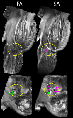 |
108 |
Diffusion tensor subspace imaging (DiTSI) delineates small
fibers and gray matter microstructure invisible to
single-encoding techniques
Elizabeth Hutchinson1,
Jean-Pierre Galons2,
Courtney Comrie1,
Seraphina Solders3,
Geidy Serrano4,
Thomas Beach4,
Vitaly Galinsky3,
and Lawrence Frank3
1Biomedical Engineering, University of Arizona, Tucson, AZ, United States, 2Medical Imaging, University of Arizona, Tucson, AZ, United States, 3Center for Scientific Computing in Imaging, University of California, San Diego, CA, United States, 4Banner Sun Health, Sun City, AZ, United States Keywords: Alzheimer's Disease, Diffusion/other diffusion imaging techniques, double diffusion encoding Double diffusion encoding (DDE) MRI provides unique sensitization to microscale anisotropy although scalar representation of DDE data is challenging. The diffusion tensor subspace imaging (DiTSI) framework generates metrics from DDE images. In this study, post-mortem human brain tissue specimens were imaged and comparative analysis found that DiTSI but not DTI was able to provide distinct contrast in gray matter regions and at the gray/white interface. Additionally, small cranial nerve fibers of the brain stem were detectable using DiTSI, but not DTI or NODDI approaches and also showed remarkable abnormalities in Alzheimer’s disease tissue that were absent for DTI. |
|
3516.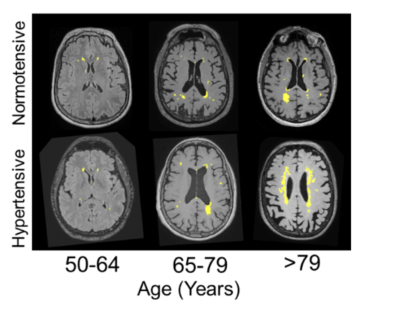 |
109 |
APOE ε4 and Hypertension: Cardiovascular Risk Accelerate the
Brain Structure Atrophy and Small Vessel Disease Pathologies in
MCI and Dementia
Aniket Aman1,
Neha Yadav1,
Arkaprava Majumdar1,
and Vivek Tiwari1
1Department of Biological Sciences, Indian Institute of Science Education and Research, Berhampur, Berhampur, India Keywords: Dementia, Aging, White Matter Hyperintensity, APOE E4, Cognition, Hypertension Transformation of brain heath with aging comprehend a series of brain structure, vascular and functional changes over time. The key to understanding the brain health in normal and pathological aging is to identify the series of early, intermediate and late events that encodes for brain health. Here, we have investigated the temporal and spatial orders of brain volumetry, small vessel pathology (white matter hyperintensity) and its kinetics with normal aging and aging with cardiovascular risks and gene defects such as APOE ε4, across subjects classified as cognitively Normal (CN), Mild Cognitive Impairment (MCI) and Dementia (DM). |
|
3517.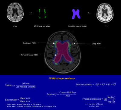 |
110 |
White matter hyperintensity shape in relation to long-term small
vessel disease progression in community-dwelling older adults
Jasmin A. Keller1,
Sigurdur Sigurdsson 2,
Mark A. van Buchem1,
Lenore J. Launer3,
Matthias J.P. van Osch1,
Vilmundur Gudnason2,4,
and Jeroen H.J.M. de Bresser1
1Department of Radiology, Leiden University Medical Center, Leiden, Netherlands, 2Icelandic Heart Association, Kopavogur, Iceland, 3Laboratory of Epidemiology and Population Science, National Institute on Aging, Bethesda, MD, United States, 4Faculty of Medicine, University of Iceland, Reykjavik, Iceland Keywords: Dementia, Aging, Cerebral small vessel disease White matter hyperintensity (WMH) shape was recently introduced as a novel small vessel disease (SVD) marker that may provide a more detailed characterization of WMH than volume alone. We aimed to investigate the association between baseline WMH shape and cerebrovascular disease progression over 5 years. A more irregular shape of periventricular/confluent and deep WMH at baseline is associated with increased progression of WMH volume. Moreover, a more irregular shape of periventricular/confluent WMH was associated with occurrence of new microbleeds and new subcortical infarcts at follow-up. Our findings indicate that a more irregular WMH shape is associated with SVD progression. |
|
3518.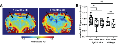 |
111 |
Quantitative Gradient Recalled Echo (qGRE) MRI enables
preatrophic neurodegeneration measurement in a mouse model of
Alzheimer’s Disease
Michal R Tomaszewski1,
Alexander L Sukstansky2,
Hyking Haley1,
Xiangjun Meng1,
Corey O Miller1,
and Dmitriy A Yablonskiy2
1Translational Imaging, Merck & Co, West Point, PA, United States, 2Mallinckrodt Institute of Radiology, Washington University, St Louis, MO, United States Keywords: Alzheimer's Disease, Animals There is an unmet need for non-invasive technique for measurement of neurodegeneration in Alzheimer’s Disease (AD). Quantitative Gradient-Recalled-Echo (qGRE) MRI showed promise to address this in patients through quantification of tissue specific R2* relaxation showing neurodegeneration before atrophy. Here qGRE is optimized and applied for the first time to measure progression of this preatrophic neurodegeneration in Tg4510 mouse model of AD. Histological neuronal density quantification and test-retest measurements validated R2* measurements. We showed significant decrease in normalized median hippocampus R2* between 3, 5 and 6 month-old Tg4510 mice. The method can be applied in AD drug discovery and model development. |
|
3519.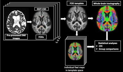 |
112 |
Fixel based analysis to investigate white matter alterations in
individuals with Mild cognitive impairment and Alzheimer’s
disease
Anjan Bhattarai 1,2,
Pauline Maillard1,
Charles DeCarli1,
and Audrey Fan1,2
1Department of Neurology, University of California Davis, Davis, CA, United States, 2Department of Biomedical Engineering, University of California Davis, Davis, CA, United States Keywords: Alzheimer's Disease, Diffusion/other diffusion imaging techniques, Fixel based analysis We investigated fibre tract-specific changes in the whole-brain white matter in individuals with Mild Cognitive Impairment (MCI) and Alzheimer’s Disease (AD) using Fixel Based Analysis (FBA). The results show significant reduction in microstructural fibre bundles in AD, in regions including splenium of the corpus callosum, fornix, and the uncinate fasciculus. FBA-derived measures, including fibre density (FD), and combination of fibre density and cross-section (FDC) demonstrated sensitivity in detecting microstructural white matter alterations in AD. These findings highlight the utility of FBA as a potential biological marker for providing valuable insights into pathophysiologic changes in AD. |
|
3520.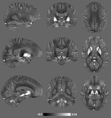 |
113 |
Development of a high-resolution magnetic susceptibility
template of the older adult brain in MIITRA space
Rasheed Abid1,
Abdur Raquib Ridwan2,
Yingjuan Wu1,
Mohammad Rakeen Niaz1,
Shengwei Zhang2,
Arnold M. Evia2,
David A. Bennett2,
and Konstantinos Arfanakis1,2
1Department of Biomedical Engineering, Illinois Institute of Technology, Chicago, IL, United States, 2Rush Alzheimer’s Disease Center, Rush University Medical Center, Chicago, IL, United States Keywords: Alzheimer's Disease, Aging, Brain, Susceptibility, Atlas Quantitative susceptibility mapping (QSM) is considered a promising tool for the detection and monitoring of disease processes that alter magnetic susceptibility in the brain of older adults. Voxel-wise and atlas-based QSM analyses require accurate spatial normalization of QSM data, which requires a high quality QSM template representative of the population. However, a QSM template of the older adult brain is not yet available. This study aimed to construct a high quality, high resolution QSM template of the older adult brain in the space of the MITRA atlas based on data from a large, diverse, community cohort of non-demented older adults. |
|
3521.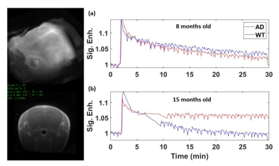 |
114 |
Measurement of blood-brain barrier leakage in AD transgenic mice
with aging: Improving sensitivity with capillary input function
Jonghyun Bae1,2,3,
Ayesha Das3,
Isabel Reyes4,
Sawwal Qayyum5,
Jin Zhang5,
Arjun Masurkar4,
and Sungheon Gene Kim5
1Vilcek Institute of Graduate Biomedical Science, NYU School of Medicine, New York, NY, United States, 2Center for Advanced Imaging Innovation and Research, Radiology, NYU School of Medicine, New York, NY, United States, 3Radiology, Weill Cornell Medical College, New York, NY, United States, 4Neurology, NYU Langone Health, New York, NY, United States, 5Weill Cornell Medical College, New York, NY, United States Keywords: Alzheimer's Disease, Aging Recent studies have suggested that the increase in blood-brain barrier (BBB) permeability is associated with both aging and the progression of the Alzheimer’s disease (AD). However, the association of the amyloid pathology with the increased BBB leakage at different disease progression is still poorly understood. In this study, we performed a cross-sectional study to investigate the BBB permeability changes in AD transgenic mice with aging. We also propose the network-aided analysis allows the scan time reduction without compromising the accuracy of the detection of the subtle permeability |
|
3522.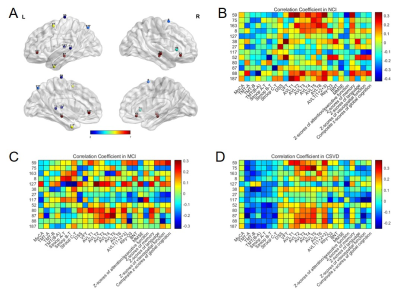 |
115 |
Anomalous Neurovascular Coupling in cerebral small vessel
disease patients related to cognitive impairment
Ying Hu1,
Yawen Sun1,
Yiming Zhang1,
and Yan Zhou1
1Renji Hospital, Shanghai Jiao Tong University School of Medicine, Shanghai, China Keywords: Dementia, Aging NVC is thought to reflect the interrelationship between nutrient demand and supply, whereby neuronal activity influences local changes in blood flow. Injuries to the NVU could be the crucial point to CSVD, but the mechanisms remain obscure. This is the first study investigating CSVD from the perspective of the NVC at the whole brain, modular and regional levels. |
|
3523.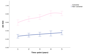 |
116 |
Dynamic Changes in Quantitative MRI-based Biomarkers of
Alzheimer’s Disease and Its Association with Cognitive Decline –
A Longitudinal Study
Xiang Fan1,2,
Yuan Cai2,
Wanting Liu2,
Lin Shi2,
and Vincent CT Mok2
1Peking University Shenzhen Hospital, Shenzhen, China, 2The Chinese University of Hong Kong, Hong Kong, SAR, China Keywords: Alzheimer's Disease, Alzheimer's Disease Alzheimer's disease (AD) resemblance atrophy index (AD-RAI) is a novel MRI-based machine-learning derived imaging biomarker for AD that is valid in detection of early AD. We selected 318 CU and MCI subjects in ADNI with four-year follow-up with 1471 serial MRI scans to assess longitudinal changes of the MRI biomarkers (i.e., AD-RAI, HV, HF, BPV, BPF) in correlation with the change in time and conversion status. We used generalized linear mixed-effects models to compute and show the predictive value. AD-RAI over time showed the highest classification accuracy for predicting converters when compared with other imaging biomarkers. |
|
3524.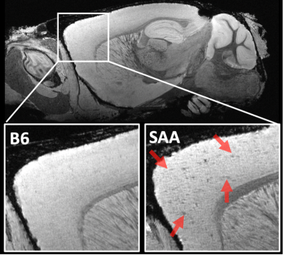 |
117 |
Microstructure and connectivity alterations in a novel mouse
model of Alzheimer’s disease
Surendra Maharjan1,
Megan Renate Jewett1,
Abigail Wallace1,
and Nian Wang1,2
1Department of Radiology and Imaging Sciences, Indiana University School of Medicine, Indianapolis, IN, United States, 2Stark Neurosciences Research Institute, Indianapolis, IN, United States Keywords: Alzheimer's Disease, Alzheimer's Disease, mice Alzheimer’s disease is a common neurodegenerative dementia. Although disease has no cure, early intervention can help to maintain normal mental function and slow down the progression of the disease. A comprehensive study incorporating diffusion tensor imaging (DTI), diffusion kurtosis imaging (DKI), neurite orientation dispersion and density imaging (NODDI), brain connectivity and fixel-based analysis (FBA) is still lacking. Hence, we aim to apply these methods in a novel SAA mouse model of Alzheimer’s disease. We found there was significant difference in cortex and hippocampus in between control B6 and SAA groups. |
|
3525.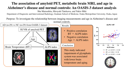 |
118 |
The association of amyloid PET, metabolic brain MRI, and age in
Alzheimer's disease and normal controls: An OASIS-3 dataset
analysis
Shu Matsushita1,
Hiroyuki Tatekawa1,
and Yukio Miki1
1Department of Diagnostic and Interventional Radiology, Osaka Metropolitan University, Osaka, Japan Keywords: Alzheimer's Disease, Alzheimer's Disease Revealing the association of amyloid PET and metabolic MRI measurements would be useful for future studies or clinical situations that evaluate Alzheimer's disease (AD). We investigated the relationship between imaging measurements and clinical information in 29 AD patients and age- and sex-matched normal controls (NCs) using OASIS-3 dataset. A significant positive correlation was found between brain temperature and index of diffusivity along the perivascular space (ALPS index) among all subjects and NCs, while a significant negative correlation was found between age and ALPS index among all subjects, AD patients, and NCs. |
|
3526.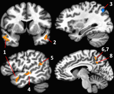 |
119 |
Within-network changes in strength of the default mode network
are related to Alzheimer’s Disease biomarkers in APOE e4
carriers
Katherine Anne Koenig1,
Sally Durgerian1,
Mark J Lowe1,
Frank DiFilippo1,
Lynn Bekris1,
James Leverenz1,
and Stephen Rao1
1The Cleveland Clinic, Cleveland, OH, United States Keywords: Alzheimer's Disease, Alzheimer's Disease, APOE The apolipoprotein E (APOE) ε4 genotype is a genetic risk factor for late onset Alzheimer’s Disease (AD). Consistent differences in network connectivity have been described in ε4 carriers (e4+). Here, we compare resting state connectivity in the default mode network between elderly, cognitively intact e4+ and non-carriers (e4-) and assess the relationship of connectivity to AD-related biomarkers. Regional changes in the e4+ group primarily consisted of weakened connectivity. Connectivity strength in the middle temporal gyrus was negatively related to amyloid PET centiloid score and p-tau in the e4+ group. |
|
3527. |
120 |
Hippocampal metabolites in patients with amnestic mild cognitive
impairment
Xin Chen1,
Tao Gong1,
and Weibo Chen2
1Shandong Provincial Hospital Affiliated to Shandong First Medical University, Jinan, China, 2Philips Healthcare, Shanghai, China Keywords: Alzheimer's Disease, Alzheimer's Disease Amnestic mild cognitive impairment (aMCI) is a precursor to Alzheimer's disease (AD). The neurometabolic changes especially the neurotransmitters in the hippocampus contributed the course of AD. In-vivo magnetic resonance spectroscopy (MRS) can be used to measure brain metabolites noninvasively. In this study, we aim to explore the changes of hippocampal metabolic changes in aMCI patients using PRESS and MEGA-PRESS. aMCI patients exhibited decreased hippocampal Glx levels from MEGA-PRESS, having low concordance with PRESS. |
|
 Back to Meeting Home
 Back to the Program-at-a-Glance
The International Society for Magnetic Resonance in Medicine is accredited by the Accreditation Council for Continuing Medical Education to provide continuing medical education for physicians.
 Back to Meeting Home
 Back to the Program-at-a-Glance
 View Presentation Video