Digital Poster
Inflammation Across Neurological Diseases II
ISMRM & ISMRT Annual Meeting & Exhibition • 03-08 June 2023 • Toronto, ON, Canada

Digital Poster
Inflammation Across Neurological Diseases II
| Computer # | |||
|---|---|---|---|
3528.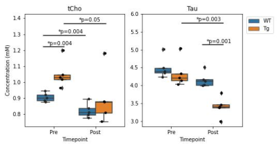 |
121 |
Inhibition of phospholipase A2 by mepacrine reduces total
choline in a rat model of Alzheimer’s disease
Colleen Bailey1,
Ved Hatolkar1,
Wendy Oakden1,2,
Margaret M Koletar1,
JoAnne McLaurin1,3,
and Jamie Near1,4
1Sunnybrook Research Institute, Toronto, ON, Canada, 2Synaptive Medical, Toronto, ON, Canada, 3Department of Laboratory Medicine and Pathobiology, University of Toronto, Toronto, ON, Canada, 4Department of Medical Biophysics, University of Toronto, Toronto, ON, Canada Keywords: Alzheimer's Disease, Preclinical, neuronal loss We examined the effect of the PLA2 inhibitor mepacrine on the levels of choline and other brain metabolites in the TgF344-AD rat model of Alzheimer's disease. Previous studies have demonstrated elevated levels of brain cholines in this model, similar to those observed in humans and attributed to increased membrane turnover. Total choline was higher in transgenic animals at baseline, but decreased following mepacrine treatment. Total choline also decreased in wildtype animals following treatment, consistent with the hypothesis that mepacrine reduces PLA2-mediated membrane turnover. Based on this finding, MRS choline measures may also serve as a biomarker for PLA2-mediated inflammatory processes. |
|
3529.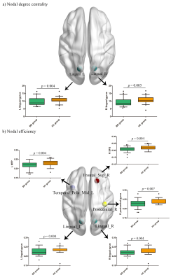 |
122 |
Association of peripheral inflammation with disrupted brain
functional network topology in bipolar disorder
Guixian Tang1,
Guanmao Chen1,
Long Qian2,
and Ying Wang1
1First Affiliated Hospital of Jinan University, Guangzhou, China, 2MR Research, GE Healthcare, Beijing, China Keywords: Neuroinflammation, fMRI (resting state) This study provided preliminary evidence of the association between disrupted brain functional network topology and neuroinflammation in BD. The current study demonstrated disrupted topological organization in the whole brain and regional connectivity was associated with inflammatory cytokines of the IL-4, IL-8 and IL-10 levels in BD. Moreover, our results indicated that higher IL-4 levels and impaired regional connectivity in the temporal pole may be associated with the severer depressive symptoms in BD. |
|
3530.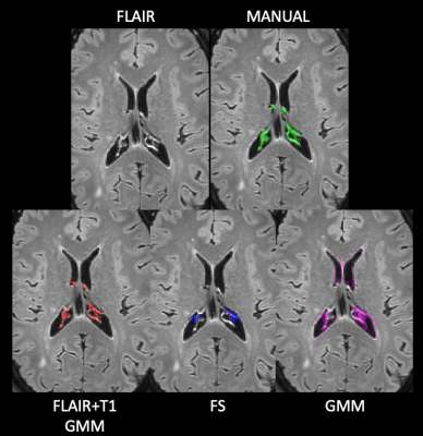 |
123 |
A Fully-Automatic Method to Segment Choroid Plexus in Multiple
Sclerosis Using Conventional MRI Sequences
Loredana Storelli1,
Elisabetta Pagani1,
Martina Rubin1,2,
Monica Margoni1,2,
Maria Assunta Rocca1,2,3,
and Massimo Filippi1,2,3,4,5
1Neuroimaging Research Unit, Division of Neuroscience, IRCCS San Raffaele Scientific Institute, Milan, Italy, 2Neurology Unit, IRCCS San Raffaele Scientific Institute, Milan, Italy, 3Vita-Salute San Raffaele University, Milan, Italy, 4Neurorehabilitation Unit, IRCCS San Raffaele Scientific Institute, Milan, Italy, 5Neurophysiology Service, IRCCS San Raffaele Scientific Institute, Milan, Italy Keywords: Multiple Sclerosis, Neuroinflammation Choroid plexus (CP) is a structure of the brain ventricular system that demonstrated an involvement in the inflammatory process of patients with multiple sclerosis (MS). However, its role in the pathophysiology of the disease needs to be further explored but its manual delineation on MRI is time-consuming. In this study, we developed a fully-automatic method to segment CP using 3DT1-weighted and FLAIR MRI sequences. The algorithm proved to be accurate, easy to implement and do not require an initial training phase on huge amount of data, being generalizable and a fast tool to potentially be included in a clinical setting. |
|
3531.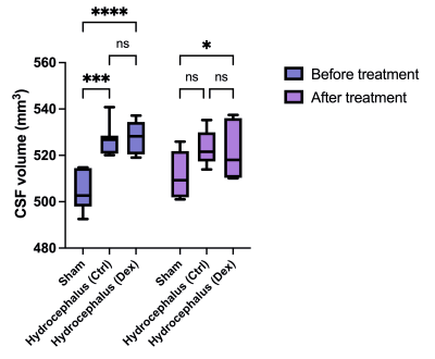 |
124 |
Dexmedetomidine Administration in a Rat Model of Kaolin-induced
Hydrocephalus
Meltem Karatas1,
Diaa K.A. Yahya2,
Merve Ayan3,
Sevda Muftuoglu3,
Kader Karli Oguz1,4,5,
and Halil Kamil Oge2
1National Magnetic Resonance Research Center (UMRAM), Bilkent University, Ankara, Turkey, 2Department of Neurosurgery, Hacettepe University, Ankara, Turkey, 3Department of Histology and Embryology, Hacettepe University, Ankara, Turkey, 4Department of Radiology, Hacettepe University, Ankara, Turkey, 5Department of Radiology, University of California, Davis, Sacramento, CA, United States Keywords: Neuroinflammation, Translational Studies, Hydrocephalus , dexmedetomidine Hydrocephalus is defined as an abnormal enlargement of cerebral ventricles due to disturbed cerebrospinal fluid (CSF) dynamics. It is characterized by neuroinflammatory processes in the brain parenchyma and CSF, with pathological findings including reactive gliosis, axonal and neuronal damage, and loss of myelin sheaths. The use of anti-inflammatory medications in hydrocephalus may be an effective treatment strategy for neuroprotection. In this study, we used a rat model of hydrocephalus induced by kaolin. We aimed to demonstrate the potential anti-inflammatory and neuroprotective effects of dexmedetomidine on hydrocephalic brains using MRI and morphometric analysis. |
|
3532.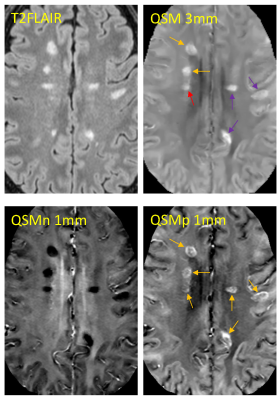 |
125 |
Quantitative susceptibility source separation improves the
reader agreement in the identification of MS paramagnetic rim
lesions
Emily Demmon1,
Susan Gauthier1,
Ilhami Kovanlikaya2,
Yi Wang2,
Alexey Dimov2,
Pascal Spincemaille2,
and Thanh Nguyen2
1Department of Neurology, Weill Cornell Medicine, New York City, NY, United States, 2Department of Radiology, Weill Cornell Medicine, New York City, NY, United States Keywords: Multiple Sclerosis, Neuroscience Multiple Sclerosis (MS) is an autoimmune disorder characterized by focal inflammatory demyelination. Paramagnetic rim lesions (PRLs) form an important subset of chronic active MS lesions that show a hypocellular core and persistent glial inflammation and continuing myelin damage at the rim on histopathology. PRLs are a critical clinical target for MS therapy. Our goal in this project is to improve overall detection of PRLs through utilization of quantitative paramagnetic source maps. |
|
3533.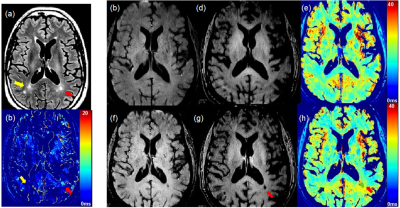 |
126 |
Novel MRI techniques for the Assessment of Activated Innate
Immunity in Progressive Multiple Sclerosis
Seong-Eun Kim1,
Trieste Francis 2,
Ka-Ho Wong 2,
John W Rose2,
J Scott MacNally1,
and M Mateo Paz Soldan 2
1UCAIR, Department of Radiology and Imaging Sciences, University of Utah, Salt Lake City, UT, United States, 2Department of Neurology, University of Utah, Salt Lake City, UT, United States Keywords: Multiple Sclerosis, Molecular Imaging, USPIO While the specific pathologic mechanisms that underpin ongoing axonal loss in progressive forms of MS are not fully characterized, ongoing innate immune activation in chronic MS lesions, particularly activated microglia and macrophages, has been proposed to be a prevailing mechanism of axonal loss in the progressive phase of disease. Ultrasmall superparamagnetic iron oxide particles have been introduced as a potential MRI biomarker for macrophage activity in focal MS lesions. This work presents a Feromoxytol enhanced MRI to measure perilesional activated microglia and macrophages in progressive MS. which is anticipated to reflect risk for ongoing loss of CNS axons and neurons.
|
|
3534.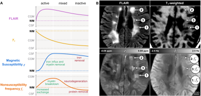 |
127 |
Nonsusceptibility frequency shift reveals subtypes of multiple
sclerosis lesions and predicts 5-year disappearance of
paramagnetic rim
Thomas Jochmann1,2,
Jack A. Reeves2,
Fahad Salman2,
Dejan Jakimovski2,
Simon Hametner3,
Maryam Mohebbi2,
Niels Bergsland2,
Bianca Weinstock-Gutman4,
Michael G. Dwyer2,
Robert Zivadinov2,5,
Jens Haueisen1,
and Ferdinand Schweser2,5
1Department of Computer Science and Automation, Technische Universität Ilmenau, Ilmenau, Germany, 2Buffalo Neuroimaging Analysis Center, Department of Neurology at the Jacobs School of Medicine and Biomedical Sciences, University at Buffalo, The State University of New York, Buffalo, NY, United States, 3Department of Neuroimmunology, Medical University of Vienna, Vienna, Austria, 4Jacobs Multiple Sclerosis Center, Department of Neurology, Jacobs School of Medicine and Biomedical Sciences, University at Buffalo, The State University of New York, Buffalo, NY, United States, 5Center for Biomedical Imaging, Clinical and Translational Science Institute, University at Buffalo, The State University of New York, Buffalo, NY, United States Keywords: White Matter, Multiple Sclerosis Multiple sclerosis lesions have different types and stages that are challenging to distinguish by conventional MRI. We recently presented DEEPOLE QUASAR, a reconstruction technique that yields nonsusceptibility frequency maps as a novel MRI contrast. In this study, we translate the novel contrast into a tool for multiple sclerosis lesion assessment. We discuss the time course of nonsusceptibility frequency throughout different lesion stages. Within different paramagnetic rim lesions that are typically indistinguishable by conventional MRI, the nonsusceptibility frequency contrast showed hypo-, iso-, and hyperintense subtypes. At a 5-year follow-up, paramagnetic rims disappeared at significantly different proportions between these subtypes. |
|
3535.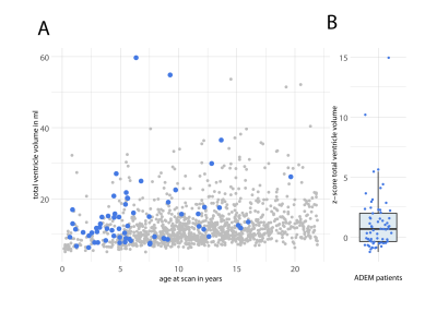 |
128 |
Whole Brain MRI Reveals that Children with Acute Disseminated
Encephalomyelitis have Enlarged Brain Ventricles at Disease
Onset
Jason Michael Millward1,2,
Elias Pilgrim1,
Matthias Baumann3,
Eva Wendel4,
Ines El-Naggar4,
Annikki Bertolini4,
Frederik Bartels5,
Carsten Finke5,
Thoralf Niendorf1,2,
Kevin Rostásy4,
and Sonia Waiczies1,2
1Berlin Ultrahigh Field Facility, Max Delbruck Center, Berlin, Germany, 2Experimental and Clinical Research Center, Charité - Universitätsmedizin Berlin, Berlin, Germany, 3Division of Paediatric Neurology, Department of Paediatrics, Medical University of Innsbruck, Innsbruck, Austria, 4Department of Paediatric Neurology, Children's Hospital Datteln, Witten/Herdecke University, Datteln, Germany, 5Department of Neurology, Charité - Universitätsmedizin Berlin, Berlin, Germany Keywords: Neuro, Neuroinflammation, acute disseminated encephalomyelitis Pediatric patients with acute disseminated encephalomyelitis (ADEM) are at risk of long-term neurocognitive consequences. We investigated a multi-center cohort of ADEM patients with serial whole brain MRI, to distinguish transient reversible expansion of brain ventricle volume (BVV) from persistent changes. Using the automated brain segmentation tool SynthSeg, we observed that ADEM patients had significantly enlarged BVV relative to sex- and age-matched pediatric control subjects, already at the first clinical presentation, before any steroid treatment. Most ADEM patients developed even greater BVV expansion over the observation period. The majority of patients recovered, though some showed persistent BVV enlargement, suggesting brain atrophy. |
|
3536.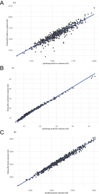 |
129 |
Evaluation of an Image Contrast-Agnostic Brain MRI Segmentation
Tool on a Heterogeneous Pediatric Clinical Cohort
Elias Pilgrim1,
Wenli Xu1,
Hampus Nils Olsson1,
Eva Wendel2,
Ines El-Naggar2,
Annikki Bertolini2,
Kevin Rostásy2,
Thoralf Niendorf1,3,
Sonia Waiczies1,3,
and Jason Michael Millward1
1Berlin Ultrahigh Field Facility, Max Delbruck Center, Berlin, Germany, 2Department of Paediatric Neurology, Children's Hospital Datteln, Witten/Herdecke University, Datteln, Germany, 3Experimental and Clinical Research Center, Charité - Universitätsmedizin Berlin, Berlin, Germany Keywords: Neuroinflammation, Segmentation, acute disseminated encephalomyelitis We evaluated the performance of the brain segmentation tool SynthSeg on a heterogeneous multi-center cohort of pediatric patients with acute disseminated encephalomyelitis. Brain MRI of the patient cohort was acquired at multiple field strengths, with a diverse range of image spatial resolutions, MR sequences, contrasts, and w/wo administration of gadolinium-based contrast agent (GBCA). SynthSeg performance was generally robust to resolution and contrast differences, especially for larger brain structures. Significant differences in some calculated volumes were detected upon pair-wise comparisons among patients with high- vs. low resolution scans, T1- vs. T2-weighed scans, and w/wo GBCA application, acquired on the same day. |
|
3537.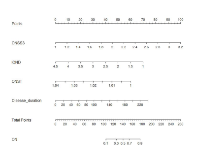 |
130 |
Novel contribution of non-enhanced MRI to neuroradiologists for
detecting neuromyelitis optica spectrum disorder-associated
optic neuritis
Jing Zhang1 and
Yali Zhao1
1Radiology, Tongji Hospital, Tongji Medical College, Huazhong University of Science and Technology, Wuhan, China Keywords: Neuroinflammation, Neuroinflammation The sensitivity of MRI in detecting optic neuritis (ON) including neuromyelitis optica spectrum disorder-associated ON (NMOSD-ON) is inadequate, and the diagnostic criteria have limitations due to inherent defects in signal intensity. In this study, we quantitatively assessed the role of intrathecal cerebrospinal fluid and diameter and curvature of anterior visual pathway in the diagnosis of NMOSD-ON by nomogram using non-enhanced MRI. The altered MRI parameters showed significant associations with visual functional system scores. Our study elucidates the novel contribution of non-enhanced MRI in the detection of NMOSD-ON, which assists to solve the problem of insufficient sensitivity of MRI for ON. |
|
3538.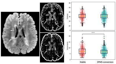 |
131 |
Brain MRI lesion heterogeneity and chronic active lesion density
identify relapsing-remitting MS 5 years before onset of
progressive disease
Yunyan Zhang1,
Zahra Hosseinpour1,
Olayinka Oladosu1,
Julia Beaumont1,
Chloe Hao1,
and Carlos Camara-Lemarroy1
1University of Calgary, Calgary, AB, Canada Keywords: Multiple Sclerosis, Brain Multiple sclerosis (MS) is a severe and heterogeneous disease. While disease worsening is common, the pattern of worsening varies between subjects. Mechanisms are unclear. This study assessed lesion heterogeneity and chronic active lesion activity in relapsing-remitting MS (RRMS) 5 years before conversion to secondary progressive MS (SPMS) as compared to stable disease. Based on brain MRI texture analysis and z-score of core versus rim diffusion in lesions, SPMS-converting patients have greater lesion damage and chronic active lesion density than stable patients 5 years prior. Combined approaches may help identify RRMS patients at high risk of worsening for early management. |
|
3539.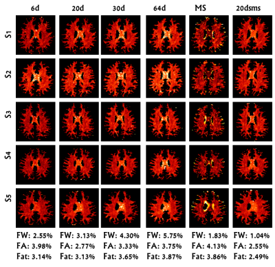 |
132 |
Reproducibility of Diffusion Microstructures Over Different
Diffusion Gradient Directionalities
Apurva Shah1,
Jitender Saini2,
Pramod Pal3,
and Madhura Ingalhalikar1
1Symbiosis Centre for Medical Image Analysis, Symbiosis International (Deemed University), Pune, India, 2Department of Radiology, National Institutes of Mental Health and Neuroscience, Bengaluru, India, 3National Institutes of Mental Health and Neuroscience, Bengaluru, India Keywords: Neuroinflammation, Brain, Diffusion Imaging, freewater, reliability Medical imaging can offer several clinically valuable biomarkers which require accuracy across the acquiring technologies. Our study investigated the variability and reliability of diffusion microstructures over different diffusion gradient direction schemes, including high angular resolution diffusion imaging sequences and a parallel slice acquisition technique. The study's results demonstrated the 20-directional parallel slice acquisition clinically feasible sequence for standard DTI analysis with less variability and more reliability in matrices, while multishell sequences are more suitable for microstructural and tractography analysis. |
|
3540.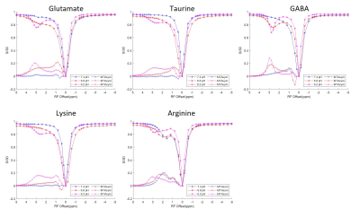 |
133 |
Biomolecular basis of changes in CEST Z-spectra of rat spinal
cord after injury
Chaoqi Mu1,2,
Jamie L Reed2,
Feng Wang2,
John C Gore2,
and Li Min Chen2
1Biomedical Engineering, Vanderbilt University, Nashville, TN, United States, 2Vanderbilt University Institute of Imaging Science & Department of Radiology and Radiological Sciences, Vanderbilt University Medical Center, Nashville, TN, United States Keywords: Spinal Cord, CEST & MT We identified molecular pools of interest using CEST imaging of rats after spinal cord injury (SCI) that correlate with post-SCI behavioral deficits, and which may indicate products of neuroinflammation. Using tissue neurochemical analysis, we isolated analytes of interest and acquired CEST Z-spectra from solutions of the chemicals to elucidate the source of the CEST pool concentration changes. Several candidates present in the tissue samples showed similar (overlapping) Z-spectra around 3.0 ppm. These results suggest that CEST imaging is not specific for identifying concentration changes of individual molecules of interest. |
|
3541.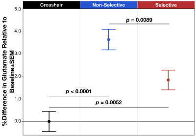 |
134 |
¹H fMRS detects changes in dorsal anterior cingulate cortex
glutamate driven by inhibitory control
Jillian M. Eichstaedt1,
Dalal Khatib2,
Phil Easter2,
David R. Rosenberg2,
Vaibhav A. Diwadkar2,
and Jeffrey A. Stanley3
1Translational Neuroscience Program, Wayne State University School of Medicine, Detroit, MI, United States, 2Wayne State University School of Medicine, Detroit, MI, United States, 3Psychiatry and Behavioral Neuroscience, Wayne State University School of Medicine, Detroit, MI, United States Keywords: Psychiatric Disorders, Spectroscopy, Inhibitory Control ¹H fMRS was used to quantify changes in dACC glutamate evoked during excitatory and inhibitory motor control. The two response modes demanded non-selective responding (motor control with excitation) or selective responding (motor control with inhibition). dACC glutamate was significantly increased (relative to a resting baseline), regardless of response mode. These functional responses (uncoupled from hemodynamics) demonstrate that ¹H fMRS can be successfully used to reveal glutamate related mechanisms of the dACC reflecting changes in the excitatory neurotransmission drive. |
|
3542.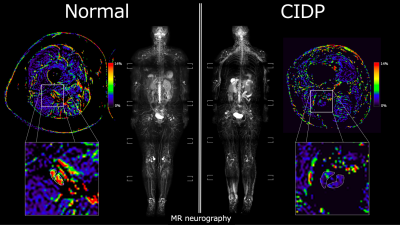 |
135 |
Inhomogeneous Magnetization Transfer (ihMT) of Peripheral Nerve
in Patients with Chronic Inflammatory Demyelinating
Polyradiculoneuropathy
Ryuna Kurosawa1,
Hajime Yokota2,
Takayuki Sada1,
Koji Matsumoto1,
Takafumi Yoda1,
Masami Yoneyama3,
Takashi Namiki3,
Guillaume Gilbert4,
Yoshitada Masuda1,
and Takashi Uno2
1Department of Radiology, Chiba university hospital, Chiba, Japan, 2Diagnostic Radiology and Radiation Oncology, Graduate School of Medicine, Chiba University, Chiba, Japan, 3Philips Japan, Tokyo, Japan, 4Philips Canada, Mississauga, ON, Canada Keywords: Neurodegeneration, CEST & MT Chronic inflammatory demyelinating polyradiculoneuropathy (CIDP) is an immune-mediated demyelinating disease. The purpose of this study is to evaluate the usefulness of ihMT in the diagnosis of CIDP by measuring myelin levels using ihMT imaging. ihMTR of the sciatic nerve of CIDP patients was significantly lower than that of the normal subjects. Our finding was compatible with previously reported pathological changes. Myelin imaging by ihMT has the potential to be used as a new biomarker in the diagnosis of CIDP. |
|
3543.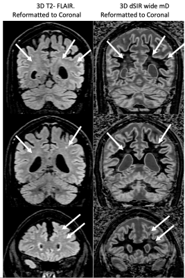 |
136 |
MRI of Neuroinflammation in the Brain following Meth Addiction
using Multiplied, Added, Subtracted and/or Divided (MASDIR)
sequences.
Paul Condron1,2,
Maryam Tayebi1,2,
Taylor Emsden1,2,
Ben Bristow1,3,
Tuta Ngārimu4,5,
Patrick McHugh1,6,
Gil Newburn1,
Davidson Taylor1,7,
Samantha Holdsworth1,2,
Daniel Cornfeld1,2,
Miriam Scadeng1,2,
and Graeme Bydder1,8
1Mātai Medical Research Institute, Gisborne, New Zealand, 2Faculty of Medical and Health Sciences & Centre for Brain Research, Auckland, New Zealand, 3Department of Neuroscience,, University of Otago, Dunedin, New Zealand, 4New Zealand P Pull, Tairāwhiti-Gisborne, New Zealand, 5Ngati Porou, Aotearoa, New Zealand, 6Tūranga Health, Tūranganui-a-kiwa, Gisborne, New Zealand, 7Ngāti Porou, Ngāti Kahungungu, Rongomaiwahine, Rongowhakaata; Tūranganui-a-Kiwa, Tairāwhiti, New Zealand, 8University of California San Diego, San Diego, CA, United States Keywords: Neuroinflammation, Drugs, brain, methamphetamine, white matter hyper intensities Keywords; Neuroinflammation, data acquisition and post processing, contrast mechanisms, white matter, drugs. MR contrast can be enhanced by exploiting the concept of tissue property filters (TPFs) by designing Multiplied, Added, Subtracted, and/or Divided Inversion Recovery (MASDIR) sequences to amplify contrast produced by small changes in white matter T1. The Divided, Subtracted IR (dSIR) sequence provides very high contrast depiction of subtle white matter changes due to disease that are not identified with standard T2-FLAIR imaging. This was demonstrated in patients with a history of methamphetamine addiction. MASDIR sequences could revolutionize the way that neuroinflammation is imaged in clinical practice. |
|
3544.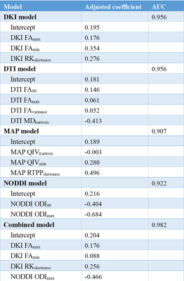 |
137 |
Histogram Analysis of Magnetic Resonance Diffusion Imaging in
Differentiating Glioma Mimicking Encephalitis from Encephalitis
Kai Zhao1,
Xiaoyue Ma1,
Ankang Gao1,
Eryuan Gao1,
Jinbo Qi1,
Peipei Wang1,
Guohua Zhao1,
Huiting Zhang2,
Guang Yang3,
Jie Bai1,
Yong Zhang1,
and Jingliang Cheng1
1Department of MRI, The First Affiliated Hospital of Zhengzhou University, Zhengzhou, China, 2Magnetic Resonance Scientific Marketing, Siemens Healthineers Ltd., Wuhan, China, 3Shanghai Key Laboratory of Magnetic Resonance, East China Normal University, Shanghai, China Keywords: Tumors, Tumor, Inflammation Diffusion/Other Diffusion Imaging Techniques Glioma Mimicking Encephalitis is difficult to be differentiated from Encephalitis by conventional MRI. In this study, 18 patients with diagnosed glioma and 15 patients with encephalitis were included. Their DWI images were processed to obtain the histogram of the parameter maps of DKI, DTI, NODDI, and MAP. We use lasso regression to fit the diagnostic models, and the diagnostic performance of different models were compared. We find no significant difference in the AUC between the single and combined diffusion lasso models. Any of the models could be individually used for differentiating glioma mimicking encephalitis from encephalitis. |
|
 Back to Meeting Home
 Back to the Program-at-a-Glance
The International Society for Magnetic Resonance in Medicine is accredited by the Accreditation Council for Continuing Medical Education to provide continuing medical education for physicians.
 Back to Meeting Home
 Back to the Program-at-a-Glance
 View Presentation Video