Digital Poster
Gray Matter I
ISMRM & ISMRT Annual Meeting & Exhibition • 03-08 June 2023 • Toronto, ON, Canada

| Computer # | |||
|---|---|---|---|
3351.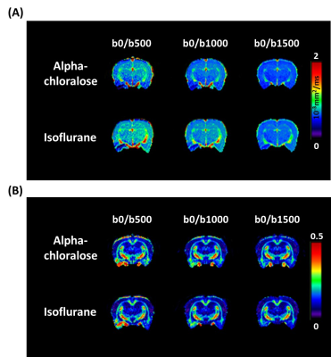 |
121 |
Anesthetic modulation of water diffusion: Insights from a
diffusion tensor imaging study
Shin-Lei Peng1,
Sheng-Min Huang2,
and Chun-Chieh Chan1
1Department of Biomedical Imaging and Radiological Science, China Medical University, Taichung, Taiwan, 2Institute of Biomedical Engineering and Nanomedicine, National Health Research Institutes, Miaoli, Taiwan Keywords: Gray Matter, Diffusion Tensor Imaging A critical step in animal diffusion tensor imaging (DTI) studies is the use of anesthetics. Understanding the influence of specific anesthesia regimes on DTI-derived parameters is imperative when comparing results between animal studies using different anesthetics. Here, the quantification of fractional anisotropy (FA) and mean diffusivity (MD) under different alpha-chloralose and isoflurane is discussed. The estimated MD under isoflurane anesthesia is higher than that under alpha-chloralose anesthesia. FA quantitation was also influenced by anesthesia regimens to varying extents, depending on the brain regions and b-values. In summary, both scanning parameters and the anesthesia regimens significantly impacted quantifications of DTI indices. |
|
3352.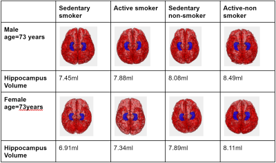 |
122 |
Lifestyle determinants of Hippocampal Volume: An AI-enabled
Volumetric Analysis on a Large Healthy Cohort.
Saurabh Garg1,
Nasrin Akbari1,
Saqib Basar1,
Thanh-Duc Nguyen1,
Sean London2,
Yosef Chodakiewitz2,
Rajpaul Attariwala1,
Mostafa Fatehi3,
and Sam Hashemi1
1Voxelwise Imaging Technology Inc, Vancouver, BC, Canada, 2Prenuvo, Vancouver, BC, Canada, 3Division of Neurosurgery, Vancouver General Hospital, Vancouver, BC, Canada Keywords: Gray Matter, Aging, Machine Learning, Artificial Intelligence The volume of the hippocampus and temporal horn of the lateral ventricle provides insight into the state of neurological health and the changes in volume are associated with conditions such as traumatic brain injury1, Alzheimer's Disease 2 and treatment response. It remains an open question what are the normal age-related changes over time in hippocampal and temporal horn volumes in an average healthy population cohort. We seek to establish these baseline parameters via AI-based MRI brain segmentation and quantification techniques applied to a large cohort of screened healthy patients. |
|
3353.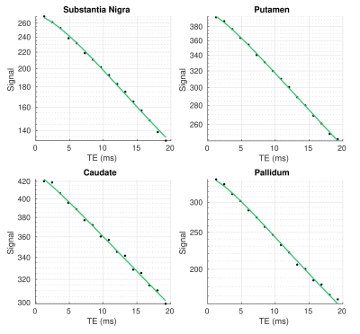 |
123 |
Non-exponential transverse relaxation in the brain’s basal
ganglia
Rita Oliveira1,
Quentin Raynaud1,
Valerij Kiselev2,
Ileana Jelescu3,
and Antoine Lutti1
1Laboratory for Research in Neuroimaging, Department of Clinical Neuroscience, Lausanne University Hospital and University of Lausanne, Lausanne, Switzerland, 2Division of Medical Physics, Department of Radiology, University Medical Center Freiburg, Freiburg, Germany, 3Department of Radiology, Lausanne University Hospital (CHUV) and University of Lausanne, Lausanne, Switzerland Keywords: Gray Matter, Relaxometry Non-exponential transverse relaxation in the brain’s basal ganglia has been investigated in theoretical studies but little evidence from in-vivo MRI data exists in support of this behaviour. Here, we provide experimental observation of non-exponential transverse relaxation in-vivo MRI data in the basal ganglia at 3T. The strongest deviations from exponential behaviour take place in the iron-rich pallidum and substantia nigra. Our results suggest that water diffusion through the inhomogeneous magnetic field induced by paramagnetic iron deposits may be at the source of non-exponential transverse relaxation in the basal ganglia. |
|
3354.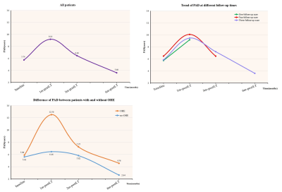 |
124 |
Dynamic evolution of the predicated brain age in liver
transplantation recipients: a longitudinal study
Zining Lu1,
Yue Cheng1,
Xianchang Zhang2,
Junhai Xu3,
and Wen Shen1
1Department of Radiology, Tianjin First Central Hospital, Tianjin, China, Tianjin, China, 2MR Collaboration, Siemens Healthcare Ltd., Beijing, China, Beijing, China, 3College of Intelligence and Computing, Tianjin University, Tianjin, China, Tianjin, China Keywords: Gray Matter, Machine Learning/Artificial Intelligence To evaluate the dynamic change in overall brain health in liver transplantation (LT) recipients, we constructed a deep learning-based brain age prediction model to measure the longitudinal changes of ‘brain age’ before and one, three, and six months after surgery. The LT recipients’ brain age showed an inverted U-shaped change pattern in the early stages after transplantation. In addition, brain aging was aggravated within one month after surgery, and the patients with a history of overt hepatic encephalopathy were particularly affected. Therefore, the age prediction model can be used to monitor the post-surgical brain function recovery trajectory in LT recipients. |
|
3355.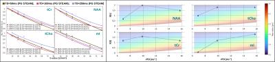 |
125 |
Time dependent diffusion and kurtosis of human brain metabolites
André Döring1,
Frank Rösler2,
Kadir Şimşek1,3,
Maryam Afzali1,4,
Roland Kreis5,6,
Derek K Jones1,
Julien Valette7,
and Marco Palombo1,3
1Cardiff University Brain Research Imaging Centre (CUBRIC), School of Psychology, Cardiff University, Cardiff, United Kingdom, 2Department of Mathematics, University of Bern, Bern, Switzerland, 3School of Computer Science and Informatics, Cardiff University, Cardiff, United Kingdom, 4Leeds Institute of Cardiovascular and Metabolic Medicine, University of Leeds, Leeds, United Kingdom, 5Magnetic Resonance Methodology, Institute of Diagnostic and Interventional Neuroradiology, University Bern, Bern, Switzerland, 6Translational Imaging Center, sitem-insel, Bern, Switzerland, 7Université Paris-Saclay, CEA, CNRS, MIRCen, Laboratoire des Maladies Neurodégénératives, Fontenay-aux-Roses, Paris, France Keywords: Gray Matter, Spectroscopy, Metabolites, Diffusion, ADC, Kurtosis, Morphology, Gray Matter, Spectroscopy This work demonstrates that metabolites diffusion and kurtosis time-dependence can be measured in vivo in the human brain using Diffusion-Weighted MR Spectroscopy (DW-MRS) and ultra-strong gradients. At short diffusion-times, DW-MRS is sensitive to cytoplasmic viscosity and short-range structures; at long diffusion-times to long-range structures. We show that modeling the diffusion-time dependence of intracellular and cell-type specific metabolites can be used to infer brain cell morphology and recover fiber radii consistent with healthy human brain histology. Furthermore, we show that water diffusion at long diffusion-times is affected by exchange between intra- and extracellular compartment, which poses challenges for microstructural modeling. |
|
3356.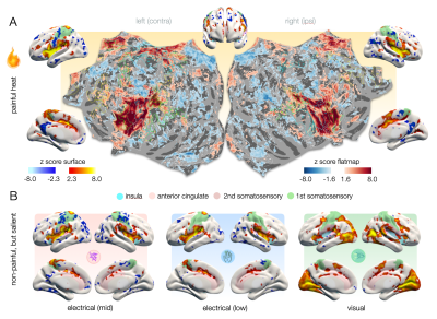 |
126 |
Where is Pain in the Brain? Revealing Neural Correlates of Pain
as Orchestration of Allostatic Domain-General Patterns via fMRI
at 7T
Henning Matthias Reimann1,
Jurjen Heij2,
Thomas Gladytz1,
and Thoralf Niendorf1,3
1Berlin Ultrahigh Field Facility (B.U.F.F.), Max Delbrück Center for Molecular Medicine, Berlin, Germany, Berlin, Germany, 2Spinoza Centre for Neuroimaging, Amsterdam, Netherlands, 3Experimental and Clinical Research Center, a joint cooperation between the Charité Medical Faculty and the Max Delbrück Center for Molecular Medicine in the Helmholtz Association, Berlin, Germany Keywords: Gray Matter, fMRI, pain, allostasis To date, fMRI has failed to distinguish an unambiguous and reliable signature that is unique to pain. Pain is accompanied by the activation of networks related to saliency processing. Employing the high sensitivity and spatial fidelity of 7T fMRI, we disentangle these overlapping representations. We apply stimuli that are perceived as painful and others that trigger saliency-related domain-general brain activity. Applying partial least square regression, we identify modality-specific characteristics in the way domain-general patterns are orchestrated relative to each other, revealing a large number of neurosignatures that are specific to the perception of pain. |
|
3357.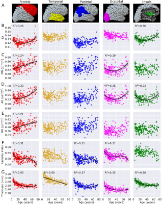 |
127 |
High resolution diffusion tensor imaging shows non-linear
trajectories in the human cortex over the healthy lifespan
J. Alejandro Acosta-Franco1,
Graham Little2,
and Christian Beaulieu1
1Biomedical Engineering, University of Alberta, Edmonton, AB, Canada, 2Computer Science, Université de Sherbrooke, Sherbrooke, QC, Canada Keywords: Gray Matter, Diffusion Tensor Imaging The brain cortex is thin and has a complex morphology making it difficult to analyze with in vivo diffusion MRI. A high-resolution 1.5 mm isotropic diffusion MRI protocol and an automated cortex segmentation methodology on the diffusion images alone was used to assess cortical diffusion changes over the healthy lifespan (n=165, 5-74 years). The diffusion metrics versus age trajectories differed across the cortical surface despite similar cortical thickness reductions with age. The most robust age changes were for axial diffusivity (quadratic U) and radiality (cubic) potentially reflecting changes in columnar microstructural organization of the cortex in typical development and aging. |
|
3358.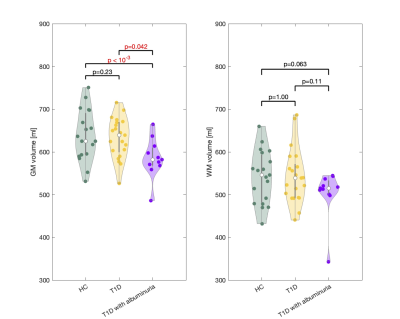 |
128 |
Patients with type 1 diabetes and albuminuria have reduced
cerebral grey matter unrelated to cerebrovascular dysfunction.
Mark Bitsch Vestergaard1,
Jens Christian Laursen2,
Niels Søndergaard Heinrich2,
Peter Rossing2,
Tine Willum Hansen2,
and Henrik Bo Wiberg Larsson1
1Clinic for Clinical Physiology and Nuclear Medicine, Rigshospitalet, Glostrup, Denmark, 2Steno Diabetes Center Copenhagen, Herlev, Denmark Keywords: Gray Matter, Diabetes, Cerebrovascular function Patients with diabetes mellitus type 1 (T1D) demonstrate brain alterations including gray matter (GM) atrophy. We examined whether a GM reduction could be related to cerebrovascular dysfunction. GM volume was measured by anatomical MRI. Cerebrovascular function was examined by measuring cerebral blood flow, oxygen metabolism and lactate concentration in response to inhalation of hypoxic air using phase-contrast MRI and MRS. T1D patients were evaluated for albuminuria as indication of vessel damages. Patients with T1D and albuminuria had reduced GM compared to patients with normal kidney function or healthy controls, however, the GM atrophy could not be related to cerebrovascular dysfunction. |
|
3359.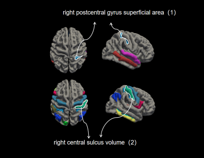 |
129 |
ROI-based morphometry brain neuroimaging reveals grey and white
matter abnormalities in type 2 and 3 juvenile SMA patients
Wanqing Shen1,
Yingqian Chen1,
Shu Su1,
Yujian Liang1,
Pei Xiang1,
Long Qian2,
and Zhiyun Yang1
1The First Affiliated Hospital of Sun Yat-Sen University, Guangzhou, China, 2ge health care, Guangzhou, China Keywords: Gray Matter, Gray Matter, SMA,FreeSurfer,cerebral cortex The study investigated brain structure changes in type 2 and 3 SMA patients and their correlation with the severity of clinical symptoms. MRI examinations were performed on 3.0T MRI scanner and 3D T1W data were analyzed using FreeSurfer.The result showed SMA patients had extensive, multifocal, symmetrical gray white matter degeneration.The postcentral gyrus is strongly associated with symptom severity in SMA patients. It might be explained by the fact that motor neurons exhibit functional alterations, leading to selective motor neuron loss. So it is crucial for brain development and cognitive development if we intervene in SMA disease in an early stage. |
|
3360.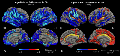 |
130 |
Mapping Cortical Diffusion Variations with Normal Aging Using
DTI: The Validity of the Tensor Interpretation
Yutong L. Sun1,2,
Jordan A. Chad2,
and J. Jean Chen1,2
1Medical Biophysics, University of Toronto, Toronto, ON, Canada, 2Rotman Research Institute, Baycrest Health Sciences, Toronto, ON, Canada Keywords: Gray Matter, Aging Aging-related changes in diffusion as measured by diffusion MRI (dMRI) of the white matter (WM) are often interpreted as being driven degeneration of myelinated axons and neurites, but the validity of this tensor (DTI) interpretation of the dMRI signal has never been tested in the cortex. In this study, age-related cortical diffusion variations were assessed using metrics derived from DTI and from the orthogonal-moment diffusion-tensor decomposition (DT-DOME) method. We demonstrate that the tensor interpretation of aging-related dMRI variations are most likely inadequate in the cortex. |
|
3361.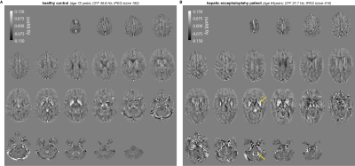 |
131 |
QSM and edited MRS reveal altered deep-brain gray matter
microstructure and neurometabolism in patients with hepatic
encephalopathy
Helge J. Zöllner1,2,3,4,
Thomas A. Thiel3,4,
Markus S. Jördens5,
Dieter Häussinger5,
Georg Oeltzschner1,2,
Markus Butz3,
Hans-Jörg Wittsack4,
Alfons Schnitzler3,
and Eric Bechler4
1The Russell H. Morgan Department of Radiology and Radiological Science, The Johns Hopkins University School of Medicine, Baltimore, MD, United States, 2Kennedy Krieger Institute, F. M. Kirby Research Center for Functional Brain Imaging, Baltimore, MD, United States, 3Institute of Clinical Neuroscience and Medical Psychology, Medical Faculty, Heinrich-Heine-University Düsseldorf, Düsseldorf, Germany, 4Department of Diagnostic and Interventional Radiology, Medical Faculty, Heinrich-Heine-University Düsseldorf, Düsseldorf, Germany, 5Department of Gastroenterology, Hepatology and Infectiology, Medical Faculty, Heinrich-Heine-University Düsseldorf, Düsseldorf, Germany Keywords: Gray Matter, Quantitative Susceptibility mapping, Hepatic encephalopathy Hepatic encephalopathy (HE) is a common neurological manifestation of liver cirrhosis characterized by altered brain microstructure and metabolism. Here, Quantitative Susceptibility Mapping (QSM) was performed in a well-characterized cohort of HE patients and correlated with metabolite estimates derived from GABA-edited MR spectroscopy. Deep-brain gray matter micro-susceptibility was significantly altered in the bilateral pallidum and dentate nuclei in HE patients. These results were closely linked to metabolite estimates and clinical metrics. |
|
3362.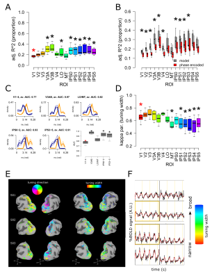 |
132 |
Estimating population motor fields (pMF) from saccadic eye
movements at high field (7T)
Alessio Fracasso1,
Katarina Moravkova1,
Jasper Fabius1,
Rosanne Timmermann1,
and Anna Gaglianese2
1University of Glasgow, Glasgow, United Kingdom, 2Lausanne University Hospital, Lausanne, Switzerland Keywords: Gray Matter, fMRI (task based), saccade, high-field, modelling Here we implemented a variation of the population receptive field model (pRF, Dumoulin et al., 2008; Fracasso et al., 2016), to model blood oxygenation level dependent (BOLD) signal and obtain estimates of saccade tuning direction and width from human PPC, to obtain estimates of the population motor fields (pMF). Saccade tuning width shows a novel organizational property of human posterior parietal cortex, unveiling a gradient from posterior to anterior PPC, with tuning width steadily increasing along the posterior-anterior axis. |
|
3363.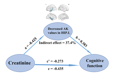 |
133 |
Abnormal cerebral micro-structures in end-stage renal disease
patients related to mild cognitive impairment
Jiahui Zheng1,
Jiankun Dai2,
Xiangxiang Wu1,
and Haifeng Shi1
1Department of Radiology, The Affiliated Changzhou NO.2 People’s Hospital of Nanjing Medical University, changzhou, China, 2GE Healthcare, MR Research China, Beijing, China Keywords: Gray Matter, Diffusion/other diffusion imaging techniques This study aimed to investigate the abnormal cerebral micro-structures related to mild cognitive impairment (MCI) and further predict individual cognitive function in end-stage renal disease (ESRD) patients undergoing maintenance hemodialysis. Specially, diffusion kurtosis imaging (DKI), mediation analysis, and the least squares support vector regression machine (LSSVRM) were utilized to conduct our study. We observed that aberrant micro-structures partially mediated the association between clinical risk factors and MCI, which is a novel insight into the progression of cognitive dysfunction. The combination of DKI metrics and clinical characteristics could be used as features to efficiently predict cognitive function associated with ESRD. |
|
3364.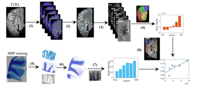 |
134 |
Cortical laminar specific microstructure and magnetic
susceptibility in ex-vivo human brain
Zhiyong Zhao1,
Zuozhen Cao1,
Qinfeng Zhu1,
Liangying Zhu1,
Sihui Li1,
Keqing Zhu2,3,
Jing Zhang2,3,
and Dan Wu1
1Key Laboratory for Biomedical Engineering of Ministry of Education, Department of Biomedical Engineering, College of Biomedical Engineering & Instrument Science, Zhejiang University, Hangzhou, China, 2China Brain Bank and Department of Neurology in Second Affiliated Hospital, Key Laboratory of Medical Neurobiology of Zhejiang Province, and Department of Neurobiology, Zhejiang University School of Medicine, Hangzhou, China, 3Department of Pathology, The First Affiliated Hospital and School of Medicine, Zhejiang University, Hangzhou, China Keywords: Gray Matter, Ex-Vivo Applications, cortical laminae, microstructure, magnetic susceptibility, human brain Although recent high-resolution functional MRI studies have revealed laminar-specific cortical activity in the human brain, the underlying structural underpinning remains unclear. The present study collected submillimeter-resolution MR images from 10 ex-vivo human hemispheres. We computed the microstructural metrics and magnetic susceptibility for each sample at different cortical depths. We found that the cortical microstructure and magnetic susceptibility showed a depth-dependent pattern, which varied in different cortical regions. The cortical profiles were then correlated with cell density and myelin content obtained from histological staining. These findings may help to understand the structural basis of laminar brain function. |
|
3365.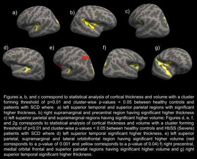 |
135 |
Analyzing Cortical Thickness and Volume in patients with Sickle
Cell Disease using 7T-MRI
Sharadhi Umesh Bharadwaj1,
Tales Santini2,
Joel D.K Disu1,
Busola Oluwole3,
Enrico M. Novelli4,5,6,
Tamer S Ibrahim7,8,9,
and Sossena Wood1,10
1Biomedical Engineering, Carnegie Mellon University, Pittsburgh, PA, United States, 2University of Pittsburgh, Pittsburgh, PA, United States, 3University of Washington/Fred Hutch, Seattle, WA, United States, 4Vascular Medicine Institute, University of Pittsburgh Medical Center, Pittsburgh, PA, United States, 5Department of Hematology/Oncology, University of Pittsburgh Medical Center, Pittsburgh, PA, United States, 6Sickle Cell Center of Excellence, University of Pittsburgh Medical Center, Pittsburgh, PA, United States, 7Department of Bioengineering, University of Pittsburgh, Pittsburgh, PA, United States, 8Department of Radiology, University of Pittsburgh, Pittsburgh, PA, United States, 9Department of Psychiatry, University of Pittsburgh, Pittsburgh, PA, United States, 10Neuroscience Institute, Carnegie Mellon University, Pittsburgh, PA, United States Keywords: Gray Matter, High-Field MRI, Sickle cell disease, cortical thickness, cortical volume The study compares cortical thickness and volume between patients with Sickle Cell Disease (SCD) and healthy controls (HC). There were no significant differences in the cortical thickness and volume of the whole brain between HC and patients with SCD. We found significant differences (p<0.05) in the temporal and parietal regions between HC and patients with SCD. We found significant differences in the parietal and frontal lobes between severe SCD (HbSS) and HC. Between patients with mild SCD (HbSC and HbSβ+thalassemia) and HC, significant differences in cortical thickness in the inferior parietal and lateral occipital regions were found. |
|
3366.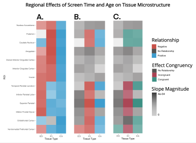 |
136 |
Developmental Ramifications of the Screen Age: Uncovering
Effects of Screen Time on Neural Maturation
Alexander S. Atalay1,
Benjamin T. Newman2,
and T. Jason Druzgal2
1Radiology and Medical Imaging, University of Virginia, East Greenwich, RI, United States, 2Radiology and Medical Imaging, University of Virginia, Charlottesville, VA, United States Keywords: Gray Matter, Adolescents, Screen Time Increased screen usage during adolescent development correlates with a complex response in the brain. Using a massive longitudinal human brain MRI dataset, we describe relationships between screen time and cellular microstructure across 13 cortical regions previously implicated in screen usage. Longitudinally, grey matter density had no relationship with screen time, and fractional anisotropy was significantly associated with screen time in only the ventromedial prefrontal cortex. However, microstructural 3T-CSD diffusion MRI metrics were significantly associated with increased screen time in 8 regions. The comparison between these results and typical age-associated development suggests potential negative implications of increased screen usage during adolescence. |
|
3367.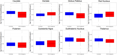 |
137 |
Investigating Regional Changes in Brain Magnetic Susceptibility
in Tanzanian Children with Sickle Cell Anaemia at 1.5 Tesla
Mitchel Lee1,
Russell Murdoch1,2,
Mboka Jakob3,
Fenella Kirkham2,
and Karin Shmueli1
1Department of Medical Physics and Biomedical Engineering, University College London, London, United Kingdom, 2Imaging and Biophysics, Developmental Neurosciences, UCL Great Ormond Street Institute of Child Health, London, United Kingdom, 3Department of Radiology & Imaging, Muhimbili University of Health and Allied Sciences, Dar Es Salaam, Tanzania Keywords: Gray Matter, Susceptibility Although sickle cell anaemia (SCA) affects the brain, causing stroke and neurocognitive complications, its pathophysiological mechanisms are poorly understood. Quantitative Susceptibility Mapping (QSM) reveals alterations in tissue composition. Therefore, we applied QSM to investigate changes in brain susceptibility in 168 SCA patients compared to 47 healthy controls scanned at 1.5 Tesla in Tanzania. We found a significant susceptibility decrease in SCA vs. controls in the caudate nucleus and globus pallidus and a significant increase in susceptibility in the red nucleus and dentate. Blood haemoglobin levels had a significant positive correlation with susceptibility in the globus pallidus, caudate nucleus and putamen. |
|
3368.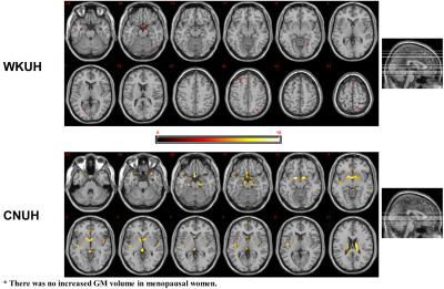 |
138 |
A multicentric validation of cerebral morphologic changes in
menopausal women: DARTEL-based voxel-based morphometry (VBM)
study
Tae-Hoon Kim1,
Youe Ree Kim2,
Chang-Won Jeong1,
ByoungRyun Kim3,
Chungsub Lee1,
SiHyeong Noh1,
DongWook Lim1,
and Young Hwan Lee2
1Medical Convergence Research Center, Wonkwang University, Iksan, Korea, Republic of, 2Radiology, Wonkwang University School of Medicine and Hospital, Iksan, Korea, Republic of, 3Obstetrics and Gynecology, Wonkwang University School of Medicine and Hospital, Iksan, Korea, Republic of Keywords: Gray Matter, Aging, menopause Among recent voxel based-morphometry (VBM) technique, DARTEL (Diffeomorphic Anatomical Registration Through Exponentiated Lie Algebra)-based VBM method can provide powerful information to understanding morphologic variations in whole brain areas. In women, several VBM studies reported unveiling the brain morphologic reductions (putamen, pallidum, hippocampus, postcentral gyrus, frontal/paritetal/temporal gyrus, and so on) after menopause. These studies provided menopause-related brain centers in postmenopausal women; however these morphologic findings might be included potentially case-sensitive results as a consequence of the enrolled population. This study was performed a multicentric study to evaluate cerebral morphological changes in menopausal women by using a DARTEL-based VBM method. |
|
3369. |
139 |
Differential Gray Matter Volume Loss in Controlled and
Uncontrolled Type 2 Diabetes Mellitus Over Time
Bhaswati Roy1,
Sarah E. Choi2,
Matthew J. Freeby3,
and Rajesh Kumar1,4,5,6
1Anesthesiology, University of California Los Angeles, Los Angeles, CA, United States, 2UCLA School of Nursing, University of California Los Angeles, Los Angeles, CA, United States, 3Medicine, University of California Los Angeles, Los Angeles, CA, United States, 4Radiology, University of California Los Angeles, Los Angeles, CA, United States, 5Bioengineering, University of California Los Angeles, Los Angeles, CA, United States, 6Brain Research Institute, University of California Los Angeles, Los Angeles, CA, United States Keywords: Gray Matter, Diabetes Patients with Type 2 diabetes mellitus (T2DM) show brain tissue changes in mood and cognitive control sites, functions that are deficient in the condition. However, the extent of brain gray matter volume loss in various sites, as well as progression of volume loss with time in controlled and uncontrolled T2DM adults are unclear. We observed higher gray matter volume loss in crucial brain areas involved in cognition and mood control in T2DM adults with uncontrolled glucose levels and accelerated progression after 6-months of follow-up. These findings indicate the need for optimal glucose management in T2DM adults. |
|
3370.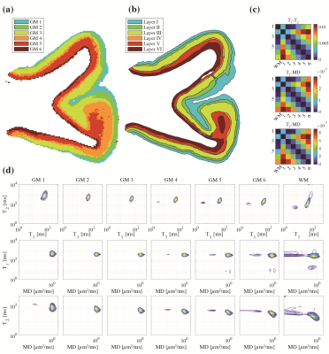 |
140 |
Mapping the individual human cortex using multidimensional MRI
and unsupervised learning
Shinjini Kundu1,
Stephanie Barsoum2,
Jeanelle Ariza3,
Amber L Nolan3,
C. Dirk Keene3,
Peter J Basser4,
and Dan Benjamini2
1The Johns Hopkins Hospital, Baltimore, MD, United States, 2National Institute on Aging, Baltimore, MD, United States, 3University of Washington, Seattle, WA, United States, 4National Institute of Child Health and Human Development, Bethesda, MD, United States Keywords: Gray Matter, Machine Learning/Artificial Intelligence Histology-based cellular composition and tissue architecture provide the biological basis for the brain’s cytoarchitectonic areas and for characterizing neuropathology. Noninvasive methods to assess cortical cyto- and myeloarchitectonic features are therefore urgently needed. In an ex vivo human brain study, we used multidimensional diffusion-relaxation MRI to investigate changes in spectral signatures with cortical depth. We designed an unsupervised segmentation procedure that captures this information and provides cortical laminar maps, which were co-registered to histological images and compared. The ability to map cortical cytoarchitectonic features noninvasively makes multidimensional MRI a promising tool for studying whole-brain cortical organization. |
|
 Back to Meeting Home
 Back to the Program-at-a-Glance
The International Society for Magnetic Resonance in Medicine is accredited by the Accreditation Council for Continuing Medical Education to provide continuing medical education for physicians.
 Back to Meeting Home
 Back to the Program-at-a-Glance
 View Presentation Video