Digital Poster
Gray Matter II
ISMRM & ISMRT Annual Meeting & Exhibition • 03-08 June 2023 • Toronto, ON, Canada

| Computer # | |||
|---|---|---|---|
3545.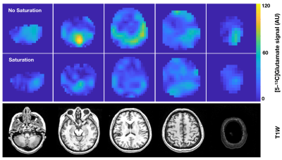 |
141 |
Assessing the effects of hyperpolarized [2-13C]lactate
saturation on detectable brain metabolism
Adam Autry1,
Sana Vaziri1,
Hsin-Yu Chen1,
Yaewon Kim1,
Duey Dang1,
Javier Villanueva-Meyer1,
Susan M Chang2,
Jennifer Clarke2,
Nancy A Oberheim-Bush2,
Robert Bok1,
Duan Xu1,
Janine M Lupo1,
Daniel B Vigneron1,
Jeremy Gordon1,
and Yan Li1
1Department of Radiology and Biomedical Imaging, University of California San Francisco, San Francisco, CA, United States, 2Department of Neurological Surgery, University of California San Francisco, San Francisco, CA, United States Keywords: Gray Matter, Hyperpolarized MR (Non-Gas), metabolism Following hyperpolarized [1-13C]pyruvate MRI studies that demonstrated insights into brain metabolism using saturation experiments, this study sought to assess whether metabolic pathways probed by [2-13C]pyruvate could similarly be interrogated. Our investigation showed that not exciting [2-13C]lactate led to a 2-fold increase in [5-13C]glutamate signal, which may indicate compartmentalized metabolism in the brain. |
|
3546.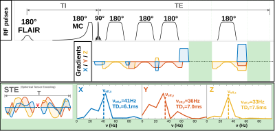 |
142 |
Chasing the Dot: Diffusion-Weighted MR Spectroscopy with
Spherical Tensor Encoding
André Döring1,
Frank Rösler2,
Kadir Şimşek1,3,
Karl Landheer4,
Roland Kreis5,6,
Wolfgang Bogner7,
and Derek K Jones1
1Cardiff University Brain Research Imaging Centre (CUBRIC), School of Psychology, Cardiff University, Cardiff, United Kingdom, 2Department of Mathematics, University of Bern, Bern, Switzerland, 3School of Computer Science and Informatics, Cardiff University, Cardiff, United Kingdom, 4Regeneron Pharmaceuticals, Inc. Tarrytown, New York, NY, United States, 5Magnetic Resonance Methodology, Institute of Diagnostic and Interventional Neuroradiology, University Bern, Bern, Switzerland, 6Translational Imaging Center, sitem-insel, Bern, Switzerland, 7High-field MR Centre, Department of Biomedical Imaging and Image-guided Therapy, Medical University of Vienna, Vienna, Austria Keywords: Gray Matter, Spectroscopy, Spherical Tensor Encoding, Diffusion, Metabolites Diffusion-weighted MR spectroscopy (DW-MRS) can measure diffusion properties of cell type-specific and intracellular metabolites. However, for advanced microstructure modeling, diffusion of brain metabolites has to be measured at specific length scales and with specific structural sensitivity. To this end, new diffusion encoding strategies have been developed, but not all have found their ways into DW-MRS. In this work, we fill this gap for spherical-tensor-encoding (STE), providing the first evidence that useful diffusion metrices of human brain metabolites can be quantified by combining DW-MRS, STE, and ultra-strong gradients. |
|
3547. |
143 |
Cerebral Blood Flow quantification in anesthetized macaque using
pseudo-continuous Arterial Spin Labeling at 3T
Leyre Garcia-Ruiz1,2,
Karol M. Córdoba3,
Daniel Jericó3,
Maite Aznárez-Sanado4,
Marta Vidorreta5,
Antonio Fontanellas3,
and Maria A. Fernández-Seara1,2
1Radiology, Clínica Universidad de Navarra, Pamplona, Spain, 2IdiSNA,Instituto de Investigación Sanitaria de Navarra, Pamplona, Spain, 3Hepatology, CIMA Universidad de Navarra, Pamplona, Spain, 4School of Education and Psychology Universidad de Navarra, Pamplona, Spain, 5Siemens Healthcare, Madrid, Spain Keywords: Gray Matter, Arterial spin labelling, Animal, Preclinical, White Matter A pseudo-continuous arterial spin labeling (pCASL) technique was implemented on a Siemens 3T Skyra for quantitative cerebral blood flow (CBF) measurements in non-human primates (rhesus monkeys). Different regions were manually segmented based on T1 weighted images. ASL images were obtained in 10 min with 2.2-mm isotropic resolution. Whole brain CBF was 32.40±14.61 ml/100g/min (n=8) for female and 44.28±24.61 ml/100 g/min for male (n=4) rhesus monkeys under ketamine and midazolam anesthesia. Gender differences of CBF among brain regions will be address in this study. |
|
3548.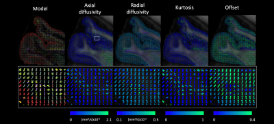 |
144 |
Fiber-specific modelling in spherical deconvolution
Alberto De Luca1 and
Alexander Leemans1
1Division Imaging and Oncology, University Medical Center Utrecht, Utrecht, Netherlands Keywords: Gray Matter, Tractography & Fibre Modelling Spherical deconvolution is commonly employed to reconstruct fiber orientations distributions with brain diffusion MRI. A key assumption in spherical deconvolution is that all brain fibers share the same microstructural properties. This is unlikely the case in white matter, but even more so in grey matter. Here, we propose a novel framework to perform spherical deconvolution with fiber specific models. As such, this framework allows to quantify diffusion properties associated to each fiber orientation. In this work, we showcase the application of this framework to investigate the organization of the brain cortex high resolution diffusion MRI data. |
|
3549.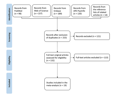 |
145 |
Mapping extraversion in brain activation patterns: a functional
neuroimaging meta-analysis of resting state studies
Qingyuan Li1,2,3,4,
Song Wang2,3,4,
Xiao Li1,
and Qiyong Gong3,5
1Department of Interventional Therapy, National Cancer Center/National Clinical Research Center for Cancer/Cancer Hospital, Chinese Academy of Medical Sciences and Peking Union Medical College, Beijing, China, 2Research Unit of Psychoradiology, Chinese Academy of Medical Sciences, Chengdu, China, 3Huaxi MR Research Center (HMRRC), Department of Radiology, West China Hospital of Sichuan University, Chengdu, China, 4Functional & Molecular Imaging Key Laboratory of Sichuan Province, West China Hospital of Sichuan University, Chengdu, China, 5Department of Radiology, West China Xiamen Hospital of Sichuan University, Xiamen, China Keywords: Gray Matter, Brain Our meta-analysis reveals that extraversion was linked with resting-state brain activity differences widely distributed across cortical and subcortical regions involved in emotion and behavioral regulation. The meta-regression results suggest an effect of gender on the association between extraversion and neural activity in the right inferior frontal gyrus. Our findings support that extraversion could lead to neural activity changes, which may be correlated with behavioral differences between extraverts and introverts. |
|
3550.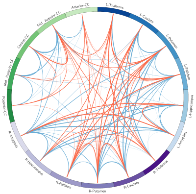 |
146 |
Brain morphological alterations in patients with lung cancer
after chemotherapy
Liu Renyuan1,
Rong Ping1,
Han Xiaowei1,
and Zhang Bing1
1Radiology, Drum Tower hospital, Nanjing, China Keywords: Gray Matter, Cancer The pathophysiological and biochemical effects of lung cancer and chemotherapy would result in abnormal alterations of brain morphometry. In particular, chemotherapy could endanger critical cognition-related subcortical nucleus, such as amygdala. |
|
3551.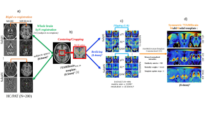 |
147 |
Segmentation of Substantia Nigra, Subthalamic and Red Nuclei
with a Multi-Modal Quantitative 7T MRI High Resolution Template
Alexandre CABANE1,2,
Arnaud LE TROTER1,2,
Benoit TESTUD1,2,
Stephan GRIMALDI1,2,
Maxim GUYE1,2,
Jean Philippe RANJEVA1,2,
and Ludovic DE ROCHEFORT1,2
1Aix Marseille Univ, CNRS, CRMBM, Marseille, France, 2AP-HM, CHU Timone, CEMEREM, Marseille, France Keywords: Gray Matter, Quantitative Susceptibility mapping, T1, high-resolution multi-modal template The segmentation of brain substructures is very useful in the characterization of alterations involved in multiple diseases. From 200 7T brain MRI scan including MP2RAGE and MGRE used to generate quantitative T1 maps (qT1), R2* and QSM volumes, a pipeline was developed to create a high-resolution multi-modal template at (400 µm)3 based on these multiple quantitative imaging modalities. Preliminary results show that multi-modality allows for a more precise parcellation of the SN, RN and STH substructures. |
|
3552.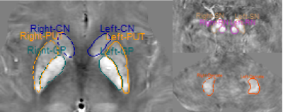 |
148 |
Altered patterns of brain iron deposition in end-stage renal
disease patients with depression
Yuan Li1,
Yuhan Jiang1,
Bingbing Gao1,
Mingrui Qu1,
Liangjie Lin2,
Qingwei Song1,
and Yanwei Miao1
1the First Affiliated Hospital of Dalian Medical University, Dalian, China, 2Philips Healthcare, Beijing, China Keywords: Gray Matter, Kidney Both depression and end-stage renal disease (ESRD) may affect the pattern of iron deposition in human brain. This study aimed to quantify the changes of brain iron deposition in ESRD patients with and without depression using quantitative susceptibility mapping, and to further explore the effect of depression on iron deposition pattern in ESRD patients. Results showed that ESRD patients had increased brain iron deposition compared with heathy controls, especially in the left putamen and the right red nucleus, and ESRD patients with depression had less increase in iron deposition than those without depression. |
|
3553.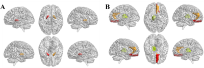 |
149 |
Reveal cortical abnormalities by quantitative synthetic MRI in
children with beta-thalassemia
Meiru Bu1,
Xi Deng1,
Lingling Shi2,
Wei Cui3,
Long Qian3,
Zisan Zeng1,
and Muliang Jiang1
1Radiology Department of the First Affiliated Hospital of Guangxi Medical University, Nanning, China, 2Hematology Department of The First Affiliated Hospital of Guangxi Medical University, Nanning, China, 3MR Research, GE Healthcare, Beijing, China, Beijing, China Keywords: Gray Matter, Blood, Beta-thalassemia Beta-thalassemia (β-TM) is a chronic blood disorder presenting with severe anemia. However, the abnormalities of microstructure in cortical regions are still unclear. Herein, we explored the difference of quantitative MRI parameters in gray matter between β-TM patients and healthy controls. The results showed that altered T1 and T2 values in specific gray matter in β-TM patients, which may be associated with alteration in brain gray matter, such as reduced myelin content and excessive iron deposition. Thus, T1 and T2 quantitative variables may be promising imaging markers for further exploring the pathophysiological mechanisms of β-TM. |
|
3554.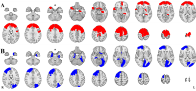 |
150 |
Altered cortical susceptibility heterogeneity in patients with
beta-thalassemia
Meiru Bu1,
Xi Deng1,
Lingling Shi2,
Wei Cui3,
Long Qian3,
Zisan Zeng1,
and Muliang Jiang1
1Radiology Department of the First Affiliated Hospital of Guangxi Medical University, Nanning, China, 2Hematology Department of The First Affiliated Hospital of Guangxi Medical University, Nanning, China, 3MR Research, GE Healthcare, Beijing, China, Beijing, China Keywords: Gray Matter, Blood, Beta-thalassemia Beta-thalassemia (β-TM) is an inherited blood disorder that causes the body to make less hemoglobin. In this study, we examined altered cortical susceptibility heterogeneity in patients with β-TM vs. healthy controls using magnetic resonance imaging (MRI). For the altered regions of susceptibility heterogeneities in the β-TM group, an increase was only found in the left hippocampus, while decreased heterogeneities were observed for all other regions located in the frontal and temporal lobes. Thus, we concluded that susceptibility heterogeneities could reveal the β-TM-affecting regions. |
|
3555.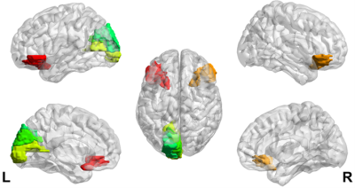 |
151 |
T2 heterogeneity of cortex in patients with beta-thalassemia
major
Xi Deng1,
Meiru Bu1,
Meiqing Wu2,
Wei Cui3,
Long Qian3,
Zisan Zeng1,
and Muliang Jiang1
1Radiology Department of the First Affiliated Hospital of Guangxi Medical University, Nanning, China, 2Hematology Department of The First Affiliated Hospital of Guangxi Medical University, Nanning, China, 3MR Research, GE Healthcare, Beijing, China, Beijing, China Keywords: Gray Matter, Blood, Beta-thalassemia; T2 heterogeneity; Beta-thalassemia (β-TM) is an inherited blood disorder with severe anemia. In this study, we investigated alterations of T2 heterogeneity in cortical regions caused by β-TM using magnetic resonance imaging (MRI). Compared with healthy controls, β-TM patients showed increased T2 heterogeneities in bilateral inferior orbitofrontal, left calcarine, left cuneus and left superior occipital lobe, while no decreased T2 heterogeneity was observed. Thus, we concluded that T2 heterogeneities could reveal the β-TM-affecting T2 alterations in brain. |
|
3556.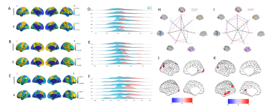 |
152 |
Transcriptional signatures underpinnings of altered large-scale
functional connectome hierarchy in patients with IBS
Jun Wang1,2,
Guangyao Liu2,3,
Pengfei Zhang1,
Wenjing Huang1,
Kai Ai4,
and Jing Zhang2,3
1Second Clinical School, Lanzhou University, Lanzhou, China, 2Department of Magnetic Resonance, Lanzhou University Second Hospital, Lanzhou, China, 3Gansu Province Clinical Research Center for Functional and Molecular Imaging, Lanzhou University Second Hospital, Lanzhou, China, 4Department of clinical science, Philips Healthcare, Xi’an, China Keywords: Gray Matter, fMRI (resting state) Functional connectivity is altered in patients with irritable bowel syndrome (IBS), but its histological basis is unknown. We used resting-state fMRI imaging combined with transcriptomics to examine changes in functional connectivity gradients in IBS patients and their corresponding gene regulatory basis. The reduced connectivity gradients and contractions in IBS patients compared to healthy controls were mainly involved in primary sensory and cross-modal structural domains and were associated with aberrant expression of genes related to neurological development. These results reveal gradient dysfunction of large-scale functional connectomes in IBS and elucidate the role of abnormal gene-function connectivity in the disease. |
|
3557.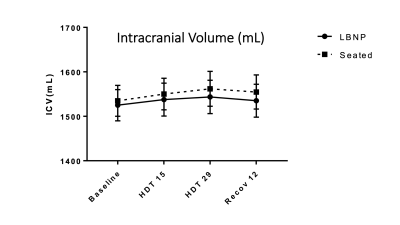 |
153 |
OPTICS STUDY: INTRACRANIAL EFFECTS OF STATIC LOWER BODY NEGATIVE
PRESSURE WITH HEAD DOWN TILT BED REST: COMPARISON TO UPRIGHT
POSTURE
Larry A. Kramer1,
Khader M Hasan1,
Lindsey Bishop2,
Xu Zhang3,
Karina Marshall-Goebel4,
Brandon Macias5,
Steven S Laurie6,
and Bryn Martin7,8
1Diagnostic Imaging and Intervention, University of Texas Health Science Center at Houston, Houston, TX, United States, 2University of Texas Health Science Center at Houston, Houston, TX, United States, 3Clinical and Translational Sciences, University of Texas Health Science Center at Houston, Houston, TX, United States, 4Karina Marshall-Goebel, NASA, Houston, TX, United States, 5NASA, Houston, TX, United States, 6KBR, Houston, TX, United States, 7Acyonetx, Moscow, ID, United States, 8Chemical and Biological Engineering, University of Idaho, Moscow, ID, United States Keywords: Blood vessels, Quantitative Imaging, Microgravity, Head down tilt, Spaceflight Associated Neuro-ocular Syndrome Lower body negative pressure applied a total of 6 hours daily during 29 days of head down tilt bed rest shows promise as a countermeasure to spaceflight acquired neuro-ocular syndrome. |
|
3558.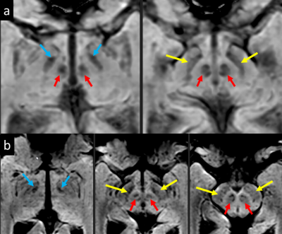 |
154 |
Visualization of Deep Brain Stimulation Targets at 0.5T
Chad Harris1,
Andrew Curtis1,
Curtis Wiens1,
Jurgen Germann2,3,
Alexandre Boutet2,3,
Andres M Lozano2,3,
and Jeff Stainsby1
1MR, Synaptive Medical, Toronto, ON, Canada, 2University Health Network, Toronto, ON, Canada, 3University of Toronto, Toronto, ON, Canada Keywords: Epilepsy, Contrast Mechanisms The utility of visualizing deep brain stimulation targets at 0.5T was explored. 3D SWI images show direct visualization of the subthalamic nuclei and surrounding iron-rich structures. With the excellent gray-white matter contrast possible at mid-field, 3D T1-weighted imaging can directly visualize the globus pallidus interna. The elevated T1 contrast produces excellent 3D T1-weighted imaging at multiple inversion times which are suitable for, and aid in, atlas-based segmentation approaches for DBS targeting. |
|
3559.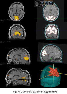 |
155 |
Stereotactic MRI-guided radiosurgery using AI resting state
networks recognition
Luis Ancari1,
Abril Vergne2,3,
María Paula Del Popolo2,3,4,
Guillermo Alvarez3,
Clara Lisazo3,
Federico Julián González1,3,
Sebastián Moguilner5,
Maximo Melchor2,3,
Trinidad González3,6,
and Daniel Fino1,3,4
1Instituto Balseiro, Universidad Nacional de Cuyo, Bariloche, Argentina, 2Universidad de Mendoza, Mendoza, Argentina, 3Fundación Escuela de Medicina Nuclear, Mendoza, Argentina, 4Fundacion Argentina para el Desarollo en Salud, Mendoza, Argentina, 5Harvard Medical School, Boston, MA, United States, 6Fundación Argentina para el Desarrollo en Salud, Mendoza, Argentina Keywords: Tumors, Radiotherapy Stereotactic radiosurgery (SRS) is a minimally invasive procedure that reduces tumor size without subjecting surrounding organs at risk (OAR) to harmful radiation levels. The delimitation of OAR in SRS planning, and the conservation of the prescribed dose in tumoral regions, can be improved by the implementation of independent component analysis to obtain resting-state fMRI networks (RSNs). In this aspect, AI plays an important role to decrease computational-processing times while increasing the method’s efficiency. One of the main solved challenges was the lack of compatibility between the Nifti protocol (RSNs format) and the treatment planning system’s protocol (coordinate system of CT-simulation). |
|
3560.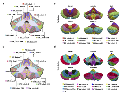 |
156 |
A Multimodal Atlas of the Human Cerebellum at 760 μm Resolution
Wenjiao Lyu1,
Ye Wu1,
Khoi Minh Huynh1,
Sahar Ahmad1,
and Pew-Thian Yap1
1Radiology and Biomedical Research Imaging Center (BRIC), The University of North Carolina at Chapel Hill, Chapel Hill, NC, United States Keywords: Multimodal, Multimodal, Cerebellum; atlas; The human cerebellum is engaged in a broad array of tasks related to motor coordination, cognition, and emotional regulation. Here, we construct a cerebellar atlas using high-resolution multimodal MRI, capturing multiple characteristics of the cerebellum, including cortical morphology, tissue microstructure, and cerebellar/cerebello-cerebral connectivity. Our atlas facilitates the understanding of the neurodevelopment and neurodegeneration of the cerebellum. |
|
3561.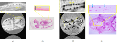 |
157 |
Brain structures of a human embryo depicted by MR microscopy
with different contrasts
Kazuki Kunieda1,
Kazuyuki Makihara1,
Shigehito Yamada2,
and Yasuhiko Terada1
1Graduate School of Science and Technology, University of Tsukuba, Tsukuba, Japan, 2Congenital Anomaly Research Center, Kyoto University Graduate School of Medicine, Kyoto, Japan Keywords: Brain Connectivity, Microstructure Brain structures of human embryos are several tens of micrometers in size, and MR microscopy is essential for elucidating human embryos. However, conventional fast T1-weighted imaging could not delineate some critical structures. In this study, T1, T2, and T2* values were measured to simulate the contrast of each tissue of the human embryo brain, and MR images were acquired at the extremely high resolution of (30 μm)3 using pulse sequences that emphasized the contrast difference in the brain structures. This technique would unveil the three-dimensional microstructure and developmental process of the brain’s central nervous system in its early stages. |
|
3562.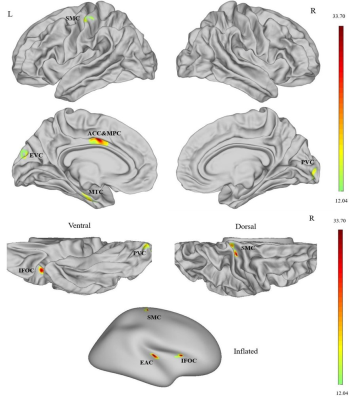 |
158 |
Cortical Sulcus Depth Alterations in Patients with Tinnitus
Before and After Sound Therapy: A Surface-Based Morphometry
Study.
Xuan Wei1
1Department of Radiology, Beijing Friendship Hospital, Capital Medical University, Beijing, China Keywords: Head & Neck/ENT, Treatment In this study, we performed brain surface-based morphometry to evaluate changes in sulcal depth after sound therapy in patients with idiopathic tinnitus. Our results showed thatsulcal depth was signiêcantly reduced in the left medial temporal cortex (MTC) and right somatosensory and motor cortex (SMC) of patients with tinnitus compared to the healthycontrols, but increased signiêcantly at 24 weeks after sound therapy. Therefore, sulcal depth in the auditory sensory regions of the brain is a potential neuroimaging biomarker forevaluating treatment eìcacy in tinnitus patients. |
|
3563.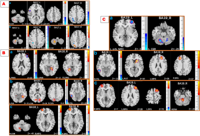 |
159 |
Disrupted Functional Connectivity of the Visual Cortex in
Dysthyroid Optic Neuropathy Due to Thyroid-Associated
Ophthalmopathy
Ping Liu1,
Gui-Hua Jiang1,
and Wan-Yi Zheng2
1Medical Imaging, Guangdong Second Pronvincal General Hospital, Guang Zhou, China, 2Jinan university, Guang Zhou, China Keywords: Head & Neck/ENT, Neuro, dysthyroid optic neuropathy,thyroid-associated ophthalmopathy Dysthyroid optic neuropathy (DON) due to thyroid-associated ophthalmopathy (TAO) is characterized by visual dysfunction and with a risk of blindness. Visual cortex is crucial for visual information, and visual impairments can affect brain activity. We investigated visual cortex functional connectivity (FC) alterations in DON and its associations with visual performance. The DON showed disrupted FC in the visual cortex, and the FC in primary rather than the secondary visual cortex associated with clinical parameters. It helps to uncover the neurological mechanisms underlying visual dysfunction and provides insight into novel therapeutic regimens to slow or prevent the neuropathy. |
|
3564.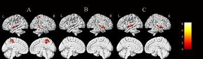 |
160 |
Maternal neural and behavioral sensitivity to baby emotional
sounds
Jiayu Lin1,
Wei Su1,
Xiaoyu Du2,
Zhenhua Sun3,
Xiang Zhang4,
Kaihua Zhang1,
and Xiaoxia Du5
1School of Psychology, Shandong Normal University, Ji’nan, Shandong, P. R. China, Jinan, China, 2The University of Melbourne, Victoria, Australia, 3Linyi University, Linyi, China, 4Shandong Provincial Hospital Affiliated to Shandong First Medical University, Jinan, China, 5Shanghai University of Sport, Shanghai, China Keywords: Head & Neck/ENT, fMRI (task based) After giving birth, women will experience a series of physiological and psychological changes. Exploring the changes in postpartum women's neurological function contributes to comprehending their cognitive and emotional processing mechanisms. Therefore, we recruited 43 new mothers and 26 nulliparous women to participate in the current fMRI study based on the baby sounds’ task. Results suggested that the brain function of new mothers showed dynamic plasticity in the temporal lobe. This study reflected the adaptive changes in new mothers' brain function toward nurturing the babies. |
|
 Back to Meeting Home
 Back to the Program-at-a-Glance
The International Society for Magnetic Resonance in Medicine is accredited by the Accreditation Council for Continuing Medical Education to provide continuing medical education for physicians.
 Back to Meeting Home
 Back to the Program-at-a-Glance
 View Presentation Video