Digital Poster
Myelin & White Matter Integrity
ISMRM & ISMRT Annual Meeting & Exhibition • 03-08 June 2023 • Toronto, ON, Canada

| Computer # | |||
|---|---|---|---|
2626.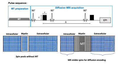 |
101 |
The effects of magnetization transfer on fast and slow diffusion
compartments in myelinated white matter
Chenyang Li1,2,
Els Fieremans1,
Dmitry S. Novikov1,
and Jiangyang Zhang1
1Department of Radiology, NYU Grossman School of Medicine, New York, NY, United States, 2Vilcek Institute of Graduate Biomedical Sciences, NYU Grossman School of Medicine, New York, NY, United States Keywords: White Matter, Contrast Mechanisms Myelin, although not directly visible in conventional diffusion MRI (dMRI) due to its short T2, still affects the dMRI signal indirectly through exchange processes with nearby water compartments. In this study, we investigated how a magnetization transfer (MT) preparation affects dMRI signals, in particular, the fast and slow diffusion compartments in myelinated white matter of ex vivo mouse brain. Our result demonstrated that the MT preparation altered the volume fractions, but not the diffusivities, of the fast and slow compartments. Examining the change in volume fractions with MT preparation can provide insights into exchange processes involving myelin and water compartments. |
|
2627.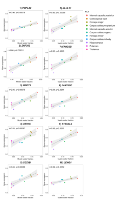 |
102 |
The myelin water imaging transcriptome: myelin water fraction
regionally varies with genes associated with oligodendrocytes
Jaimie Lee1,2,
Hanwen Liu3,
Cornelia Laule2,4,5,6,
Catrina Loucks1,7,8,
and John Kramer1,2
1Department of Pharmacology, Anesthesiology, and Therapeutics, University of British Columbia, Vancouver, BC, Canada, 2International Collaboration on Repair Discoveries, Vancouver, BC, Canada, 3Department of Medicine, University of British Columbia, Vancouver, BC, Canada, 4Department of Radiology, University of British Columbia, Vancouver, BC, Canada, 5Department of Pathology and Laboratory Medicine, University of British Columbia, Vancouver, BC, Canada, 6Department of Physics and Astronomy, University of British Columbia, Vancouver, BC, Canada, 7Division of Translational Therapeutics, Department of Pediatrics, University of British Columbia, Vancouver, BC, Canada, 8BC Children's Hospital Research Institute, Vancouver, BC, Canada Keywords: White Matter, Relaxometry, myelin, transcriptome, myelin water fraction, myelin water imaging, gene expression, microstructure Our objective for this study was to determine the relationship between regional variations in myelin water imaging (MWI) and gene expression – a “MWI transcriptome”. Regional variations in myelin water fraction (MWF) from a normative MWF atlas were examined in a correlation analysis with gene expression data retrieved from the Allen Human Brain Atlas. Genes that significantly covaried with MWF were involved in lipid metabolism and binding, and were enriched for myelin terms (e.g., oligodendrocytes). This preliminary work highlights the utility of an imaging transcriptomic approach and further supports MWI as a robust in vivo measure of myelin. |
|
2628.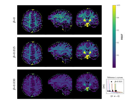 |
103 |
Myelin water fraction mapping using relaxation spectra from
steady-state gradient echo imaging with partial RF spoiling
Tony Stöcker1,2,
Rüdiger Stirnberg1,
Difei Wang1,
Philipp Ehses1,
and Eberhard Pracht1
1German Center for Neurodegenerative Diseases (DZNE), Bonn, Germany, 2Department of Physics & Astronomy, University of Bonn, Bonn, Germany Keywords: White Matter, Modelling, Myelin A new approach for myelin water fraction (MWF) mapping is presented, based on gradient echo signals with small RF spoiling phase increments. Partially spoiled gradient echo data with 20 different phase increments ($$$|\Delta\phi|\le20^\circ$$$) were efficiently acquired with a custom skipped-CAIPI 3D-EPI sequence. The data is well-suited for a novel joint-fitting approach of $$$T_1$$$- and $$$T_2$$$-spectra, from which high-quality whole-brain MWF maps are derived. MWF-maps show qualitatively good agreement with published data in the white matter (WM). Additionally, the approach seems to provide robust estimates in regions with small MWF, such as the gray matter (GM). |
|
2629.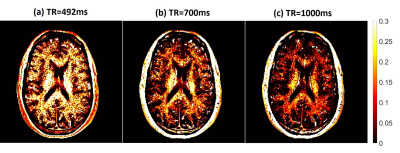 |
104 |
TR effect on Myelin Water Imaging
Jing Zhang1,
Suchandrima Banerjee2,
and Alexander L. MacKay3
1Global MR Applications & Workflow, GE Healthcare, Vancouver, BC, Canada, 2Global MR Applications & Workflow, GE Healthcare, Menlo Park, CA, United States, 3Department of Radiology, University of British Columbia, Vancouver, BC, Canada Keywords: White Matter, White Matter, myelin water imaging Myelin water fraction (MWF) is conventionally measured using the T2 decay curve. For a spin-echo based sequence, the repetition time (TR) is limited by SAR. Most MWF studies have been carried out at 3T; but a 1.5T system has lower power deposition, which enables MWF imaging at lower TRs. Shorter TR will also reduce the total scan time. In this work, we performed myelin water imaging on a 1.5T system to investigate the effect of shorter TR on this technique. This work demonstrated increased MWF in human brain in vivo with decreasing TR. |
|
2630. |
105 |
Myelin Water Atlas Template derived from Quantitative Parameter
Mapping
Yuki Kanazawa1,
Shun Kitano1,
Masafumi Harada1,
Yo Taniguchi2,
Yuki Matsumoto1,
Hiroaki Hayashi3,
Kosuke ito2,
Yoshitaka Bito2,
and Akihiro Haga1
1Tokushima University, Tokushima, Japan, 2FUJIFILM Healthcare Corporation, Tokyo, Japan, 3Kanazawa University, Kanazawa, Japan Keywords: White Matter, Relaxometry We generated a myelin water atlas template derived from QPM. Imaging data were acquired using a multi-gradient-echo sequence for QPM. All the data in the 48 white matter regions measured in the volume-of-interest were plotted, and quadratic polynomial equations of each region were derived from the relationship between R1·R2* and the two-component model-MWF. As a result, the relationship between R1·R2* and MWF showed a strong significant correlation for all of the white matter regions (R2 ≥ 0.963, P < 0.0001). Our myelin water atlas template derived from QPM can be used as a reference to demonstrate areas of demyelinating disease. |
|
2631.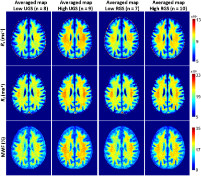 |
106 |
Associations between gait speeds and myelin content in
cognitively unimpaired adults using multicomponent relaxometry
Mary E. Faulkner1,
John P. Laporte1,
Elango Palchamy2,
Zhaoyuan Gong1,
Curtis Triebswetter1,
Matthew Kiely1,
M.A.B.S. Akhonda1,
Luigi Ferrucci2,
Richard G. Spencer1,
and Mustapha Bouhrara1
1Laboratory of Clinical Investigation, National Institute on Aging, Baltimore, MD, United States, 2Translational Gerontology Branch, National Institute on Aging, Baltimore, MD, United States Keywords: White Matter, Relaxometry Abnormal gait speed is a reliable indicator of the progression of age-related neurodegenerations. However, the association between gait speed and myelin content remains unclear. We used multicomponent MR relaxometry of myelin water fraction (MWF) and longitudinal and transverse (R1 and R2) relaxation rates to investigate the association between cerebral myelination and usual or rapid gait speeds in cognitively unimpaired adults spanning a wide age range. Our results indicate that lower myelin content is associated with lower gait speed across several while matter structures. |
|
2632.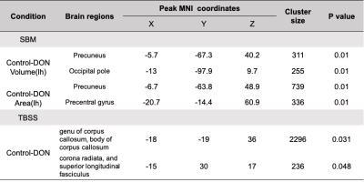 |
107 |
Cortical thickness and white matter integrity abnormalities in
dysthyroid optic neuropathy
Mengsha Zou1,
Hongzhang Zhu1,
Yunzhu Wu2,
and Zhiyun Yang1
1Department of Radiology, The First Affiliated Hospital of Sun Yat-Sen University, 58th, The Second Zhongshan Road, Guangzhou, China, Guangzhou, China, 2MR Scientific Marketing, Siemens Healthineers Ltd., Shanghai, China, Shanghai, China Keywords: White Matter, Brain A pilot study to examine the structural MRI features of patients with dysthyroid optic neuropathy (DON) as compared to normal controls, and correlate pathologic MRI changes to clinical symptoms of the disease. DON patients show reduced cortical volume in precuneus and occipital pole, decrease area in precuneus and precentral gyrus, and widespread decreased FA value, mainly in genu of corpus callosuma and body of corpus callosum. Meanwhile, the FA of was associated with visual acuity. Our study provides a new insight of pathological mechanism of DON. |
|
2633.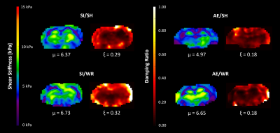 |
108 |
Sensitivity of magnetic resonance elastography to white matter
alterations in a rat model of Fetal Alcohol Spectrum Disorders
Katrina A. Milbocker1,
L. Tyler Williams2,
Ian F. Smith1,
Diego A. Caban-Rivera2,
Samuel Kurtz3,4,
Matthew D.J. McGarry5,
Elijah Van Houten4,
Curtis L. Johnson1,2,
and Anna Y. Klintsova1
1Dept. of Psychological & Brain Sciences, University of Delaware, Newark, DE, United States, 2Dept. of Biomedical Engineering, University of Delaware, Newark, DE, United States, 3Laboratorie de Mécanique et Génie Civil, CNRS, Université de Montpellier, Montpellier, France, 4Département de Génie Mécanique, Université de Sherbrooke, Sherbrooke, QC, Canada, 5Thayer School of Engineering, Dartmouth College, Hanover, NH, United States Keywords: White Matter, Preclinical Magnetic resonance elastography (MRE) produces spatially-resolved maps of brain tissue mechanical properties by estimating parameters, such as stiffness and viscosity, via inverse solution of the underlying equations of motion. When measured in the brain, estimated properties from MRE detect effects of disease or interventions with high fidelity and relate to functional outcomes, making it a potentially invaluable technique in neuroradiology. Application of MRE in white matter (WM) tracts is limited. To evaluate the sensitivity of MRE to WM alterations, this study compared values of total brain stiffness and damping ratio derived from MRE scanning of rats with impaired WM development. |
|
2634.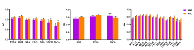 |
109 |
Comparative evaluation of DKI and DTI in detecting white matter
microstructural alterations in early-blind adolescents
Zhifeng Zhou1,
Long Qian2,
Gangqiang Hou1,
Wentao Jiang1,
and Xia Liu1
1Shenzhen Mental Health Center/Shenzhen Kangning Hospital, Shenzhen, China, 2GE Healthcare, Beijing, China Keywords: White Matter, Adolescents Blind people is a natural bio-model for the investigation of neural plasticity. Diffusion-based MRI techniques are powerful probes for characterizing the effects of disease and neural development on tissue microstructure. This study compared diffusion Kurtosis Imaging (DKI) metrics and diffusion tensor imaging (DTI) metrics to explore white matter microstructural changes and neural plasticity in early-blind adolescents (EBAs). The results demonstrate microstructural complexity reduction and coexistence of neural reorganization and compensatory development process induced by visual deprivation in EBAs. And the DKI metrics are more sensitive in detecting changes in crossing fibers and pathology and development of disease than DTI metrics. |
|
2635.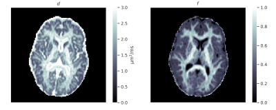 |
110 |
Microstructural neuroimaging using spherical convolutional
neural networks
Leevi Kerkelä1,
Kiran Seunarine2,
Filip Szczepankiewicz3,
and Chris A. Clark1
1UCL Great Ormond Street Institute of Child Health, University College London, London, United Kingdom, 2Great Ormond Street Hospital, London, United Kingdom, 3Clinical Sciences Lund, Lund University, Lund, Sweden Keywords: White Matter, Machine Learning/Artificial Intelligence We present a novel framework for estimating microstructural parameters of compartment models using recently developed orientationally invariant spherical convolutional neural networks and efficiently simulated training data. The networks were trained to predict the ground-truth parameter values from simulated noisy data and applied on imaging data acquired in a clinical setting to generate microstructural maps. Our network could estimate model parameters more accurately than conventional non-linear least squares or a multi-layer perceptron applied on powder-averaged data (i.e., the spherical mean technique). |
|
2636.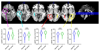 |
111 |
Bright light therapy increases myelin density in posterior
thalamic radiation in young adults with subthreshold depression:
An ihMT MRI study
Guanmao Chen1,
Guixian Tang1,
Long Qian2,
and Ying Wang1
1First Affiliated Hospital of Jinan University, Guangzhou, China, 2MR Research, GE Healthcare, Beijing, China Keywords: White Matter, Magnetization transfer Subthreshold depression (SD) is a significant risk indicator of major depressive episodes. This study used the inhomogeneous magnetization transfer (ihMT) technique to probe myelin abnormalities and its response to bright light therapy (BLT) in SD. The findings of this study suggest the macromolecular disruption of myelin in the posterior thalamic radiation, sagittal stratum, and uncinate fasciculus in the early stages of depression. Furthermore, the myelin impairments in the PTR could be reversed by BLT, thus suggesting they might be used as the potential neural target for BLT in SD. |
|
2637.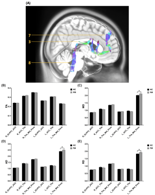 |
112 |
Alterations in white matter integrity of the pain modulatory
system in patients with fibromyalgia
Yu-Ting Huang1,
Ting-Chun Lin2,
Yao-Wen Liang2,
You-Yin Chen2,3,
Jiunn-Horng Kang4,5,6,
and Yu-Chun Lo3
1Department of Medicine, Taipei Medical University, Taipei, Taiwan, 2Department of Biomedical Engineering, National Yang Ming Chiao Tung University, Taipei, Taiwan, 3Ph.D. Program in Medical Neuroscience, College of Medical Science and Technology, Taipei Medical University, Taipei, Taiwan, 4College of Biomedical Engineering, Taipei Medical University, Taipei, Taiwan, 5Department of Physical Medicine and Rehabilitation, Taipei Medical University Hospital, Taipei, Taiwan, 6Professional Master Program in Artificial Intelligence in Medicine, College of Medicine, Taipei, Taiwan Keywords: White Matter, Diffusion Tensor Imaging The relationship between abnormal pain sensation and alterations in the central nervous system of fibromyalgia patients is unknown. In this study, diffusion tensor imaging was used to systematically investigate the alterations in white matter tracts of the pain modulatory pathways. The findings revealed the altered integrity of spinothalamic tract-thalamus, thalamus-insula tracts, tracts connected thalamus, midbrain and pons, were associated with the physical and psychological dysfunction in patients with fibromyalgia. |
|
2638. |
113 |
Automatic detection of cognitive impairment in patients with
White matter hyperintensity based on deep learning and radiomics
of MRI
Junbang Feng1,
Qingqing Zheng2,
Yuwei Xia3,
Shi Feng 3,
Qing Zhou3,
Hang Yin1,
Shike Wang2,
and Chuanming Li1
1Medical Imaging Department, Chongqing University Central Hospital, Chongqing, China, 2The Second Affiliated Hospital of Chongqing Medical University, Chongqing, China, 3Shanghai United Imaging Intelligence, Co., Ltd., Shanghai, China Keywords: White Matter, Machine Learning/Artificial Intelligence White matter hyperintensity (WMH) is common in the aging brain, which is associated with cognitive decline and dementia. At present, there is still no objective method for early detection of cognitive impairment from these populations. In this study, deep learning and radiomics techniques were used to automatically segment and extract the characteristics of WMH and other regional brain tissues, and models were established to detect mild cognitive impairment. |
|
2639.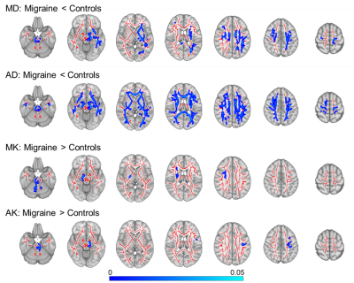 |
114 |
Episodic menstrual migraine patients exhibit white matter
microstructural changes compared to hormonal controls
Ana R Fouto1,
Rita G Nunes1,
Irene Guadilla1,2,
Amparo Ruiz-Tagle1,
Inês Esteves1,
Gina Caetano1,
Nuno A Silva3,
Pedro Vilela4,
Raquel Gil-Gouveia5,6,
and Patrícia Figueiredo1
1Institute for Systems and Robotics - Lisboa and Department of Bioengineering, Instituto Superior Técnico, Universidade de Lisboa, Lisbon, Portugal, 2Universidad Autónoma de Madrid, Madrid, Spain, 3Learning Health, Hospital da Luz, Lisbon, Portugal, 4Imaging Department, Hospital da Luz, Lisbon, Portugal, 5Neurology Department, Hospital da Luz, Lisbon, Portugal, 6Center for Interdisciplinary Research in Health, Universidade Católica Portuguesa, Lisbon, Portugal Keywords: White Matter, Diffusion/other diffusion imaging techniques, Migraine Migraine is one of the most prevalent brain disorders worldwide. Although features extracted from diffusion MRI have been suggested to hold potential as disease biomarkers, research outputs remain inconsistent across studies. We investigated voxelwise microstructural alterations in episodic menstrual migraine patients (interictal phase) and appropriate hormonal controls (post-ovulation) by comparing diffusion-tensor and diffusion-kurtosis imaging metrics. Moreover, we extracted histogram measures (median, peak height, width, and value for each metric); and we evaluated their relationship with clinical factors (disease duration, attack frequency and pain intensity). Several metrics revealed significant differences between groups, indicating that they may be potential disease biomarkers. |
|
2640.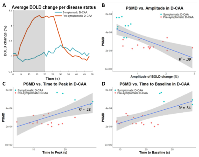 |
115 |
THE RELATIONSHIP BETWEEN VASCULAR REACTIVITY AND MICROSTRUCTURAL
WHITE MATTER INTEGRITY IN DUTCH-TYPE CEREBRAL AMYLOID ANGIOPATHY
Manon Roxanne Schipper1,
Naomi Vlegels2,3,
Sabine Voigt1,4,
Thijs W. van Harten1,
Alberto de Luca2,5,
Ingeborg Rasing4,
Geert Jan Biessels2,
Matthias J.P. van Osch1,
Marianne A.A. van Walderveen1,
and Marieke J.H. Wermer4
1Radiology, Leiden University Medical Center, Leiden, Netherlands, 2Neurology and Neurosurgery, University Medical Center Utrecht, Utrecht, Netherlands, 3LMU Klinikum, München, Germany, 4Neurology, Leiden University Medical Center, Leiden, Netherlands, 5Image Sciences Institute, University Medical Center Utrecht, Utrecht, Netherlands Keywords: White Matter, Diffusion Tensor Imaging, PSMD Vascular reactivity is an early marker of Cerebral Amyloid Angiopathy (CAA), but its relation to structural brain damage remains unclear. We aimed to study this relationship in Dutch-type CAA (D-CAA; genetic form of CAA): vascular reactivity and microstructural white matter integrity were studied through visually stimulated fMRI and ‘Peak width Skeletonized Mean Diffusivity (PSMD)’. Reduced BOLD amplitude, delayed time to peak, and delayed time to baseline were significantly related to a higher PSMD (β=-1.15e-02, β =1.08e-05, β=8.64e-06, respectively). These results indicate that vascular reactivity and microstructural white matter integrity may deteriorate hand-in-hand in D-CAA. |
|
2641. |
116 |
Remodeling of White Matter Caused by Sickle Cell Disease
Clio Gonzalez-Zacarias1,
SoYoung Choi2,
Anand A. Joshi1,
Richard M. Leahy1,
and John C. Wood3
1Ming Hsieh Department of Electrical and Computer Engineering, University of Southern California, Los Angeles, CA, United States, 2Institute of Imaging Science, Vanderbilt University, Nashville, TN, United States, 3Department of Pediatrics and Radiology, Children's Hospital Los Angeles, USC, Los Angeles, CA, United States Keywords: White Matter, Diffusion/other diffusion imaging techniques, Sickle Cell Disease Sickle cell disease (SCD) is characterized by the presence of different degrees (mild, moderate, or severe) of chronic anemia (quantified by hemoglobin values). Alterations in WM were assessed by performing a voxel-wise analysis in the fractional anisotropy (FA, i.e., overall directionality of water diffusion), mean diffusivity (MD) and their kurtosis analogous (FAK and MK) maps. Surprisingly, when controlling for log-age, sex and hemoglobin, MK showed bigger derangements in the watershed areas than other measurements like FA and FAK despite of controlling for Hb measurements. |
|
2642.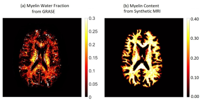 |
117 |
Comparison of Myelin Water Imaging from Multi-echo T2 Decay
Curve and Myelin Content from Synthetic MRI
Jing Zhang1,
David Shin2,
and Suchandrima Banerjee2
1Global MR Applications & Workflow, GE Healthcare, Vancouver, BC, Canada, 2Global MR Applications & Workflow, GE Healthcare, Menlo Park, CA, United States Keywords: White Matter, Brain, myelin water imaging, Synthetic MRI, Myelin Imaging myelin has long been a target in neuroimaging. Several MR techniques have been developed for in vivo measurement of myelin content. Myelin water fraction (MWF) from multi-echo T2 decay curve has been shown to be a reliable marker for myelin. Myelin content using quantitative synthetic MRI (SyMRI) has also been used to quantify myelin in the brain. In this work, we performed 3D GRASE and SyMRI (using multi-contrast 3D gradient echo) to estimate the myelin correlations between these two techniques. This work will provide guidance for future studies using myelin imaging techniques. |
|
2643.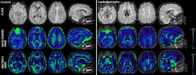 |
118 |
Two clinically feasible myelin water imaging methods (MCR-DIMWI
and METRICS) can differentiate patients with a leukodystrophy
from controls.
Menno D Stellingwerff1,
Murtadha L Al-Saady1,
Kwok-Shing Chan2,
Adam Dvorak3,
José P Marques2,
Shannon Kolind3,
Marjo S van der Knaap1,
and Petra JW Pouwels4
1Department of Pediatric Neurology, Amsterdam UMC, Amsterdam, Netherlands, 2Donders Institute for Brain, Cognition and Behaviour, Radboud University, Nijmegen, Netherlands, 3Department of Physics and Astronomy, University of British Columbia, Vancouver, BC, Canada, 4Department of Radiology and Nuclear Medicine, Amsterdam UMC, Amsterdam, Netherlands Keywords: White Matter, Genetic Diseases, Leukodystrophy Imaging biomarkers are needed for studying white matter (WM) diseases. Myelin water imaging (MWI) uses multi-compartment relaxometry to estimate myelin content, and is promising for use in leukodystrophies. We applied MCR-DIMWI and METRICS, two novel whole-brain MWI techniques, to a cohort of 9 leukodystrophy patients and 15 controls. Myelin water fractions (MWFs) from both techniques correlated well. In patients, MWF was decreased. For both techniques region-specific MWFs and relaxation metrics could differentiate patients from controls. They are promising for use in the context of leukodystrophies; additional studies are required to further explore potential clinical application. |
|
2644. |
119 |
Quantification of myelination in Children with
Attention-deficit/hyperactivity Disorder: A Comparative
Assessment with Synthetic MRI and DTI
liping lin1,
Yingqian chen1,
Yan Dai1,
yan zi1,
Mengsha zou1,
Long qian2,
Meina Liu3,
Hongyu zhang3,
Zhiyun yang1,
and Shu su1
1Department of Radiology, First Affiliated Hospital, Sun Yat-sen University, Guangzhou, China, Guangzhou, China, 2Department of Biomedical Engineering, College of Engineering, Peking University, Beijing, Beijing, China, 3Department of Pediatric, First Affiliated Hospital, Sun Yat-sen University, Guangzhou, China, Guangzhou, China Keywords: White Matter, White Matter, attention-deficit/hyperactivity disorder, Synthetic MRI, myelin volume fraction, myelin volume, diffusion tensor imaging, Children. Evaluation of myelin content is crucial for attention-deficit/hyperactivity disorder (ADHD) and other neurodevelopmental disorders. Diffusion tensor imaging (DTI) is a usual tool to assess white matter structural change in ADHD but it’s indirectly. Synthetic MRI–based (SyMRI-based) method, as a suitable quantitative technique, can investigate myelin content through quantifying whole-brain myelin volume fraction (MVF) and myelin volume (MYV). We aim to evaluate myelin estimation using SyMRI–based method and compared it with established DTI metrics in ADHD children. |
|
2645. |
120 |
Quantitative synthetic MRI reveals white matter alterations in
patients with beta-thalassemia
Xi Deng1,
Meiru Bu1,
Meiqing Wu2,
Wei Cui3,
Long Qian3,
Zisan Zeng1,
and Muliang Jiang1
1Radiology Department of the First Affiliated Hospital of Guangxi Medical University, Nanning, China, 2Hematology Department of The First Affiliated Hospital of Guangxi Medical University, Nanning, China, 3MR Research, GE Healthcare, Beijing, China, Beijing, China Keywords: White Matter, Blood, Beta-thalassemia Beta-thalassemia (β-TM) is a genetically haematological disorder leading to reduced production of hemoglobin. Yet its alterations in white matter (WM) microstructure remain unclear. The present study aimed to analyze quantitative MRI parameters of WM in β-TM patients using synthetic MRI. The results showed that T1, T2 and T1/T2 ratio of widespread WM areas were affected by β-TM, which may be related to iron deposition and decrease of myelin concentration. Thus, we concluded that WM alterations in β-TM patients can be revealed by quantitative MRI parameters. |
|
 Back to Meeting Home
 Back to the Program-at-a-Glance
The International Society for Magnetic Resonance in Medicine is accredited by the Accreditation Council for Continuing Medical Education to provide continuing medical education for physicians.
 Back to Meeting Home
 Back to the Program-at-a-Glance
 View Presentation Video